Chinese Journal of Tissue Engineering Research ›› 2016, Vol. 20 ›› Issue (39): 5859-5866.doi: 10.3969/j.issn.2095-4344.2016.39.013
Previous Articles Next Articles
Single iliac screw and dual iliac screws and titanium mesh cage fixation in the reconstruction of lumbosacral defects with finite element analysis
Ma Liang1, Guo Wei-chun1, Xu Yong-tao2
- 1Department of Orthopedic Surgery, Renmin Hospital, Wuhan University, Wuhan 430060, Hubei Province, China; 2Department of Orthopedics, Jingzhou Central Hospital, Jingzhou 434020, Hubei Province, China
-
Revised:2016-07-10Online:2016-09-23Published:2016-09-23 -
Contact:Guo Wei-chun, M.D., Chief physician, Department of Orthopedic Surgery, Renmin Hospital, Wuhan University, Wuhan 430060, Hubei Province, China -
About author:Ma Liang, Studying for doctorate, Attending physician, Department of Orthopedic Surgery, Renmin Hospital, Wuhan University, Wuhan 430060, Hubei Province, China
CLC Number:
Cite this article
Ma Liang, Guo Wei-chun, Xu Yong-tao. Single iliac screw and dual iliac screws and titanium mesh cage fixation in the reconstruction of lumbosacral defects with finite element analysis[J]. Chinese Journal of Tissue Engineering Research, 2016, 20(39): 5859-5866.
share this article
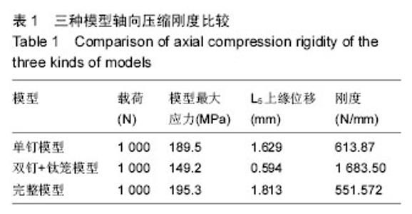
2.1 加载轴向载荷后的受力情况分析 在约束臼顶,在L2上缘施加1 000 N的轴向载荷后,完整模型及2种方法重建模型L5上缘参考点的位移,模型最大应力值,以及轴向压缩刚度如下表。在轴向压缩刚度方面:双钉+钛笼支撑模型>单钉模型>完整模型;L5上缘最大应力:完整模型>双钉+钛笼支撑组模型>单钉模型,见表1。"

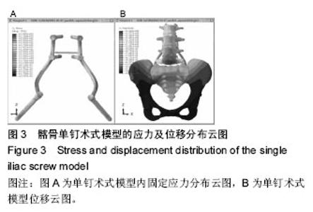
2.2 通过输出位移云图及应力云图分析2种手术方式模型的整体位移趋势及应力分布特点 从单钉模型应力图上来看应力集中在髂骨钉和椎弓根钉间的连接棒上,最大值为189.5 MPa。整体来看,应力主要集中在整个钉棒系统上,椎体前柱应力很小,也说明了钉棒系统起到了很好的支撑作用。钉棒系统的应力主要集中在钉棒连接处以及髂骨钉与椎弓根的连接棒上,此处容易疲劳断裂(图3)。"
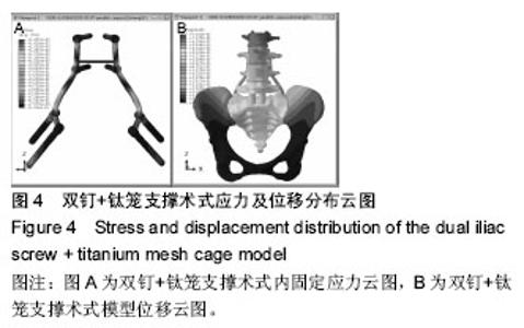
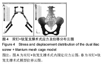
从双钉+钛笼支撑模型应力图上可以看到应力同样集中在钉棒连接处,应力最大值出现在右侧髂骨钉棒连接处,最大值为149.2 MPa,较单钉系统189.5 MPa明显减小。说明此种手术方式可以很有效的降低钉棒系统的应力。 从模型位移云图,可以看出位移最大值出现在剩余的L5椎体上,最大位移值为0.594 mm,较单钉系统1.629 mm明显减小,说明双钉+钛笼系统固定较单钉系统的压缩刚度为大,起到了很好的支撑作用(图4)。"
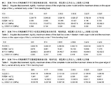
2.3 在加载弯曲载荷下模型的角位移及弯曲刚度 对两种模型在前屈,后伸,左侧屈,右侧屈,左旋转,右旋转情况施加7 N•m的弯曲载荷,记录各弯曲载荷下的角位移,计算弯曲刚度,记录模型的最大应力值及L5上缘最大应力值见表2-4。双钉+钛笼重建模型在前屈,后伸,左侧屈,右侧屈,左旋转,右旋转情况下的角位移和弯曲刚度,模型最大应力值及L5上缘最大应力值如表3。"


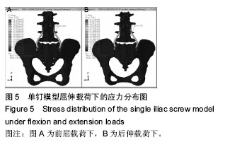
完整模型的角位移及弯曲刚度,模型最大应力,L5上缘最大应力值如表4。 在施加6个方向的7 N/m弯曲载荷下,在6个方向的弯曲刚度上,双钉+钛笼支撑模型>单钉模型>完整模型,说明双钉+钛笼支撑模型最稳定。模型的最大应力值比较,单钉模型>双钉+钛笼支撑模型>完整模型,说明单钉固定时,内固定钉棒系统承受的应力最大,在前屈时最大应力为113.013 MPa,后伸时最大应力值为113.677 MPa。说明单钉模型由于没有椎体前方的支撑,所以在前屈及后伸时不是很稳定,钉棒承受了很大的应力(图5)。"

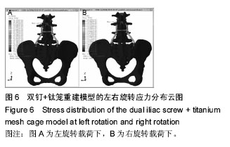
双钉+钛笼支撑模型最大刚度值是左右侧屈情况下,说明双钉+钛笼支撑有很好的侧方稳定性,模型最大应力是在旋转工况下,应力点也是在连接棒上,说明旋转载荷时,双钉+钛笼系统承受应力较大(图6)。"
| [1] Uvaraj NR, Bosco A, Gopinath NR. Global Reconstruction for Extensive Destruction in Tuberculosis of the Lumbar Spine and Lumbosacral Junction: A Case Report. Global Spine J. 2015;5(4):e17-e21. [2] Xu Z, Wang X, Shen X, et al. One-stage lumbopelvic fixation in the treatment of lumbosacral junction tuberculosis. Eur Spine J. 2015;24(8):1800-1805. [3] Varga PP, Szoverfi Z, Lazary A. Surgical resection and reconstruction after resection of tumors involving the sacropelvic region. Neurol Res. 2014;36(6):588-596. [4] Bederman SS, Shah KN, Hassan JM, et al.Surgical techniques for spinopelvic reconstruction following total sacrectomy: a systematic review. Eur Spine J. 2014;23(2):305-319. [5] Le VH, Heckmann N, Jain N, et al.Biomechanical evaluation of supplemental percutaneous lumbo-sacro-iliacscrewsfor spinopelvic fixation following total sacrectomy. J Spinal Disord Tech. 2015; 28(4):E181-185. [6] Guo LX, Wang ZW, Zhang YM, et al. Material property sensitivity analysis on resonant frequency characteristics of the human spine. J Appl Biomech. 2009;25(1):64-72. [7] Rohlmann A , Zander T, Schmidt H , et al. Analysis of the influence of disc degeneration on the mechanical behaviour of a lumbar motion segment using the finite element method. J Biomech. 2006; 39(13):2484-2490. [8] 于滨生,郑召民,庄新明,等.髂骨双钉在腰-髂重建结构中的生物力学优势[J].中华骨科杂志,2010,30(6):589-593. [9] 李全,张治宇,郑龙坡,等. 骶骨次全切除术后骨盆有限元模型的建立及验证[J].中国组织工程研究与临床康复, 2008,12(44):8649-8652. [10] Li JH, Zhang ZH, Shi T, et al. Surgical treatment of lumbosacral tuberculosis by one-stage debridement and anterior instrumentation with allograft through an extraperitoneal anterior approach. J Orthop Surg Res. 2015;10:62. [11] 高延征,余正红,高坤,等.腰骶结核不同手术方式的选择及疗效分析[J].中华骨科杂志,2014,34(2):143-148. [12] Varga PP, Szövérfi Z, Lazary A. Surgical treatment of primary malignant tumors of thesacrum. Neurol Res. 2014;36(6):577-587. [13] Zang J, Guo W, Yang R, et al. Is total en bloc sacrectomy using a posterior-only approach feasible and safe for patients with malignant sacral tumors? J Neurosurg Spine. 2015;22(6):563-570. [14] Phukan R, Herzog T, Boland PJ,et al.How Does the Level of Sacral Resection for Primary Malignant Bone Tumors Affect Physical and Mental Health, Pain, Mobility, Incontinence, and Sexual Function? Clin Orthop Relat Res.2016; (3): 687-696. [15] Ooi A, Foo L, Tan BK, et al. Massive sacral chordoma resection and reconstruction with a combination of pedicled and free flaps. J Reconstr Microsurg. 2015; 31(1): 76-78. [16] KayaniB , Hanna SA , Sewell MD, et al.A review of the surgical management of sacral chordoma. Eur J Surg Oncol. 2014;40(11): 1412-1420. [17] Dudda M, Hoffmann M, Schildhauer TA. Sacrum fractures and lumbopelvic instabilities in pelvic ring injuries: classification and biomechanical aspects. Unfallchirurg. 2013;116(11):972-978. [18] Wangtaphan W, Oo M, Paholpak P, et al.Traumatic lumbosacral spondyloptosis treated five months after injury occurrence: a case report. Spine. 2012;37(22): E1410-1414. [19] Sullivan MP, Smith HE, Schuster JM, et al. Spondylopelvic dissociation.Orthop Clin North Am. 2014;45(1):65-75. [20] Dalbayrak S, Yaman O, Ayten M, et al.Surgical treatment in sacral fractures and traumatic spinopelvic instabilities. Turk Neurosurg. 2014;24(4):498-505. [21] Schroeder GD, Kepler CK, Mba MD, et al. Axial interbody arthrodesis of the L5-S1 segment: a systematic review of the literature. J Neurosurg Spine. 2015;23(3):314-319. [22] Whang PG, Sasso RC, Patel VV, et al.Comparison of axial and anterior interbody fusions of the L5-S1 segment: a retrospective cohort analysis.J Spinal Disord Tech. 2013;26(8):437-443. [23] Mobbs RJ, Loganathan A, Yeung V, et al. Indications for anterior lumbar interbody fusion. Orthop Surg. 2013; 5(3):153-163. [24] Yoshihara H. Surgical options for lumbosacral fusion: biomechanical stability, advantage, disadvantage and affecting factors in selecting options. Eur J Orthop Surg Traumatol. 2014;24Suppl 1:S73-82. [25] Roetman B, Schildhauer TA. Lumbopelvic stabilization for bilateral lumbosacral instabilities: indications and techniques. Unfallchirurg. 2013; 116(11):991-999. [26] Zeng ZY, Zhang JQ, Song YX, et al. Combination of percutaneous unilateral translaminar facet screw fixation and interbody fusion for treatment of lower lumbar vertebra diseases: a follow-up study. Orthop Surg. 2014; 6(2):110-117. [27] Pola E, Rossi B, Nasto LA, et al.Surgical treatment of tuberculous spondylodiscitis. Eur Rev Med Pharmacol Sci. 2012;16 Suppl 2:79-85. [28] Bederman SS, Hassan JM, Shah KN, et al.Fixation techniques for complex traumatic transverse sacral fractures: a systematic review.Spine.2013;38(16): E1028-1040. [29] Kato M, Taneichi H, Suda K. Advantage of Pedicle Screw Placement Into the Sacral Promontory (Tricortical Purchase) on Lumbosacral Fixation. J Spinal Disord Tech. 2015;28(6):E336-342. [30] Kim JH, Horton W, Hamasaki T, et al.Spinal instrumentation for sacral-pelvic fixation: a biomechanical comparison between constructs ending with either S2 bicortical, bitriangulated screws or iliac screws.J Spinal Disord Tech. 2010;23(8):506-512. [31] Néron JB, Gadéa F, Fournier J, et al.Lumbosacral arthrodesis for neuromuscular scoliosis using a simplified Jackson technique.Orthop Traumatol Surg Res. 2013;99(7):845-851. [32] Zwingmann J, Hauschild O, Bode G, et al. Malposition and revision rates of different imaging modalities for percutaneous iliosacral screw fixation following pelvic fractures: a systematic review and meta-analysis.Arch Orthop Trauma Surg. 2013;133(9):1257-1266. [33] Ilyas H, Place H, Puryear A. A Comparison of Early Clinical and Radiographic Complications of Iliac Screw Fixation Versus S2Alar Iliac (S2AI) Fixation in the Adult and Pediatric Populations.J Spinal Disord Tech. 2015; 28(4):E199-205. [34] Baek SW, Park YS, Ha KY, et al. The analysis of spinopelvic parameters and stability following long fusions with S1, S2 or iliacfixation. Int Orthop. 2013; 37(10):1973-1980. [35] Park SA, Kwak DS, You SL. Entry zone ofiliac screwfixation to maintain proper entry width andscrewlength. Eur Spine J. 2015;24 (11): 2573-2579. [36] Fridley J, Fahim D, Navarro J, et al. Free-hand placement of iliac screws for spinopelvic fixation based on anatomical landmarks: technical note. Int J Spine Surg. 2014;8. doi: 10.14444/1003. eCollection 2014. [37] Santos ER, Sembrano JN, Mueller B, et al. Optimizing iliac screw fixation: a biomechanical study on screw length, trajectory, and diameter. J Neurosurg Spine. 2011;14(2):219–225. [38] Wang T, Liu H, Zheng Z, et al.Biomechanical effect of 4-rod technique on lumbosacral fixation: an in vitro human cadaveric investigation.Spine. 2013;38(15): E925-929. [39] Amaritsakul Y, Chao CK, Lin J. Multiobjective optimization design of spinal pedicle screws using neural networks and genetic algorithm: mathematical models and mechanical validation. Comput Math Methods Med. 2013;2013:462875. [40] Zhao Y, Zhang S, Sun T, et al. Mechanical comparison between lengthened and short sacroiliac screws in sacral fracture fixation: afinite element analysis. Orthop TraumatolSurg Res. 2013;99(5):601-606. |
| [1] | Shi Bin, An Jing, Chen Long-gang, Zhang Nan, Tian Ye . Influencing factors for pain after total knee arthroplasty [J]. Chinese Journal of Tissue Engineering Research, 2017, 21(7): 993-997. |
| [2] | Wang Xian-xun. Impact of local compression cryotherapy combined with continuous passive motion on the early functional recovery after total knee arthroplasty [J]. Chinese Journal of Tissue Engineering Research, 2017, 21(7): 998-1003. |
| [3] | Yuan Wei, Zhao Hui, Ding Zhe-ru, Wu Yu-li, Wu Hai-shan, Qian Qi-rong. Association between psychological resilience and acute mental disorders after total knee arthroplasty [J]. Chinese Journal of Tissue Engineering Research, 2017, 21(7): 1015-1019. |
| [4] | Chen Qun-qun, Qiao Rong-qin, Duan Rui-qi, Hu Nian-hong, Li Zhao, Shao Min. Acu-Loc®2 volar distal radius bone plate system for repairing type C fracture of distal radius [J]. Chinese Journal of Tissue Engineering Research, 2017, 21(7): 1025-1030. |
| [5] | Huang Xiang-wang, Liu Hong-zhe. A new low elastic modulus of beta titanium alloy Ti2448 spinal pedicle screw fixation affects thoracic stability: biomechanical analysis [J]. Chinese Journal of Tissue Engineering Research, 2017, 21(7): 1031-1035. |
| [6] | Xie Qiang. Three-dimensional finite element model for biomechanical analysis of stress in knee inversion and external rotation after posterior cruciate ligament rupture [J]. Chinese Journal of Tissue Engineering Research, 2017, 21(7): 1036-1040. |
| [7] | He Ze-dong, Zhao Jing, Chen Liang-yu, Li Ke, Weng Jie. Multilevel finite element analysis on the biological tribology damage of water on bone tissue [J]. Chinese Journal of Tissue Engineering Research, 2017, 21(7): 1041-1045. |
| [8] | Jiang Zi-wei, Huang Feng, Cheng Si-yuan, Zheng Xiao-hui, Sun Shi-dong, Zhao Jing-tao, Cong Hai-chen,Sun Han-qiao, Dong Hang. Design and finite element analysis of digital splint [J]. Chinese Journal of Tissue Engineering Research, 2017, 21(7): 1052-1056. |
| [9] | Wang Fei, Liu Zhi-bin, Tao Hui-ren, Zhang Jian-hua, Li Chang-hong, Cao Qiang, Zheng Jun, Liu Yan-xiong, Qu Xiao-peng. Clinical efficacy of preoperative osteotomy designs using paper-cut technology versus photoshop software for ankylosing spondylitis with kyphosis [J]. Chinese Journal of Tissue Engineering Research, 2017, 21(7): 1057-1063. |
| [10] | Li Hui, Ma Jun-yi, Ma Yuan, Zhu Xu . Establishment of a three-dimensional finite element model of ankylosing spondylitis kyphosis [J]. Chinese Journal of Tissue Engineering Research, 2017, 21(7): 1069-1073. |
| [11] | Ling Guan-han, Ou Zhi-xue, Yao Lan, Wen Li-chun, Wang Guo-xiang, Lin Heng-feng. Establishment of simulating three-dimensional model of China-Japan Friendship Hospital Classification for L type osteonecrosis of the femoral head [J]. Chinese Journal of Tissue Engineering Research, 2017, 21(7): 1074-1079. |
| [12] | Fu Wei-min, Wang Ben-jie. Assessing the degree of necrotic femoral head, and association of blood supply with pathlogical changes: study protocol for a diagnostic animal trial [J]. Chinese Journal of Tissue Engineering Research, 2017, 21(7): 1086-1091. |
| [13] | Zhang Wen-qiang, Ding Qian, Zhang Na. Associations between alpha angle and herniation pit on oblique axial magnetic resonance imaging in asymptomatic hip joints of adults [J]. Chinese Journal of Tissue Engineering Research, 2017, 21(7): 1098-1103. |
| [14] | Sun Xiao-xin1, Zhou Wei2, Zuo Shu-ping3, Liu Hao1, Song Jing-feng1, Liang Chun-yu1. Morphological characteristics for the magnetic resonance imaging assessment of discoid lateral meniscal tears in children [J]. Chinese Journal of Tissue Engineering Research, 2017, 21(7): 1104-1109. |
| [15] | Lin Han-wen, Wen Jun-mao, Huang Chao-yuan, Zhou Chi, Tang Hong-yu. Correlation between the changes in lower limb power line and pain area in the knee osteoarthritis patients: imaging evaluation [J]. Chinese Journal of Tissue Engineering Research, 2017, 21(7): 1110-1114. |
| Viewed | ||||||
|
Full text |
|
|||||
|
Abstract |
|
|||||