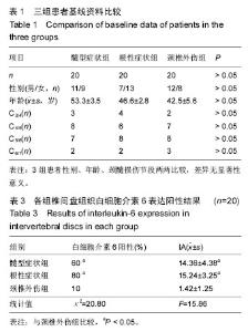
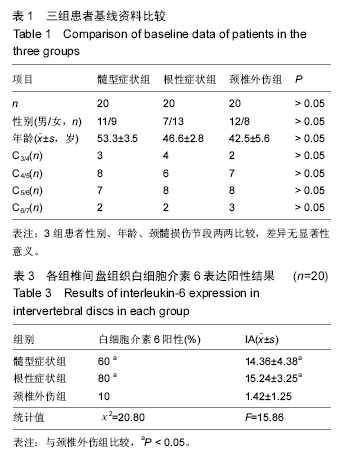
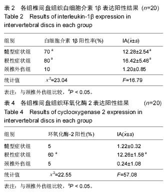
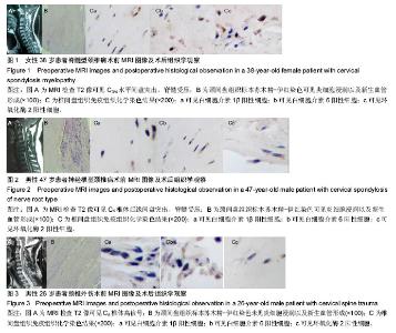
| [1] Smith M, Davis MA, Stano M, et al.Aging baby boomers and the rising cost of chronic back pain: Secular trend analysis of longitudinal Medical Expenditures Panel Survey data for years 2000 to 2007.J Manipulative Physiol Ther.2013;36(1):2-11.
[2] Son S, Kim WK,Lee SG, et al.A comparison of the clinical outcomes of decompression alone and fusion in elderly patients with two-level or more lumbar spinal stenosis. J Korean Neurosurg Soc.2013;53(1): 19-25.
[3] 李强,赵建民,张元智,等.腰椎退变黄韧带中炎性细胞因子的表达[J].中国脊柱脊髓杂志,2011,21(1):63-66.
[4] Takahashi N, Kikuchi S, Shubayev VI, et al. TNF-alpha and phosphorylation of ERK in DRG and spinal cord: insights into mechanisms of sciatica. Spine. 2006;31(5): 523-529.
[5] 梁逸仙,周光辉,赵丽,等.电针联合COX-2抑制剂治疗腰椎间盘突出症的临床疗效及对IL-6和IL-8表达的影响[J]. 微循环学杂志,2015,20(2):61-63.
[6] Kaymaz M,Borcek AO,Emmez H,et al.Effectiveness of single posterior decompressive laminectomy in symptomatic lumbar spinal stenosis: a retrospective study. Turk Neurosurg. 2012;22(4):430-434.
[7] 王靖,史立君,滕红林,等.兔椎间盘生理屏障损伤后髓核组织辅助性T细胞1/辅助性T细胞2平衡漂移状态的研究[J].中华实验外科杂志,2016,33(03):727-729.
[8] 杨剑,康建平,冯大雄,等.IL-1和IL-6的表达增强且与盘源性下腰痛改良日本骨科学会(mJOA)评分负相关[J].细胞与分子免疫学杂志,2016,32(01):88-91.
[9] 苏世先,刘前前,江美林.脑脊液及血清疼痛物质相关指标与腰椎间盘突出的关系研究[J].临床和实验医学杂志,2016, (03):232-234.
[10] 姜世峰,申才良.腰椎Modic改变中炎性细胞因子的效应[J].中国组织工程研究,2012,16(17):3213-3217.
[11] Ozaktay AC, Kallakuri S, Takebayashi T,et al. Effects of interleukin-1 beta, interleukin-6, and tumor necrosis factor on sensitivity of dorsal root ganglion and peripheral receptive fields in rats. Eur Spine J. 2006; 15(10):1529-1537.
[12] 孙中仪,田纪伟.炎性因子与椎间盘退变的研究进展[J].中华医学杂志,2013,93(41):3322-3324.
[13] 张威.多种炎性因子在椎间盘突出的炎症机制中的作用研究进展[J].医学综述,2013,19(20):3685-3688.
[14] 宋艳照,邓小柱.腰椎间盘髓核组织中TNF-α、IL-1、IL-6的检测及意义[J].山东医药,2009,49(50):87-88.
[15] William CW, Jamie B, Thomas JG, et al. Degenerative lumbar spinal stenosis : an evidence-based clinical guide line for the diagnosis and treatment of degenerative lumbar spinalstenosis. Spine. 2008;2: 305-310.
[16] 钱胜君,张宁,陈维善.炎症因子与椎间盘退变[J].国际骨科学杂志,2009,30(4):247-248,265.
[17] 刘继巍,项玉萍.腰椎间盘突出类型与其疼痛程度相关性的临床观察[J].中国实用医刊,2016,43(6):126-127.
[18] 张琰,孟阳,赵卫东,等.腰椎管内静脉血清中炎性因子与腰椎管狭窄的关系[J].中国组织工程研究,2014,18(26): 4229-4235.
[19] Nakamura T, Okada T, Endo M, et al. Angiopoietin-like protein 2 induced by mechanical stress accelerates degeneration and hypertrophy of the ligamentum flavum in lumbar spinal canal stenosis. PLoS One. 2014;9(1):e85542.
[20] 李威,唐勇,刘天懿,等. IL-10和TGF-β介导的生物学治疗犬椎间盘退变的体外实验研究[J]。中国骨与关节杂志, 2015, 4(3):230-234.
[21] 杨允学,刘飞,刘超.白细胞介素1β对人胶质细胞瘤细胞内环氧合酶-2系统的调节机制[J].中华医学杂志, 2015,95(9): 697-700.
[22] 夏婷,李双庆.炎症相关骨质疏松症的发病机制[J].中国骨质疏松杂志,2015,(01):117-120.
[23] 何主强,殷莉,黄姗姗.环氧化酶-2及细胞因子在创伤后应激障碍患者中的表达变化及意义[J].实用医学杂志, 2015, (19): 3166-3168.
[24] Podichetty VK. The aging spine: the role of inflammatory mediators in intervertebral disc degeneration. Cell Mol Biol (Noisy-le-grand). 2007;53(5):4-18.
[25] Gronbald M, Virri J, Tolonen J, et al. A Controlled immunohistochemical study of inflammatory cells in disc herniation tissue. Spine. 1994;19:2744.
[26] Gruber-Schoffnegger D, Drdla-Schutting R, Hönigsperger C,et al. Induction of thermal hyperalgesia and synaptic long-term potentiation in the spinal cord lamina I by TNF-αand IL-1β is mediated by glial cells. J Neurosci. 2013;33(15):6540-6551.
[27] 赵洪宇,蒋梦会,曹久妹,等.IL-6/STAT3通路对THP-1单核细胞环氧化酶2表达的影响[J].中国动脉硬化杂志,2016, 24(3):251-255,260
[28] Kang JD, Stefanovic Racic M, McIntyre LA, et al. Toward abiochemical understanding of human intervertebral disc degenera tion and herniation. Contributions of nitric oxide, in ter leukins, prostaglandi n E2, and matrix metalloproteinases. Spine,2012, 22(10):1065-1073. |