Chinese Journal of Tissue Engineering Research ›› 2016, Vol. 20 ›› Issue (35): 5237-5243.doi: 10.3969/j.issn.2095-4344.2016.35.010
Previous Articles Next Articles
Finite element analysis of distal femoral locking plate and minimally invasive internal fixation system in different motion states
Hao Ting1, Wang Xing-guo1, Li Xiao-he2
- 1Department of Orthopedic Trauma, Second Affiliated Hospital of Inner Mongolia Medical University, Hohhot 010059, Inner Mongolia Autonomous Region, China; 2Department of Human Anatomy, College of Basic Medical Sciences, Inner Mongolia Medical University, Hohhot 010059, Inner Mongolia Autonomous Region, China
-
Revised:2016-06-16Online:2016-08-26Published:2016-08-26 -
Contact:Wang Xing-guo, Chief physician, Department of Orthopedic Trauma, Second Affiliated Hospital of Inner Mongolia Medical University, Hohhot 010059, Inner Mongolia Autonomous Region, China -
About author:Hao Ting, M.D., Associate chief physician, Department of Orthopedic Trauma, Second Affiliated Hospital of Inner Mongolia Medical University, Hohhot 010059, Inner Mongolia Autonomous Region, China -
Supported by:the National Natural Science Foundation of China, No. 81460330; the Natural Science Foundation of Inner Mongolia Autonomous Region, No. 2013MS11107
CLC Number:
Cite this article
Hao Ting, Wang Xing-guo, Li Xiao-he. Finite element analysis of distal femoral locking plate and minimally invasive internal fixation system in different motion states[J]. Chinese Journal of Tissue Engineering Research, 2016, 20(35): 5237-5243.
share this article

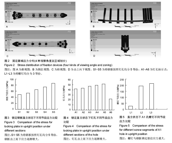
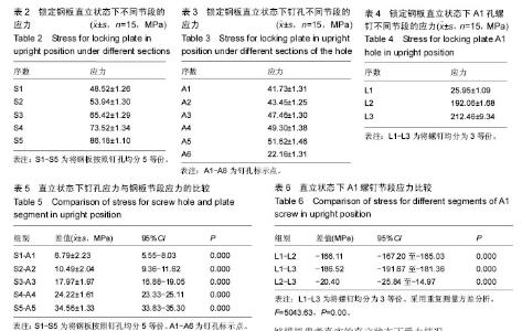
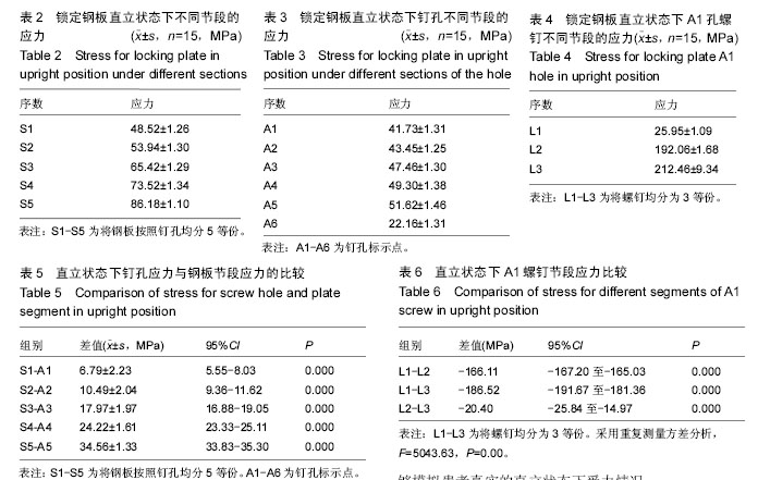
2.1 股骨远端骨折锁定钢板固定有限元模型 利用三维重建软件Mimics 16.01和有限元软件Ansys成功建立股骨远端骨折锁定钢板固定有限元模型。共有43 536个单元,41 256个节点,建成后的三维有限元模型与实体组织有较好的几何相似性。 2.2 股骨远端骨折锁定钢板固定有限元模型有限元模型的验证 试验的模型是根据Shih等[8]已通过验证的建模方法而建立的,所以本模型符合要求。经与以往试验模型加载相同应力后,其应力分布一致。 2.3 股骨远端骨折锁定钢板固定有限元模型有限元不同部位应力 随着钢板节段逐渐向下(S1-S5),其应力逐渐增大,应力与节段序数Spearman相关系数为0.966,P < 0.001(表2,图3);A1-A5随着螺钉序数增加,其应力逐渐增大,应力与节段序数Spearman相关系数为0.895,P < 0.001,但A6却突然减小(表3,图4)。由应力分布云图可见(图2),除了A1钉孔螺钉各节段应力分布不均外,其余均分布均匀,A1螺钉3个节段间由重复测量方差分析结果可知,3个节段间差异均有显著性意义,由螺钉尖到末端,其应力逐渐增大,应力与节段序数Spearman相关系数为0.913,P < 0.001(表4,图5)。 相应节段间钉孔与钢板应力比较,钉孔周围应力与钢板节段应力间差异均有显著性意义,且钢板应力大于相应节段钉孔应力(表5,6)。"
"
| [1] Höntzsch D, Blauth M, Attal R,et al.Angle-stable fixation of intramedullary nails using the Angular Stable Locking System® (ASLS). Oper Orthop Traumatol. 2011;23(5):387-396. [2] Ehlinger M, Adam P, Arlettaz Y,et al. Minimally-invasive fixation of distal extra-articular femur fractures with locking plates: limitations and failures. Orthop Traumatol Surg Res. 2011;97(6):668-674.16:28 2016-9-6 [3] Nayak RM, Koichade MR, Umre AN,et al.Minimally invasive plate osteosynthesis using a locking compression plate for distal femoral fractures. J Orthop Surg (Hong Kong).2011;19(2):185-190. [4] Malviya A, Reed MR, Partington PF. Acute primary total knee arthroplasty for peri-articular knee fractures in patients over 65 years of age.Injury.2011;42(11):1368-1371. [5] Lopes JB, Figueiredo CP, Caparbo VF,et al. Osteoporotic fractures in the Brazilian community- dwelling elderly: prevalence and risk factors. J Clin Densitom. 2011;14(3):359-366. [6] [6] Weil YA, Rivkin G, Safran O,et al. The outcome of surgically treated femur fractures associated with long-term bisphosphonate use. J Trauma. 2011;71(1): 186-190. [7] Hierholzer C, von Rüden C, Pötzel T, et al. Outcome analysis of retrograde nailing and less invasive stabilization system in distal femoral fractures: A retrospective analysis. Indian J Orthop. 2011;45(3): 243-250. [8] Shih KS, Hsu CC, Hsu TP. A biomechanical investigation of the effects of static fixation and dynamization after interlocking femoral nailing: A finite element study. J Trauma. 2012;72(2):E46-E53. [9] Salas C, Mercer D, DeCoster TA, et al. Experimental and probabilistic analysis of distal femoral periprosthetic fracture: a comparison of locking plate and intramedullary nail fixation. Part A: experimental investigation. Comput Methods Biomech Biomed Engin. 2011;14(2):157-164. [10] Han QT, Wang YJ, Tang HW. Biomechanical comparison of three methods of internal fixation for distal femoral fractures. Zhongguo Gu Shang. 2010; 23(8):601-604. [11] Efstathopoulos N, Nikolaou VS, Xypnitos FN, et al.Investigation on the distal screw of a trochanteric intramedullary implant (Fi-nail) using a simplified finite element model. Injury. 2010;41(3):259-265. [12] Chang CY, Rupp JD, Reed MP, et al.Predicting the effects of muscle activation on knee, thigh, and hip injuries in frontal crashes using a finite-element model with muscle forces from subject testing and musculoskeletal modeling. Stapp Car Crash J. 2009; 53:291-328. [13] Bougherara H, Zdero R, Miric M, et al.The biomechanics of the T2 femoral nailing system: a comparison of synthetic femurs withfinite element analysis. Proc Inst Mech Eng H. 2009;223(3):303-314. [14] Chen SH, Yu TC, Chang CH, et al. Biomechanical analysis of retrograde intramedullary nail fixation in distal femoral fractures. Knee. 2008;15(5):384-389. [15] Mann KA, Lee J, Arrington SA, et al.Predicting distal femur bone strength in a murine model of tumor osteolysis. Clin Orthop Relat Res. 2008;466(6): 1271-1278. [16] Shih KS, Tseng CS, Lee CC, et al.Influence of muscular contractions on the stress analysis of distal femoral interlocking nailing. Clin Biomech(Bristol, Avon). 2008;23(1):38-44. [17] Mahaisavariya B, Sitthiseripratip K, Suwanprateeb J. Finite element study of the proximal femur with retained trochanteric gamma nail and after removal of nail. Injury. 2006;37(8):778-785. [18] Cegoñino J, García Aznar JM, Doblaré M, et al. A comparative analysis of different treatments for distal femur fractures using the finite element method. Comput Methods Biomech Biomed Engin. 2004;7(5): 245-256. [19] Dubov A, Kim SY, Shah S, et al.The biomechanics of plate repair of periprosthetic femur fractures near the tip of a total hip implant: the effect of cable-screw position. Proc Inst Mech Eng H. 2011;225(9):857-865. [20] Shah S, Kim SY, Dubov A, et al.The biomechanics of plate fixation of periprosthetic femoral fractures near the tip of a total hip implant: cables, screws, or both? Proc Inst Mech Eng H. 2011;225(9):845-856. [21] Wilson LJ, Richards CJ, Irvine D, et al. Irvine D,Risk of periprosthetic femur fracture after anterior cortical bone windowing: a mechanical analysis of short versus long cemented stems in pigs. Acta Orthop. 2011;82(6): 674-678. [22] Chen SH, Tai CL, Yu TC, et al. Modified fixations for distal femur fractures following total knee arthroplasty: a biomechanical and clinical relevance study. Knee Surg Sports Traumatol Arthrosc. 2016. [Epub ahead of print] [23] Filardi V.The healing stages of an intramedullary implanted tibia: A stress strain comparative analysis of the calcification process.J Orthop. 2015;12(Suppl 1):S51-61. [24] Yoshimoto K, Nakashima Y, Nakamura A, et al. Neck fracture of femoral stems with a sharp slot at the neck: biomechanical analysis.J Orthop Sci. 2015;20(5):881-887. [25] Li L, Yu M, Ma R, et al.Initial Stability of Subtrochanteric Oblique Osteotomy in Uncemented Total Hip Arthroplasty: A Preliminary Finite Element Study. Med Sci Monit. 2015;21:1969-1975. [26] Märdian S, Schaser KD, Duda GN, et al. Working length of locking plates determines interfragmentary movement in distal femur fracturesunder physiological loading. Clin Biomech (Bristol, Avon). 2015;30(4): 391-396. [27] Zhang J, Ebraheim N, Li M, et al.External fixation using locking plate in distal tibial fracture: a finite element analysis.Eur J Orthop Surg Traumatol. 2015;25(6): 1099-1104. [28] Leonidou A, Moazen M, Lepetsos P, et al. The biomechanical effect of bone quality and fracture topography on locking plate fixation in periprosthetic femoral fractures. Injury. 2015;46(2):213-217. [29] Mahaisavariya B, Chantarapanich N, Riansuwan K, et al. Prevention of excessive medialisation of trochanteric fracture by a buttress screw: a novel methodand finite element analysis. J Med Assoc Thai. 2014;97 Suppl 9:S127-132. [30] Martelli S, Pivonka P, Ebeling PR. Femoral shaft strains during daily activities: Implications for atypical femoral fractures. Clin Biomech (Bristol, Avon). 2014; 29(8):869-876. [31] Tan CO, Battaglino RA, Doherty AL, et al.Adiponectin is associated with bone strength and fracture history in paralyzed men with spinal cord injury.Osteoporos Int. 2014;25(11):2599-2607. [32] Sun ZH, Liu YJ, Li H. Femoral stress and strain changes post-hip, -knee and -ipsilateral hip/knee arthroplasties: a finiteelement analysis. Orthop Surg. 2014;6(2):137-144. [33] Tran TN, Warwas S, Haversath M, et al.Experimental and computational studies on the femoral fracture risk for advanced core decompression.Clin Biomech (Bristol, Avon). 2014;29(4):412-417. [34] Kimura G, Onishi E, Kawanabe K, et al.Characteristic loosening of the distally straight cylindrical femoral stem. J Orthop Sci. 2014;19(3):437-442. [35] Shih KS, Hsu CC, Hsu TP, et al. Biomechanical analyses of static and dynamic fixation techniques of retrograde interlocking femoralnailing using nonlinear finite element methods.Comput Methods Programs Biomed. 2014;113(2):456-464. [36] Edwards WB, Schnitzer TJ, Troy KL.Bone mineral and stiffness loss at the distal femur and proximal tibia in acute spinal cord injury.Osteoporos Int. 2014;25(3): 1005-1015. [37] Erhardt JB, Khoo PP, Stoffel KK, et al.Periprosthetic fractures around polished collarless cemented stems: the effect of stem design on fracture pattern.Hip Int. 2013;23(5):459-464. [38] Chen SH, Chiang MC, Hung CH, et al.Finite element comparison of retrograde intramedullary nailing and locking plate fixation with/without an intramedullary allograft for distal femur fracture following total knee arthroplasty.Knee. 2014;21(1):224-231. |
| [1] | Yuan Wei, Zhao Hui, Ding Zhe-ru, Wu Yu-li, Wu Hai-shan, Qian Qi-rong. Association between psychological resilience and acute mental disorders after total knee arthroplasty [J]. Chinese Journal of Tissue Engineering Research, 2017, 21(7): 1015-1019. |
| [2] | Chen Qun-qun, Qiao Rong-qin, Duan Rui-qi, Hu Nian-hong, Li Zhao, Shao Min. Acu-Loc®2 volar distal radius bone plate system for repairing type C fracture of distal radius [J]. Chinese Journal of Tissue Engineering Research, 2017, 21(7): 1025-1030. |
| [3] | Huang Xiang-wang, Liu Hong-zhe. A new low elastic modulus of beta titanium alloy Ti2448 spinal pedicle screw fixation affects thoracic stability: biomechanical analysis [J]. Chinese Journal of Tissue Engineering Research, 2017, 21(7): 1031-1035. |
| [4] | Xie Qiang. Three-dimensional finite element model for biomechanical analysis of stress in knee inversion and external rotation after posterior cruciate ligament rupture [J]. Chinese Journal of Tissue Engineering Research, 2017, 21(7): 1036-1040. |
| [5] | He Ze-dong, Zhao Jing, Chen Liang-yu, Li Ke, Weng Jie. Multilevel finite element analysis on the biological tribology damage of water on bone tissue [J]. Chinese Journal of Tissue Engineering Research, 2017, 21(7): 1041-1045. |
| [6] | Jiang Zi-wei, Huang Feng, Cheng Si-yuan, Zheng Xiao-hui, Sun Shi-dong, Zhao Jing-tao, Cong Hai-chen,Sun Han-qiao, Dong Hang. Design and finite element analysis of digital splint [J]. Chinese Journal of Tissue Engineering Research, 2017, 21(7): 1052-1056. |
| [7] | Wang Fei, Liu Zhi-bin, Tao Hui-ren, Zhang Jian-hua, Li Chang-hong, Cao Qiang, Zheng Jun, Liu Yan-xiong, Qu Xiao-peng. Clinical efficacy of preoperative osteotomy designs using paper-cut technology versus photoshop software for ankylosing spondylitis with kyphosis [J]. Chinese Journal of Tissue Engineering Research, 2017, 21(7): 1057-1063. |
| [8] | Li Hui, Ma Jun-yi, Ma Yuan, Zhu Xu . Establishment of a three-dimensional finite element model of ankylosing spondylitis kyphosis [J]. Chinese Journal of Tissue Engineering Research, 2017, 21(7): 1069-1073. |
| [9] | Ling Guan-han, Ou Zhi-xue, Yao Lan, Wen Li-chun, Wang Guo-xiang, Lin Heng-feng. Establishment of simulating three-dimensional model of China-Japan Friendship Hospital Classification for L type osteonecrosis of the femoral head [J]. Chinese Journal of Tissue Engineering Research, 2017, 21(7): 1074-1079. |
| [10] | Fu Wei-min, Wang Ben-jie. Assessing the degree of necrotic femoral head, and association of blood supply with pathlogical changes: study protocol for a diagnostic animal trial [J]. Chinese Journal of Tissue Engineering Research, 2017, 21(7): 1086-1091. |
| [11] | Zhang Wen-qiang, Ding Qian, Zhang Na. Associations between alpha angle and herniation pit on oblique axial magnetic resonance imaging in asymptomatic hip joints of adults [J]. Chinese Journal of Tissue Engineering Research, 2017, 21(7): 1098-1103. |
| [12] | Sun Xiao-xin1, Zhou Wei2, Zuo Shu-ping3, Liu Hao1, Song Jing-feng1, Liang Chun-yu1. Morphological characteristics for the magnetic resonance imaging assessment of discoid lateral meniscal tears in children [J]. Chinese Journal of Tissue Engineering Research, 2017, 21(7): 1104-1109. |
| [13] | Lin Han-wen, Wen Jun-mao, Huang Chao-yuan, Zhou Chi, Tang Hong-yu. Correlation between the changes in lower limb power line and pain area in the knee osteoarthritis patients: imaging evaluation [J]. Chinese Journal of Tissue Engineering Research, 2017, 21(7): 1110-1114. |
| [14] | Zhang Yun-ge, Song Ke-guan. Periprosthetic osteolysis induced by wear particles: research progress of calcineurin/activated T cell nuclear factor signaling pathway [J]. Chinese Journal of Tissue Engineering Research, 2017, 21(7): 1115-1122. |
| [15] | Liu Wei, Huang Jian. Applied research and progress of three-dimensional printing technology in joint replacement [J]. Chinese Journal of Tissue Engineering Research, 2017, 21(7): 1123-1130. |
| Viewed | ||||||
|
Full text |
|
|||||
|
Abstract |
|
|||||