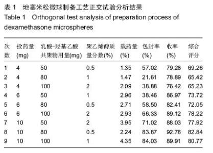
| [1] Tabesh H, Amoabediny G, Nik NS, et al. The role of biodegradable engineered scaffolds seeded with Schwann cells for spinal cord regeneration. Neurochem Int. 2009; 54(2):73-83.
[2] 王涛丽,顾兵,李华南,等.急性脊髓损伤后的炎症反应及其抗炎治疗[J].中国药理学通报,2015,(4):452-457.
[3] 冯云枝,凌天牖,吴汉江,等.地塞米松对成纤维细胞增殖及表达细胞间粘附分子-1的影响[J].中国免疫学杂志,2002,18(6): 423-425.
[4] 许剑荣,郑学毅,唐绍生,等.地塞米松对瘢痕疙瘩成纤维细胞基质金属蛋白酶1调控的研究[J].皮肤性病诊疗学杂志,2008,15(6): 310-313.
[5] Jain S, Datta M. Oral extended release of dexamethasone: Montmorillonite–PLGA nanocomposites as a delivery vehicle. Appl Clay Sci. 2015;104:182-188.
[6] Donzelli E, Salvadè A, Mimo P, et al. Mesenchymal stem cells cultured on a collagen scaffold: In vitro osteogenic differentiation. Arch Oral Biol. 200;52(1):64-73.
[7] Shen YH, Shoichet MS, Radisic M. Vascular endothelial growth factor immobilized in collagen scaffold promotes penetration and proliferation of endothelial cells.Acta Biomater. 2008;4(3):477-489.
[8] Zhong S, Teo WE, Zhu X, et al. An aligned nanofibrous collagen scaffold by electrospinning and its effects on in vitro fibroblast culture. J Biomed Mater Res A. 2006;79(3): 456-463.
[9] Bosnakovski D, Mizuno M, Kim G, et al. Chondrogenic differentiation of bovine bone marrow mesenchymal stem cells (MSCs) in different hydrogels: influence of collagen type II extracellular matrix on MSC chondrogenesis. Biotechnol Bioeng. 2006;93(6):1152-1163.
[10] Rhee SH, Choi JY, Kim HM. Preparation of a bioactive and degradable poly(epsilon -caprolactone)/silica hybrid through a sol-gel method.Biomaterials. 2002;23(24):4915-4921.
[11] 孙彬.新型静电纺丝技术制备功能微纳米纤维及其应用[D].青岛:青岛大学,2014.
[12] 郭廓.真空冷冻干燥技术在生物制药方面的应用[J].生物技术世界,2015,(3):58-60.
[13] 黄小舟,成晓岚,罗宇燕,等.初乳搅拌速率对复乳法制备载蛋白微球性质的影响[J].中山大学学报:自然科学版,2015,3(3): 119-124.
[14] 赵莉,何晨光,高永娟,等.PLGA的不同组成对支架材料性能的影响研究[J].中国生物工程杂志,2008,28(5):22-28.
[15] Wang J, Wang J, Lu P, et al. Local delivery of FTY720 in PCL membrane improves SCI functional recovery by reducing reactive astrogliosis. Biomaterials. 2015;62:76-87.
[16] Altinova H, Möllers S, Führmann T, et al. Functional improvement following implantation of a microstructured, type-I collagen scaffold into experimental injuries of the adult rat spinal cord. Brain Res. 2014;1585:37-50.
[17] Pawar K, Cummings BJ, Thomas A, et al. Biomaterial bridges enable regeneration and re-entry of corticospinal tract axons into the caudal spinal cord after SCI: Association with recovery of forelimb function. Biomaterials. 2015;65:1-12.
[18] Madigan NN, McMahon S, O'Brien T, et al. Current tissue engineering and novel therapeutic approaches to axonal regeneration following spinal cord injury using polymer scaffolds. Respir Physiol Neurobiol. 2009;169(2):183-199.
[19] Shi Q, Gao W, Han X, et al. Collagen scaffolds modified with collagen-binding bFGF promotes the neural regeneration in a rat hemisected spinal cord injury model. Sci China Life Sci. 2014;57(2):232-240.
[20] Ghasemi-Mobarakeh L, Prabhakaran MP, Morshed M, et al. Electrospun poly(epsilon-caprolactone)/gelatin nanofibrous scaffolds for nerve tissue engineering. Biomaterials. 2008; 29(34):4532-4539.
[21] Yang F, Murugan R, Wang S, et al. Electrospinning of nano/micro scale poly(L-lactic acid) aligned fibers and their potential in neural tissue engineering. Biomaterials. 2005; 26(15): 2603-2610.
[22] Kabu S, Gao Y, Kwon BK, et al. Drug delivery, cell-based therapies, and tissue engineering approaches for spinal cord injury.J Control Release. 2015;219:141-154.
[23] Popat KC, Leoni L, Grimes CA, et al. Influence of engineered titania nanotubular surfaces on bone cells. Biomaterials. 2007; 28(21):3188-3197.
[24] Brammer KS, Choi C, Frandsen CJ, et al. Comparative cell behavior on carbon-coated TiO2 nanotube surfaces for osteoblasts vs. osteo-progenitor cells. Acta Biomater. 2011; 7(6):2697-2703.
[25] Brammer KS, Oh S, Cobb CJ, et al. Improved bone-forming functionality on diameter-controlled TiO(2) nanotube surface. Acta Biomater. 2009;5(8):3215-3223.
[26] Das K, Bose S, Bandyopadhyay A. TiO2 nanotubes on Ti: Influence of nanoscale morphology on bone cell-materials interaction. J Biomed Mater Res A. 2009;90(1):225-237.
[27] Miyauchi T, Yamada M, Yamamoto A, et al. The enhanced characteristics of osteoblast adhesion to photofunctionalized nanoscale TiO2 layers on biomaterials surfaces. Biomaterials. 2010;31(14):3827-3839.
[28] Isa ZM, Schneider GB, Zaharias R, et al. Effects of fluoride-modified titanium surfaces on osteoblast proliferation and gene expression. Int J Oral Maxillofac Implants. 2006; 21(2):203-211.
[29] Brammer KS, Choi C, Frandsen CJ, et al. Hydrophobic nanopillars initiate mesenchymal stem cell aggregation and osteo-differentiation. Acta Biomater. 2011;7(2):683-690.
[30] Yu WQ, Jiang XQ, Zhang FQ, et al. The effect of anatase TiO2 nanotube layers on MC3T3-E1 preosteoblast adhesion, proliferation, and differentiation. J Biomed Mater Res A. 2010; 94(4):1012-1022.
[31] Kommireddy DS, Sriram SM, Lvov YM, et al. Stem cell attachment to layer-by-layer assembled TiO2 nanoparticle thin films. Biomaterials. 2006;27(24):4296-4303. |