Chinese Journal of Tissue Engineering Research ›› 2014, Vol. 18 ›› Issue (16): 2493-2498.doi: 10.3969/j.issn.2095-4344.2014.16.006
Previous Articles Next Articles
Multiwalled carbon nanotubes improve the morphology of the femoral head of a rabbit model of steroid-induced necrosis of femoral head
Qi Chao1, Wang Xiao-jun1, Wang Xiao-qiang2, Feng Xin1, Zhang Xiao-sheng1, Zhao Xia1, Yu Teng-bo1
- 1Department of joint Surgery, Affiliated Hospital of Medical College, Qingdao University, Qingdao 266003, Shandong Province, China; 2Shouguang Municipal Hospital, Shouguang 262700, Shandong Province, China
-
Revised:2014-03-05Online:2014-04-16Published:2014-04-16 -
Contact:Yu Teng-bo, M.D., Chief physician, Professor, Department of joint Surgery, Affiliated Hospital of Medical College, Qingdao University, Qingdao 266003, Shandong Province, China -
About author:Qi Chao, M.D., Attending physician, Department of Joint Surgery, Affiliated Hospital of Medical College, Qingdao University, Qingdao 266003, Shandong Province, China Wang Xiao-jun, Studying for master’s degree, Department of Joint Surgery, Affiliated Hospital of Medical College, Qingdao University, Qingdao 266003, Shandong Province, China Qi Chao and Wang Xiao-jun contributed equally to this work.
CLC Number:
Cite this article
Qi Chao, Wang Xiao-jun, Wang Xiao-qiang, Feng Xin, Zhang Xiao-sheng, Zhao Xia, Yu Teng-bo. Multiwalled carbon nanotubes improve the morphology of the femoral head of a rabbit model of steroid-induced necrosis of femoral head[J]. Chinese Journal of Tissue Engineering Research, 2014, 18(16): 2493-2498.
share this article
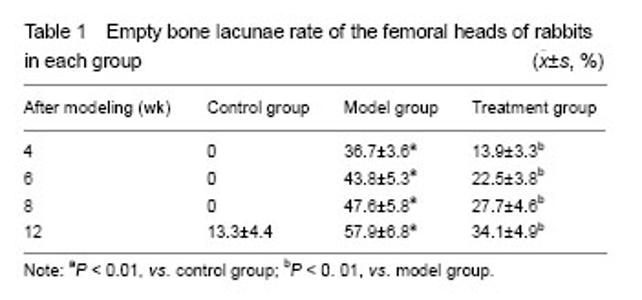
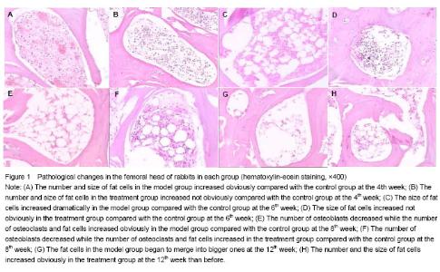
Quantitative analysis of experimental animals Rabbits in the treatment group had no obvious side reactions and biocompatibility problems. All of the 36 rabbits in the treatment group survived until the expected dates. Multiwalled carbon nanotubes improve the pathological pattern of the rabbit models of SNFH No necrotic bone collapses were found through gross observation in the three groups when the femoral heads were cut along the coronal plane. Four weeks after hormone injection, the number of empty bone lacunae in subchondral area and trabeculae in the model group significantly increased than that in the control group, the trabeculae began to have a small amount of thinner fractures, extensive pimelosis in the bone marrow was detectable, fat cells in the bone marrow became bigger and microvascular thrombosis appeared, while there was no positive histopathological manifestation in the treatment group. At the 6th and 8th week, the model group appeared to have the following changes: necrotic bone appeared in the subchondral area, parts of the trabeculae were fractured, the number of osteoblasts and blood vessels decreased, the number of osteoclasts and empty bone lacunae increased and the empty bone lacunae distributed focally, a large number of bone marrow and blood vessels became necrotic. The treatment group appeared to have the following changes: the bone trabeculae became thinner, the number of osteoblasts and blood vessels decreased while the number of osteoclasts and empty bone lacunae increased compared with the control group, intraosseous vessels were slightly damaged. Twelve weeks after hormone injection, the necrosis situation of bone in the subchondral area became even worse and the number of empty bone lacunae increased further more in the model group, while in the treatment group, the number of empty bone lacunae increased further, parts of the trabeculae were fractured, small pieces of necrotic bone marrow appeared. The number of osteoblasts decreased while the number of osteoclasts and fat cells increased obviously in the model group (Table 1). Due to the use of multiwalled carbon nanotubes, the number of osteoblasts decreased while the number of osteoclasts and fat cells increased in the treatment group, but they were not as worse as the model group. As osteoblasts, osteoclasts and bone fat cells were all differentiated from BMSCs, the results proved that multiwalled carbon nanotubes can inhibit the differentiation of BMSCs to osteoclasts and bone fat cells and promote the differentiation of BMSCs to osteoblasts. And this may be the mechanism of SNFH (Figure 1)."

"
| [1] Bao SY, Zhao GQ, Bi LQ. Changes in apoptosis related gene expression following alendronate intervention in the early srage of steroid-induced necrosis of the femoral head. Zhongguo Zuzhi Gongcheng Yanjiu yu Linchuang Kangfu. 2008;12(46):9095-9099
[2] Glueck CJ, Freiberg RA, Sieve L, et al. Enoxaparin prevents progression of stages I and II osteonecrosis of the hip. Clin Orthop Relat Res. 2005;(435):164-170.
[3] Bank I, Libourel EJ, Middeldorp S, et al. Elevated levels of FVIII:C within families are associated with an increased risk for venous and arterial thrombosis. J Thromb Haemost. 2005; 3(1):79-84.
[4] Iuchi T, Akaike M, Mitsui T, et al. Glucocorticoid excess induces superoxide production in vascular endothelial cells and elicits vascular endothelial dysfunction. Circ Res. 2003; 92(1):81-87.
[5] Drescher W, Li H, Lundgaard A, et al. Endothelin-1-induced femoral head epiphyseal artery constriction is enhanced by long-term corticosteroid treatment. J Bone Joint Surg Am. 2006;88 Suppl 3:173-179.
[6] Hu ZM, Wang HB, Li ZG, et al. Change of TNF-α and VEGF on glucocorticoid-induced avascular necrosis of femoral head in rabbits. Zhongguo Jiaoxing Waike Zazhi. 2006;14(12): 912-914.
[7] Kabata T, Matsumoto T, Yagishita S, et al. Vascular endothelial growth factor in rabbits during development of corticosteroid-induced osteonecrosis: a controlled experiment. J Rheumatol. 2008;35(12):2383-2390.
[8] Varoga D, Drescher W, Pufe M, et al. Differential expression of vascular endothelial growth factor in glucocorticoid-related osteonecrosis of the femoral head. Clin Orthop Relat Res. 2009;467(12):3273-3282.
[9] Murata M, Kumagai K, Miyata N, et al. Osteonecrosis in stroke-prone spontaneously hypertensive rats: effect of glucocorticoid. J Orthop Sci. 2007;12(3):289-295.
[10] Kitajima M, Shigematsu M, Ogawa K, et al. Effects of glucocorticoid on adipocyte size in human bone marrow. Med Mol Morphol. 2007;40(3):150-156.
[11] Nishida K, Yamamoto T, Motomura G, et al. Pitavastatin may reduce risk of steroid-induced osteonecrosis in rabbits: a preliminary histological study. Clin Orthop Relat Res. 2008; 466(5):1054-1058.
[12] Zanello LP, Zhao B, Hu H, et al. Bone cell proliferation on carbon nanotubes. Nano Lett. 2006;6(3):562-567.
[13] Elias KL, Price RL, Webster TJ. Enhanced functions of osteoblasts on nanometer diameter carbon fibers. Biomaterials. 2002;23(15):3279-3287.
[14] Price RL, Waid MC, Haberstroh KM, et al. Selective bone cell adhesion on formulations containing carbon nanofibers. Biomaterials. 2003;24(11):1877-1887.
[15] Abarrategi A, Gutiérrez MC, Moreno-Vicente C, et al. Multiwall carbon nanotube scaffolds for tissue engineering purposes. Biomaterials. 2008;29(1):94-102.
[16] Green DE, Longtin JP, Sitharaman B. The effect of nanoparticle-enhanced photoacoustic stimulation on multipotent marrow stromal cells. ACS Nano. 2009;3(8): 2065-2072.
[17] Supronowicz PR, Ajayan PM, Ullmann KR, et al. Novel current-conducting composite substrates for exposing osteoblasts to alternating current stimulation. J Biomed Mater Res. 2002;59(3):499-506.
[18] Li X, Gao H, Uo M, et al. Effect of carbon nanotubes on cellular functions in vitro. J Biomed Mater Res A. 2009;91(1): 132-139.
[19] Narita N, Kobayashi Y, Nakamura H, et al. Multiwalled carbon nanotubes specifically inhibit osteoclast differentiation and function. Nano Lett. 2009;9(4):1406-1413.
[20] Li Z, Liao W, Zhao Q, et al. Angiogenesis and bone regeneration by allogeneic mesenchymal stem cell intravenous transplantation in rabbit model of avascular necrotic femoral head. J Surg Res. 2013;183(1):193-203.
[21] The Ministry of Science and Technology of the People’s Republic of China. Guidance Suggestions for the Care and Use of Laboratory Animals. 2006-09-30.
[22] Yeh CH, Chang JK, Wang YH, et al. Ethanol may suppress Wnt/beta-catenin signaling on human bone marrow stroma cells: a preliminary study. Clin Orthop Relat Res. 2008;466(5): 1047-1053.
[23] Bekler H, Uygur AM, Gökçe A, et al. The effect of steroid use on the pathogenesis of avascular necrosis of the femoral head: an animal model. Acta Orthop Traumatol Turc. 2007; 41(1):58-63.
[24] Moraes LA, Paul-Clark MJ, Rickman A, et al. Ligand-specific glucocorticoid receptor activation in human platelets. Blood. 2005;106(13):4167-4175.
[25] O'Brien CA, Jia D, Plotkin LI, et al. Glucocorticoids act directly on osteoblasts and osteocytes to induce their apoptosis and reduce bone formation and strength. Endocrinology. 2004; 145(4):1835-1841.
[26] Yun SI, Yoon HY, Jeong SY, et al. Glucocorticoid induces apoptosis of osteoblast cells through the activation of glycogen synthase kinase 3beta. J Bone Miner Metab. 2009; 27(2):140-148.
[27] Kogianni G, Mann V, Ebetino F, et al. Fas/CD95 is associated with glucocorticoid-induced osteocyte apoptosis. Life Sci. 2004;75(24):2879-2895.
[28] Weinstein RS, Manolagas SC. Apoptosis in glucocorticoid-induced bone disease. Curr Opin Endocrinol Diabetes. 2005;12(3):219-223.
[29] Hofbauer LC, Gori F, Riggs BL, et al. Stimulation of osteoprotegerin ligand and inhibition of osteoprotegerin production by glucocorticoids in human osteoblastic lineage cells: potential paracrine mechanisms of glucocorticoid-induced osteoporosis. Endocrinology. 1999; 140(10):4382-4389.
[30] Okada Y, Tanikawa T, Iida T, et al. Vascular injury by glucocorticoid; involvement of apoptosis of endothelial cells. Clin Calcium. 2007;17(6):872-877.
[31] Tsuji M, Ikeda H, Ishizu A, et al. Altered expression of apoptosis-related genes in osteocytes exposed to high-dose steroid hormones and hypoxic stress. Pathobiology. 2006; 73(6):304-309.
[32] Ferrari P, Schroeder V, Anderson S, et al. Association of plasminogen activator inhibitor-1 genotype with avascular osteonecrosis in steroid-treated renal allograft recipients. Transplantation. 2002;74(8):1147-1152.
[33] Ekmekci Y, Keven K, Akar N, et al. Thrombophilia and avascular necrosis of femoral head in kidney allograft recipients. Nephrol Dial Transplant. 2006;21(12):3555-3558.
[34] Hirata T, Fujioka M, Takahashi KA, et al. ApoB C7623T polymorphism predicts risk for steroid-induced osteonecrosis of the femoral head after renal transplantation. J Orthop Sci. 2007;12(3):199-206.
[35] Hirata T, Fujioka M, Takahashi KA, et al. Low molecular weight phenotype of Apo(a) is a risk factor of corticosteroid-induced osteonecrosis of the femoral head after renal transplant. J Rheumatol. 2007;34(3):516-522.
[36] Wang S, Wei M, Han Y, et al. Roles of TNF-alpha gene polymorphisms in the occurrence and progress of SARS-Cov infection: a case-control study. BMC Infect Dis. 2008;8:27.
[37] Glueck CJ, Freiberg RA, Boppana S, et al. Thrombophilia, hypofibrinolysis, the eNOS T-786C polymorphism, and multifocal osteonecrosis. J Bone Joint Surg Am. 2008;90(10): 2220-2229.
[38] Asano T, Takahashi KA, Fujioka M, et al. ABCB1 C3435T and G2677T/A polymorphism decreased the risk for steroid-induced osteonecrosis of the femoral head after kidney transplantation. Pharmacogenetics. 2003;13(11): 675-682.
[39] Han N, Yan Z, Guo CA, et al. Effects of p-glycoprotein on steroid-induced osteonecrosis of the femoral head. Calcif Tissue Int. 2010;87(3):246-253.
[40] Tokuhara Y, Wakitani S, Oda Y, et al. Low levels of steroid-metabolizing hepatic enzyme (cytochrome P450 3A) activity may elevate responsiveness to steroids and may increase risk of steroid-induced osteonecrosis even with low glucocorticoid dose. J Orthop Sci. 2009;14(6):794-800.
[41] Masada T, Iwakiri K, Oda Y, et al. Increased hepatic cytochrome P4503A activity decreases the risk of developing steroid-induced osteonecrosis in a rabbit model. J Orthop Res. 2008;26(1):91-95.
[42] Sekiya I, Larson BL, Vuoristo JT, et al. Adipogenic differentiation of human adult stem cells from bone marrow stroma (MSCs). J Bone Miner Res. 2004;19(2):256-264.
[43] Dennis JE, Charbord P. Origin and differentiation of human and murine stroma. Stem Cells. 2002;20(3):205-214.
[44] Yin L, Li YB, Wang YS. Dexamethasone-induced adipogenesis in primary marrow stromal cell cultures: mechanism of steroid-induced osteonecrosis. Chin Med J (Engl). 2006;119(7):581-588.
[45] Wang BL, Sun W, Shi ZC, et al. Decreased proliferation of mesenchymal stem cells in corticosteroid-induced osteonecrosis of femoral head. Orthopedics. 2008;31(5):444.
[46] Liu J, Sun ZY, Cao L. Effect of dexamethasone on biological characteristics of bone marrow stromal stem cells. Zhonghua Guke Zazhi. 2003;23(11):691-693.
[47] Tamura K, Nakajima S, Hirota Y, et al. Genetic association of a polymorphism of the cAMP-responsive element binding protein-binding protein with steroid-induced osteonecrosis after kidney transplantation. J Bone Miner Metab. 2007;25(5): 320-325.
[48] Urbaniak JR, Seaber AV, Chen LE. Assessment of ischemia and reperfusion injury. Clin Orthop Relat Res. 1997;(334): 30-36.
[49] Xue YS, Shi SS, Li YF, et al. The change in bone morphogenetic protein-2 during experimental steroid-induced necrosis of femoral head. Zhonghua Shiyan Waike Zazhi. 2000; 17(5):455-456.
[50] Matsui M, Saito S, Ohzono K, et al. Experimental steroid-induced osteonecrosis in adult rabbits with hypersensitivity vasculitis. Clin Orthop Relat Res. 1992;(277): 61-72.
[51] Soto KF, Carrasco A, Powell TG, et al. Proceedings of the first TMS symposium on biological materials science. Mater Sci Eng C. 2006;26(8):1421-1427.
[52] Jia G, Wang H, Yan L, et al. Cytotoxicity of carbon nanomaterials: single-wall nanotube, multi-wall nanotube, and fullerene. Environ Sci Technol. 2005;39(5):1378-1383.
[53] Lobo AO, Antunes EF, Palma MBS, et al. Biocompatibility of multi-walled carbon nanotubes grown on titanium and silicon surfaces. Mater Sci Eng C. 2008;28(4):532-538.
[54] MacDonald RA, Laurenzi BF, Viswanathan G, et al. Collagen-carbon nanotube composite materials as scaffolds in tissue engineering. J Biomed Mater Res A. 2005;74(3): 489-496.
[55] Singh R, Pantarotto D, Lacerda L, et al. Tissue biodistribution and blood clearance rates of intravenously administered carbon nanotube radiotracers. Proc Natl Acad Sci U S A. 2006;103(9):3357-3362.
[56] Tian F, Cui D, Schwarz H, et al. Cytotoxicity of single-wall carbon nanotubes on human fibroblasts. Toxicol In Vitro. 2006;20(7):1202-1212.
[57] Nagai H, Okazaki Y, Chew SH, et al. Diameter and rigidity of multiwalled carbon nanotubes are critical factors in mesothelial injury and carcinogenesis. Proc Natl Acad Sci U S A. 2011;108(49):E1330-1338.
[58] Nimmagadda A, Thurston K, Nollert MU, et al. Chemical modification of SWNT alters in vitro cell-SWNT interactions. J Biomed Mater Res A. 2006;76(3):614-625.
[59] Kagan VE, Tyurina YY, Tyurin VA, et al. Direct and indirect effects of single walled carbon nanotubes on RAW 264.7 macrophages: role of iron. Toxicol Lett. 2006;165(1):88-100.
[60] Wörle-Knirsch JM, Pulskamp K, Krug HF. Oops they did it again! Carbon nanotubes hoax scientists in viability assays. Nano Lett. 2006;6(6):1261-1268. |
| [1] | Song Qing-qing, Yu Ling-fan, Xu Li-li, Yang Nai-long. Effect of different concentrations of uric acid on the neural differentiation of human bone marrow mesenchymal stem cells [J]. Chinese Journal of Tissue Engineering Research, 2014, 18(6): 847-852. |
| Viewed | ||||||
|
Full text |
|
|||||
|
Abstract |
|
|||||