Chinese Journal of Tissue Engineering Research ›› 2013, Vol. 17 ›› Issue (20): 3626-3634.doi: 10.3969/j.issn.2095-4344.2013.20.003
Previous Articles Next Articles
Rabbit articular chondrocyte dedifferentiation during in vitro expansion
Xu Lei, Ye Zhao-yang, Zhou Yan, Tan Wen-song
- State Key Laboratory of Bioreactor Engineering, East China University of Science and Technology, Shanghai 200237, China
-
Received:2012-09-29Revised:2012-11-19Online:2013-05-14Published:2013-05-14 -
Contact:Ye Zhao-yang, Ph.D., Associate professor, State Key Laboratory of Bioreactor Engineering, East China University of Science and Technology, Shanghai 200237, China zhaoyangye@ecust.edu.cn -
About author:Xu Lei★, Master, State Key Laboratory of Bioreactor Engineering, East China University of Science and Technology, Shanghai 200237, China xulei502211964@yahoo.com.cn -
Supported by:by the Scientific Research Foundation for the Returned Overseas Chinese Scholars, State Education Ministry
CLC Number:
Cite this article
Xu Lei, Ye Zhao-yang, Zhou Yan, Tan Wen-song. Rabbit articular chondrocyte dedifferentiation during in vitro expansion [J]. Chinese Journal of Tissue Engineering Research, 2013, 17(20): 3626-3634.
share this article
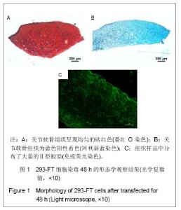
2.1 兔软骨组织的组织学鉴定结果 实验中削取兔膝关节表层软骨组织,制备组织切片,进行组织学鉴定。图1A番红O染色分离的关节软骨组织呈现均匀的砖红色,表明组织中存在大量的糖胺聚糖,同时可以观察到软骨陷窝的存在;图1B为阿利新蓝染色,关节软骨组织为蓝色阳性着色,同样证实了软骨组织特征胞外基质的存在;图1C是针对Ⅱ型胶原的免疫荧光染色,结果表明,组织样品中分布了大量的Ⅱ型胶原。这些结果证实所获组织为单一透明软骨组织,未见参杂的软骨下骨组织。"

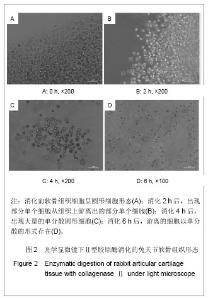
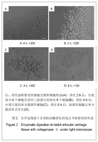
2.2 酶消化法分离的兔软骨细胞形态 见图2。"
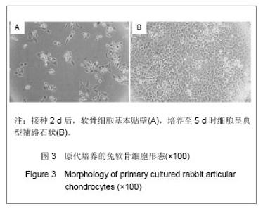
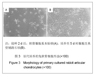
2.3 原代培养的兔软骨细胞形态 从软骨组织分离所得的细胞(P0)经冻存复苏,接种2 d后,基本上完全贴壁,见图3A,而且细胞多呈圆形或者多角形,继续培养至5 d时细胞基本达到100%汇合,细胞呈典型铺路石状,见图3B。"
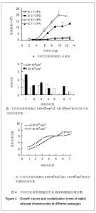
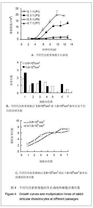
2.4 不同代次兔软骨细胞生长及增殖变化 在24孔板中,将不同代次的软骨细胞同时以5 000/cm2浓度接种,在不同时间点消化计数,绘制生长曲线。如图4A所示,P1代细胞的增殖能力最好,呈现明显的对数生长期;P3代软骨细胞的生长速率明显下降,而且迟滞期明显延长,但依然表现出典型的对数生长期。相比较而言,P5和P7代的细胞生长能力有限,在13 d的培养时间内基本没有增殖。同样如图4B,C所示,实验选取了两种细胞接种浓度(1.8×10⁴/cm2和3.6× 104/cm2),并从P1代开始连续传代,计算每代细胞到达100%汇合时其倍增次数。可以发现,倍增次数从P1代至P2代迅速下降,其后基本维持在1左右;累积倍增次数的增长趋势随着培养代次的延长而趋于缓和。在2种细胞接种浓度的条件下,尽管细胞的倍增次数差别不大,但仍然可以观察到累积倍增次数在较高细胞浓度的条件下P6代后才出现了平台,而低浓度下平台出现在P4代。见图4。"
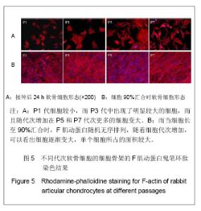
2.5 传代过程中兔软骨细胞的形态 利用鬼笔环肽染色方法,表征F肌动蛋白的分布,可以间接地考察细胞形态。如图5所示,接种24 h后,P1代细胞较小,形态多呈圆形或多角形,而P3代中出现了明显较大的细胞,而且随代次增加在P5和P7代次更多的细胞变大,同时部分细胞形态呈现狭长的有纤维样变化,见图5A;而当细胞长至90%汇合时,见图5B,同样P1代的细胞较小,而且可以发现F肌动蛋白随机无序排列,随着细胞代次增加,可以看出细胞逐渐变大,单个细胞所占的面积较大,更加重要的变化是F肌动蛋白逐步出现有序的排列,P7代细胞表现得尤为明显。"

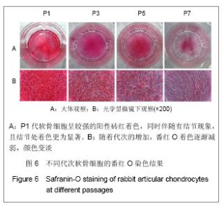
2.6 传代过程中兔软骨细胞的基质分泌情况 从图6的番红O染色结果可以看出,P1代软骨细胞呈较强的阳性砖红着色,同时伴随有结节现象,且结节处着色更为显著,见图6A,显微镜下放大观察可以看到细胞内外均匀着色,见图6B;但是随着代次的增加,番红O着色逐渐减弱,颜色变淡,特别是P7代细胞,番红O着色较浅而且分布不均。"

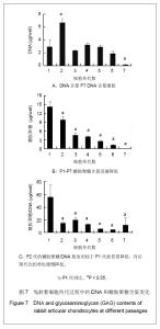
糖胺聚糖和DNA定量分析的结果如图7所示。可以看到,细胞100%汇合后,由于细胞大小和DNA合成活性的差异,各个代次的DNA含量并不完全相同,P2代DNA含量最高(P < 0.05),表明其DNA合成活性较高,而P7代则远远低于代次较低细胞的DNA含量,见图7A,这主要是因为P7代的细胞已经显著大于代次低的细胞,使得细胞的绝对数量较低。图7B则表示随着代次的增加,兔软骨细胞的糖胺聚糖分泌量迅速降低(P < 0.05);进一步将糖胺聚糖平均到单位DNA量上,如图7C所示,P2代的糖胺聚糖/DNA数值相较于P1代就显著低(P < 0.05),而后随代次的增加缓慢降低。"


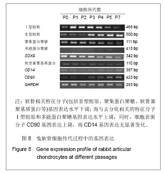
2.7 传代过程中兔软骨细胞的基因表达 将原代细胞在3.6×10⁴/cm2的浓度接种于平皿中,在细胞长至90%汇合时,收集细胞,以同样浓度传代培养,同时提取每代细胞的RNA样品,进行半定量PCR分析,结果如图8所示。"
| [1] Redman SN, Oldfield SF, Archer CW. Current strategies for articular cartilage repair. Eur Cell Mater. 2005;9:23-32; discussion 23-32.[2] Benya PD, Shaffer JD. Dedifferentiated chondrocytes reexpress the differentiated collagen phenotype when cultured in agarose gels. Cell. 1982;30(1):215-224.[3] Schnabel M, Marlovits S, Eckhoff G, et al. Dedifferentiation-associated changes in morphology and gene expression in primary human articular chondrocytes in cell culture. Osteoarthritis Cartilage. 2002;10(1):62-70.[4] Zhang Y, Cai G, Liu W, et al. Zhonghua Zhengxing Waike Zazhi. 2007;23(4):331-334.张艳,柴岗,刘伟,等.体外培养过程中去分化的人软骨细胞基因表达谱的变化[J].中华整形外科杂志,2007,23(4):331-334.[5] Karlsen TA, Shahdadfar A, Brinchmann JE. Human primary articular chondrocytes, chondroblasts-like cells, and dedifferentiated chondrocytes: differences in gene, microRNA, and protein expression and phenotype. Tissue Eng Part C Methods. 2011;17(2):219-227.[6] Barlic A, Drobnic M, Malicev E, et al. Quantitative analysis of gene expression in human articular chondrocytes assigned for autologous implantation. J Orthop Res. 2008;26(6): 847-853.[7] Wang L, Verbruggen G, Almqvist KF, et al. Flow cytometric analysis of the human articular chondrocyte phenotype in vitro. Osteoarthritis Cartilage. 2001;9(1):73-84.[8] Diaz-Romero J, Gaillard JP, Grogan SP, et al. Immunophenotypic analysis of human articular chondrocytes: changes in surface markers associated with cell expansion in monolayer culture. J Cell Physiol. 2005;202(3):731-742.[9] Chu CR, Szczodry M, Bruno S. Animal models for cartilage regeneration and repair. Tissue Eng Part B Rev. 2010;16(1): 105-115.[10] Ren B, Li HF, Zuo XA, et al. Hebei Beifang Yixue Xuebao Yixue Ban. 2010;27(2):8-11.任保,李慧芳,左喜爱,等.兔关节软骨细胞的原代培养和去分化现象[J].河北北方医学学报:医学版,2010,27(2):8-11.[11] Xia Q, Wang WD, Wei Z, et al. Zhongguo Yishi Zazhi. 2007; 9(9):1174-1177.夏青,王卫东,魏振,等.兔关节软骨细胞的体外培养及鉴定[J].中国医师杂志,2007,9(9):1174-1177.[12] Kim YJ, Sah RL, Doong JY, et al. Fluorometric assay of DNA in cartilage explants using Hoechst 33258. Anal Biochem. 1988;174(1):168-176.[13] Enobakhare BO, Bader DL, Lee DA. Quantification of sulfated glycosaminoglycans in chondrocyte/alginate cultures, by use of 1,9-dimethylmethylene blue. Anal Biochem. 1996;243(1): 189-191.[14] Gosset M, Berenbaum F, Thirion S, et al. Primary culture and phenotyping of murine chondrocytes. Nat Protoc. 2008;3(8): 1253-1260.[15] Sasazaki Y, Seedhom BB, Shore R. Morphology of the bovine chondrocyte and of its cytoskeleton in isolation and in situ: are chondrocytes ubiquitously paired through the entire layer of articular cartilage? Rheumatology (Oxford). 2008;47(11): 1641-1646.[16] Giovannini S, Diaz-Romero J, Aigner T, et al. Population doublings and percentage of S100-positive cells as predictors of in vitro chondrogenicity of expanded human articular chondrocytes. J Cell Physiol. 2010;222(2):411-420.[17] Tew SR, Murdoch AD, Rauchenberg RP, et al. Cellular methods in cartilage research: primary human chondrocytes in culture and chondrogenesis in human bone marrow stem cells. Methods. 2008;45(1):2-9.[18] Li H. Shanxi Zhigong Yixueyuan Xuebao. 2008;18(1):15-17.李昊.不同年龄兔软骨细胞培养和形态学研究[J].山西职工医学院学报,2008,18(1):15-17.[19] Schulze-Tanzil G, de Souza P, Villegas Castrejon H, et al. Redifferentiation of dedifferentiated human chondrocytes in high-density cultures. Cell Tissue Res. 2002;308(3):371-379.[20] Martin I, Jakob M, Schafer D, et al. Quantitative analysis of gene expression in human articular cartilage from normal and osteoarthritic joints. Osteoarthritis Cartilage. 2001;9(2): 112-118.[21] Diaz-Romero J, Nesic D, Grogan SP, et al. Immunophenotypic changes of human articular chondrocytes during monolayer culture reflect bona fide dedifferentiation rather than amplification of progenitor cells. J Cell Physiol. 2008;214(1):75-83.[22] The Ministry of Science and Technology of the People’s Republic of China. Guidance Suggestions for the Care and Use of Laboratory Animals. 2006-09-30. |
| [1] | Wu Xun, Meng Juanhong, Zhang Jianyun, Wang Liang. Concentrated growth factors in the repair of a full-thickness condylar cartilage defect in a rabbit [J]. Chinese Journal of Tissue Engineering Research, 2021, 25(8): 1166-1171. |
| [2] | Ma Zetao, Zeng Hui, Wang Deli, Weng Jian, Feng Song. MicroRNA-138-5p regulates chondrocyte proliferation and autophagy [J]. Chinese Journal of Tissue Engineering Research, 2021, 25(5): 674-678. |
| [3] | Xie Chongxin, Zhang Lei. Comparison of knee degeneration after anterior cruciate ligament reconstruction with or without remnant preservation [J]. Chinese Journal of Tissue Engineering Research, 2021, 25(5): 735-740. |
| [4] | Gao Kun, Chen Dayu, Zhang Yong, Liu Weidong, Sun Shufen, Lai Wenqiang, Ma Dujun, Wu Yihong, Lin Zhanpeng, Jiang Yinglu, Yu Weiji. Achyranthes bidentata alcohol extract inhibits extracellular matrix degradation of the cartilage by regulating synovial fibroblast exosomes [J]. Chinese Journal of Tissue Engineering Research, 2021, 25(23): 3636-3640. |
| [5] | Tong Jie, Liao Ying, Chen Zhengyu, Sun Guanghua . Osteoarthritic chondrocyte autophagy and regulation of mitogen-activated protein kinase signaling pathway [J]. Chinese Journal of Tissue Engineering Research, 2021, 25(20): 3246-3251. |
| [6] | Liu Xing, Wei Xiaohan, Deng Jie, Li Zhongming . Preparing a blunt contusion model of rabbit skeletal muscle under different blow strengths [J]. Chinese Journal of Tissue Engineering Research, 2021, 25(2): 196-200. |
| [7] | Mu Yufeng, Wei Lina, Wu Yong, Shao Anliang, Chen Liang, Qu Shuxin, Xu Liming. Development and evaluation of alpha-galactosyl antigen-deficient rabbit model [J]. Chinese Journal of Tissue Engineering Research, 2021, 25(2): 281-285. |
| [8] | Cao Yang, Zhang Junping, Peng Li, Ding Yi, Li Guanghui. Isolation and culture of rabbit aortic endothelial cells and biological characteristics [J]. Chinese Journal of Tissue Engineering Research, 2021, 25(19): 3000-3003. |
| [9] | Chen Pu, Ruan Anmin, Zhou Jun, Zhang Xiaozhe, Ma Yufeng, Zong Chenzhong, Wang Qingpu. Effect of Tongluo Analgesic Gel on cartilage inflammation and degeneration in a rabbit model of knee osteoarthritis [J]. Chinese Journal of Tissue Engineering Research, 2021, 25(17): 2670-2675. |
| [10] | Li Shao, Liang Yongkang, Gao Yi, Peng Qing. Establishment and functional in vitro characteristics of three-dimensional collagen HepaRG microsphere [J]. Chinese Journal of Tissue Engineering Research, 2021, 25(16): 2541-2547. |
| [11] | Fu Liwei, Yang Zhen, Li Hao, Gao Cangjian, Sui Xiang, Liu Shuyun, Guo Quanyi . New strategies and problems of affinity peptide in cartilage tissue engineering [J]. Chinese Journal of Tissue Engineering Research, 2021, 25(16): 2569-2574. |
| [12] | Wu Yukun, Han Jie, Wen Shuaibo. Mechanism of Runx2 gene in fracture healing [J]. Chinese Journal of Tissue Engineering Research, 2021, 25(14): 2274-2279. |
| [13] | Zhang Chuanhui, Li Jianjun, Yang Jun. Effects of dynamic pressure on the proliferation and mechanical properties of rabbit adipose mesenchymal stem cells transfected with insulin-like growth factor-1 [J]. Chinese Journal of Tissue Engineering Research, 2021, 25(13): 1982-1987. |
| [14] | Ye Dou, , Ma Xuexia , Guan Qian, , Luan Zuo , Yang Yinxiang , Wang Zhaoyan , Wang Qian , He Ying , Yao Ruiqin. Proportion and morphological characteristics of human oligodendrocyte precursor cells in different cell culture vessels [J]. Chinese Journal of Tissue Engineering Research, 2021, 25(1): 44-49. |
| [15] | Xu Guofeng, Li Xuebin, Tang Yifan, Zhao Yin, Zhou Shengyuan, Chen Xiongsheng, Jia Lianshun. The role of autophagy in ossification of the human ligamentum flavum [J]. Chinese Journal of Tissue Engineering Research, 2020, 24(8): 1174-1181. |
| Viewed | ||||||
|
Full text |
|
|||||
|
Abstract |
|
|||||