Chinese Journal of Tissue Engineering Research ›› 2026, Vol. 30 ›› Issue (36): 9505-9518.doi: 10.12307/2026.903
Previous Articles Next Articles
Three-dimensional finite element analysis of the biomechanical effects on surrounding tissues following crown-by-crown treatment of impacted mandibular third molars
Li Shenghao1, Zhang Borui1, Ma Yufeng1, 2
- 1School of Stomatology, Shanxi Medical University, Taiyuan 030000, Shanxi Province, China; 2Department of Stomatology, The Second Hospital of Shanxi Medical University, Taiyuan 030000, Shanxi Province, China
-
Received:2025-10-15Revised:2026-03-07Online:2026-12-28Published:2026-05-22 -
Contact:Ma Yufeng, Chief physician, School of Stomatology, Shanxi Medical University, Taiyuan 030000, Shanxi Province, China; Department of Stomatology, The Second Hospital of Shanxi Medical University, Taiyuan 030000, Shanxi Province, China -
About author:Li Shenghao, MS, Physician, School of Stomatology, Shanxi Medical University, Taiyuan 030000, Shanxi Province, China

CLC Number:
Cite this article
Li Shenghao, Zhang Borui, Ma Yufeng, . Three-dimensional finite element analysis of the biomechanical effects on surrounding tissues following crown-by-crown treatment of impacted mandibular third molars[J]. Chinese Journal of Tissue Engineering Research, 2026, 30(36): 9505-9518.
share this article
Add to citation manager EndNote|Reference Manager|ProCite|BibTeX|RefWorks
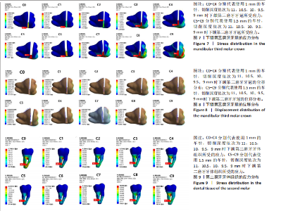
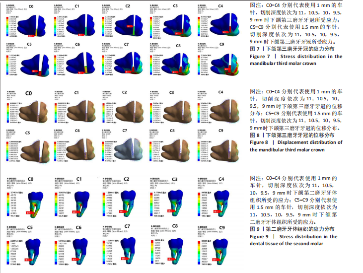
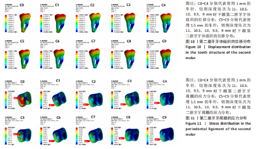
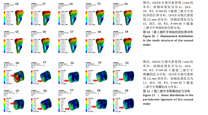
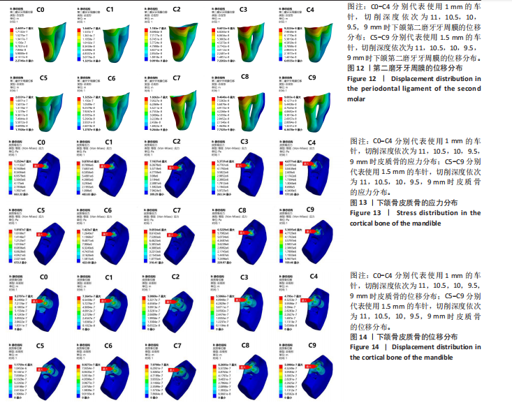
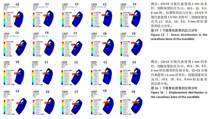
2.1 不同切削宽度与切削深度所产生的应力与位移分析 2.1.1 下颌第三磨牙的应力和位移分析 如图7所示,10组截冠模型的应力集中区主要分布在牙冠切削槽底部及附近、近中边缘嵴及附近、颊沟、舌沟、近中颊尖等区域。采用粗车针和细车针进行截冠后,二者的应力分布区域相似。 在10组模型牙冠断端的两侧,分别施加35 N的力,均垂直于表面,以模拟临床上使用牙挺断裂牙冠的操作后,C0-C9的最大应力集中区域均位于牙冠切削槽底部,C0(最大Von Mises应力值=792.81 MPa)、C1(最大Von Mises应力值=348.14 MPa)、C5(最大Von Mises应力值=1 332.34 MPa)、C6(最大Von Mises应力值=336.06 MPa)、C7(最大Von Mises应力值=341.25 MPa)均超过了牙体组织的最大屈服强度250 MPa[22-23],且最大应力集中区域均位于牙冠切削槽底部的远中,该应力传递路径完整,符合使牙冠断裂的生物力学特性,可以有效避免牙冠底部形成阶梯状残冠。细车针组的C2(最大Von Mises应力值=244.67 MPa)、C3(最大Von Mises应力值=168.06 MPa)、C4 (最大Von Mises应力值=84.55 MPa)牙冠切削槽底部最大应力值呈递减趋势,且其应力分布区域存在向牙冠切削槽底部的近中移动的趋势,说明该应力传递路径可能受阻,牙冠可能形成不完全断裂,有形成阶梯状残冠的风险,进而影响牙冠的取出。而粗车针组C9的最大应力分布区域位于牙冠切削槽底部的近中,同样存在牙冠不完全断裂的风险。 随着切削深度的减小,细车针组和粗车针粗的第三磨牙牙冠所受的最大Von Mises应力值呈非线性递减趋势,其中细车针组的C0-C1和粗车针组的C5-C6(即切削深度从11 mm减少至10.5 mm)的递减趋势最为显著,呈现骤降趋势,分别为56.09%和74.78%。 相同切削深度下,C5组对比C0组、C7组对比C2组产生的最大应力值增加了39.47%-68.01%,C6组对比C1组、C8组对比C3组、C9组对比C4组产生的最大应力值减少了3.47%-39.85%。 如图8所示,10组截冠模型的位移分布特点均为从牙冠切削槽顶部向底部及近远中逐级递减,符合生物力学能耗递减的分布模式。10种截冠模型的位移方向与牙冠断裂路径相一致,符合牙冠断裂的生物力学特性。随着切削深度的减小,两种车针的最大位移值呈非线性递减趋势,其中C0-C1和C5-C6,即切削深度从11 mm降低至10.5 mm的递减趋势最为明显,呈骤降趋势,分别降低了68.86%和70.77%。相同切削深度下,采用粗车针比采用细车针的牙冠最大位移值增加了17.84%-33.48%。 2.1.2 下颌第二磨牙牙体组织及牙周膜的应力和位移分析 如图9所示,在10组模型牙冠断端的两侧,分别施加35 N的力,均垂直于表面,以模拟临床上使用牙挺断裂牙冠的操作。10组模型的第二磨牙牙体组织的应力集中区主要分布在第二磨牙的根分叉处、近远中根融合部,该分布特点提示临床上拔除阻生牙时,应注意邻牙的保护和支点位置的选择,避免对第二磨牙牙根处产生损伤。同时,第二磨牙牙根的应力分布与牙周膜的生物学特性密切相关,根分叉区和近远中根融合部呈现最大Von Mises应力值,而牙颈部接近第三磨牙的部位因牙周膜的弹性作用,未出现应力集中。 粗车针组和细车针组的第二磨牙牙体组织最大Von Mises应力值出现区域相似,均位于第二磨牙近远中根融合部,且随着切削深度的减小,最大Von Mises应力值逐步减少,其中细车针组的C0-C1和粗车针组的C5-C6(即切削深度从11 mm减少至10.5 mm)的递减趋势最为显著,分别减少了27.82%和31.28%。相同切削深度下,粗车针组产生的最大Von Mises应力值均小于细车针组,粗车针组产生的最大Von Mises应力值相较细车针组减少了15.06%-20.84%。 如图10所示,10组模型的第二磨牙主要位移中心相似,均位于第二磨牙牙冠远中边缘嵴和远中根尖处,且呈现向周围向近中逐步减少的趋势,在第二磨牙近中根的根中1/2达到最小位移。10种模型的最大位移处均位于第二磨牙牙冠的远中边缘嵴处。随着切削深度的减小,两种车针的最大位移呈非线性递减趋势,其中C0-C1组和C5-C6组的递减趋势较为显著,分别为27.82%和31.28%。相同切削深度下,粗车针组产生的最大位移较细车针组减少了3.21%-10.15%。 如图11所示,10组模型的主要应力分布区域相似,集中在第二磨牙牙颈部、远中根的远中面上1/3、远中根远中面的中1/3、根尖部等区域。C5组和C0组的最大Von Mises应力值主要集中在第二磨牙远中根远中面的中1/3处,分别为0.036 MPa和0.030 MPa,呈向周围递减趋势。而C1-C4组和C6-C9组的最大应力区域则位于第二磨牙远中颊颈部,呈向周围递减趋势。随着切削深度的减少,粗车针组和细车针组所产生的最大Von Mises应力值均呈非线性递减的趋势,其中细车针组的C0-C1和粗车针组的C5-C6(即切削深度从11 mm减少至10.5 mm)的递减趋势最为显著,分别为28.55%和17.22%。使用粗车针比使用细车针产生的最大Von Mises应力值减少了1.04%-16.19%。 如图12所示,10组模型的主要位移分布区域相似,集中在第二磨牙远中颈部、远中根的下1/3处,并呈现向周围递减趋势。C0、C1、C5、C6的最大位移处在第二磨牙远中根的下1/3处,C2、C3、C4、C7、C8、C9的最大位移处在第二磨牙的远中颊颈部。随着切削深度的减小,粗车针组和细车针组的最大位移值均呈非线性递减趋势,其中细车针组的C0-C1和粗车针组的C5-C6(即切削深度从11 mm减少至10.5 mm)的递减趋势最为显著,分别为33.01%和34.82%。相同切削深度下,粗车针组比细车针组所产生的最大位移值减少了1.42%-19.23%。 2.1.3 下颌骨的应力和位移分析 如图13所示,10组模型皮质骨的应力集中区域相似,主要集中在下颌第三磨牙牙冠周围、颊舌侧骨板及根尖区域。10组模型皮质骨的最大Von Mises应力值区域相似,均位于下颌第三磨牙牙冠颊侧骨板。随着切削深度的减少,粗车针组和细车针组所产生的皮质骨最大Von Mises应力值呈非线性减小趋势,其中细车针组的C1(9.88 MPa)-C2(7.16 MPa)和粗车针组的C6(14.23 MPa)-C7(9.69 MPa),即切削深度从10.5 mm减小至10 mm的递减趋势最为显著,分别为27.48%和31.91%。相同切削深度下,粗车针组产生的皮质骨最大Von Mises应力值均大于细车针组。相同切削深度下,使用粗车针比使用细车针产生的皮质骨最大Von Mises应力值增加了14.80%-45.22%。 如图14所示,10组模型的皮质骨主要位移分布区域相似,主要分布在下颌第三磨牙牙冠下方骨板和颊侧骨板,并呈现向周围减小趋势。10组模型的最大位移区均位于下颌第三磨牙牙冠颊侧骨板,与图所示皮质骨最大Von Mises应力值区域相对应。随着切削深度的减小,10组模型的皮质骨位移呈非线性递减趋势,其中细车针组的C0(0.928 μm)-C1(0.736 μm)和粗车针组的C5(1.18 μm)-C6(0.893 μm),即切削深度从11 mm减少至10.5 mm的递减最为显著,分别为20.69%和24.32%。相同切削深度下,粗车针组产生的皮质骨位移均大于细车针组,增加了6.16%-26.73%。 如图15所示,10组模型松质骨的主要应力集中区域相似,均位于下颌第三磨牙牙冠颊侧骨和牙冠下方的牙槽窝处。C0、C1、C2、C3、C5、C6的最大Von Mises应力值区域位于下颌第三磨牙牙冠下方的牙槽窝处,C4、C7、C8、C9的最大Von Mises应力值区域位于下颌第三磨牙牙冠颊侧骨处。随着切削深度的减小,粗车针和细车针所产生的松质骨最大Von Mises应力值呈非线性递减趋势,其中细车针组的C0(32.58 MPa)-C1(14.74 MPa)和粗车针组的C5(28.94 MPa)-C6(12.60 MPa),即切削深度从11 mm减少至10.5 mm的递减趋势最为显著,分别为54.76%和57.13%。相同切削深度下,粗车针组所产生的松质骨最大Von Mises应力值均小于细车针组。使用粗车针比使用细车针所产生的松质骨最大Von Mises应力值区域减小了1.50%-19.82%。"
"

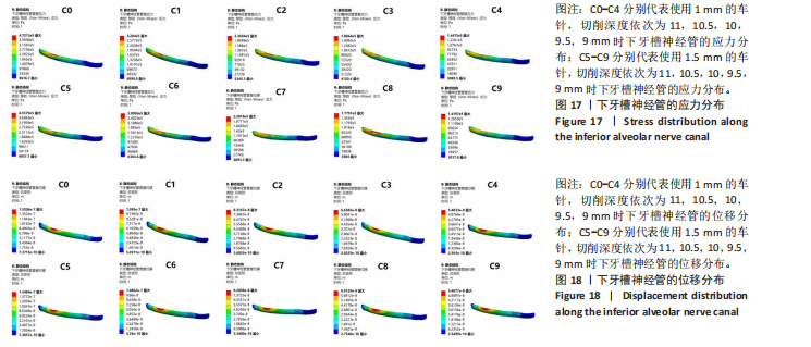
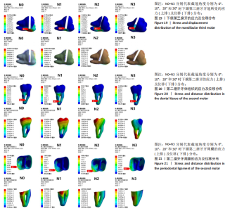
如图16所示,10组模型的松质骨最大位移处均位于下颌第三磨牙牙冠下方的牙槽窝处,并呈现向周围递减趋势。如图所示C0、C1、C2、C3、C5、C6的松质骨最大Von Mises应力值区域与最大位于区域相对应。随着切削深度的减小,松质骨的位移呈逐渐递减趋势,其中细车针组的C0(15.15 μm)-C1(7.42 μm) 和粗车针组的C5(19.04 μm)-C6(8.44 μm),即切削深度从11 mm减少至10.5 mm时的递减趋势最为显著,分别为51.02%和55.67%。相同切削深度下,粗车针组所产生的松质骨位移均大于细车针组。相同切削深度下,使用粗车针比使用细车针所产生的松质骨位移增加了1.70%-25.67%。 2.1.4 下牙槽神经管的应力和位移分析 如图17所示,下牙槽神经管与牙冠下方对应区域呈现出最大应力集中,该应力集中区域与上方的松质骨的位移(如图16所示)和第三磨牙牙冠的位移(如图8所示)相一致,说明在10组模型牙冠断端的两侧分别施加35 N的力(均垂直于断端表面)以模拟临床上使用牙挺断裂牙冠的操作后,神经管上部的松质骨和牙冠对其产生了挤压作用。随着切削深度的减小,下牙槽神经管所受的最大Von Mises应力值呈现出非线性递减趋势,其中C0(0.47 MPa)-C1(0.32 MPa)和C5(0.46 MPa)-C6(0.31 MPa),即切削深度从11 mm递减至10.5 mm的递减趋势最为显著,分别为31.91%和32.61%。相同切削深度下,粗车针组所产生下牙槽神经管的最大Von Mises应力值均小于细车针组。使用粗车针所产生的下牙槽神经管最大Von Mises应力值比使用细车针减少了1.11%-3.80%。 如图18所示10组模型的下牙槽神经管的最大位移区域与最大Von Mises应力值区域(如图17所示)相对应,呈现向周围递减趋势。下牙槽神经管的位移与下颌第三磨牙牙冠位移(如图8所示)和松质骨位移(如图16所示)相对于,说明了挤压作用的存在。随着切削深度的减小,下牙槽神经管的位移呈递减趋势,其中细车针组的C0(1.52 μm)-C1(1.10 μm)和粗车针组的C5(1.55 μm)-C6(1.09 μm),即在切削深度11 mm减小至10.5 mm时递减趋势最为明显,分别为27.63%和29.68%。在相同切削深度下,粗车针所产生的下牙槽神经管最大位移C5(1.55 μm)大于细车针组中的C0(1.52 μm),其余均小于细车针组。 2.2 不同切削角度所产生的应力与位移分析 2.2.1 下颌第三磨牙应力及位移分析 如图19所示,N0的应力集中区主要分布在牙冠切削槽底部及附近、近中边缘嵴及附近、颊沟、舌沟、近中颊尖等区域,N1、N2、N3的应力集中区主要分布在牙冠切削槽底部及附近。 N0、N1的最大应力集中区在牙冠切削槽底部颊侧偏远中处,而N2、N3的最大应力集中区在牙冠切削槽底部的近中处。N0(最大Von Mises应力值=341.25 MPa)、N1(最大Von Mises应力值=275.44 MPa)均超过了牙体组织的最大屈服强度250 MPa,且最大应力集中区域均位于牙冠切削槽底部的远中,该应力传递路径完整,符合使牙冠断裂的生物力学特性,可以有效避免牙冠底部形成阶梯状残冠。N2(最大Von Mises应力值=196.81 MPa)、N3(最大Von Mises应力值=137.73 MPa)牙冠切削槽底部最大应力值呈递减趋势,且其最大应力分布区域位于牙冠切削槽底部的近中,说明该应力传递路径可能受阻,牙冠可能形成不完全断裂,有形成阶梯状残冠的风险,进而影响牙冠的取出。同时,N3的牙冠切削槽底部近远中向出现应力中断带,说明在该负载情况下,牙冠可能形成不完全断裂。 随着切削角度的增大,下颌第三磨牙牙冠所受的最大Von Mises应力值呈非线性递减趋势,其中N2(196.81 MPa)-N3(137.73 MPa)的递减趋势最为显著,约为30.02%。 如图19所示,4组截冠模型的位移分布特点均为从牙冠切削槽顶部向底部及近远中逐级递减,符合生物力学能耗递减的分布模式。4组模型的最大位移处均位于牙冠远中边缘嵴处。4组截冠模型的位移方向与牙冠断裂路径相一致,符合牙冠断裂的生物力学特性。随着切削角度从0°增加到10°时,牙冠的最大位移值从115.31 μm增加至148.48 μm。但随着切削角度从10°增加到20°和30°时,牙冠的最大位移值呈现非线性递减趋势,裂纹扩展路径向根方偏移。其中N2(124.71 μm)-N3(49.16 μm)的递减趋势最为显著,呈骤降趋势,约为60.58%。 2.2.2 第二磨牙牙体及牙周膜的应力和位移分析 如图20所示,在4组模型牙冠断端的两侧,分别施加35 N的力,均垂直于表面,以模拟临床上使用牙挺断裂牙冠的操作。4组模型的第二磨牙牙体组织的应力集中区主要分布在第二磨牙的根分叉处、近远中根融合部,该分布特点提示临床上拔除阻生牙时,应注意邻牙的保护和支点位置的选择,避免对第二磨牙牙根处产生损伤。同时,第二磨牙牙根的应力分布与牙周膜的生物学特性密切相关,根分叉区和近远中根融合部呈现应力集中区,而牙颈部接近第三磨牙的部位因牙周膜的弹性作用,未出现应力集中。 4组模型的第二磨牙牙体组织最大Von Mises应力值出现区域相似,均位于第二磨牙近远中根融合部。且随着切削角度从0°增加到10°时,第二磨牙牙根的最大Von Mises应力值骤降,从0.12 MPa骤降到0.026 MPa,降低了约78.33%。随着切削角度从10°增加到20°时,第二磨牙牙根的最大Von Mises应力值持续骤降,降低了约65.58%。而随着切削角度从20°增加到30°时,第二磨牙牙根的最大Von Mises应力值骤增,增加了约63.9倍。 如图20所示,N0、N1、N2的第二磨牙最大位移中心相似,均位于第二磨牙牙冠远中边缘嵴,且呈现向周围向近中逐步减少的趋势。N3的第二磨牙最大位移中心第二磨牙根尖1/3的远中,并呈现向周围递减趋势。随着切削角度的增大,第二磨牙的牙根位移呈现先减小后增大的趋势:在切削角度从0°增加至10°时,第二磨牙牙体组织位移减少了约60.17%;在切削角度从10°增加至20°时,第二磨牙牙体组织位移减少了约48.60%,在切削角度从20°增加到30°时,第二磨牙牙根的最大位移增加了约2.48倍。 如图21所示,4组模型的最大应力集中区域相似,集中在第二磨牙牙颈部。在N0-N2组中,随着切削角度从0°(N0)依次递增至10°(N1)、20°(N2)时,最大Von Mises应力值呈非线性递减:N0(0.023 MPa)、N1(0.013 MPa)、N2(0.0017 MPa),最大Von Mises应力值的降低与切削角度呈负相关。而当切削角度从20°(N2)增加到30°(N3)时,最大Von Mises应力值骤增,增加了约14.21倍。"

"

"
"

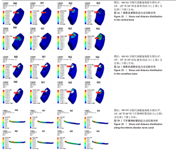
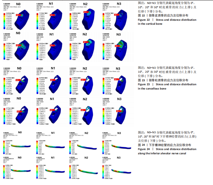
如图21所示,N0、N1、N2的最大位移中心均位于第二磨牙远中牙颈部,并呈现向周围递减趋势。N3的最大位移中心则位于远中根的根中1/3部,呈现向周围递减趋势。在N0-N2组中,随着切削角度从0°(N0)依次递增至10°(N1)、20°(N2)时,最大位移呈非线性递减:N0(0.11 μm)、N1(0.051 μm)、N2(0.027 μm)。 而当切削角度从20°(N2)增加到30°(N3)时,最大位移值骤增,增加了约3.79倍。 2.2.3 颌骨的应力和位移分析 如图22所示,N0、N1、N3的皮质骨最大应力集中区均位于下颌第三磨牙颊侧骨板,N2的最大应力集中区位于下颌第三磨牙远中骨板。随着切削角度从0°(N0)增大到10°(N1),皮质骨的最大Von Mises应力值从9.69 MPa增加到71.16 MPa;随着切削角度从10°(N1)增大到20°(N2),皮质骨的最大Von Mises应力值从71.16 MPa减少至5.89 MPa:随着切削角度从20°(N2)增大到30°(N3),皮质骨的最大Von Mises应力值从5.89 MPa增大至42.21 MPa。 如图22所示,N0的皮质骨最大位移中心位于下颌第三磨牙牙冠颊侧骨板,N1、N3的皮质骨最大位移中心均位于牙冠下方骨板、N2的皮质骨最大位移中心位于牙冠远中骨板。随着切削角度从0°(N0)增大到10°(N1),皮质骨的最大位移值从0.071 μm增加到3.01 μm;随着切削角度从10°(N1)增大到20°(N2),皮质骨的最大位移值从3.01 μm减少至0.50 μm:随着切削角度从20°(N2)增大到30°(N3),皮质骨的最大位移值从0.50 μm增大至1.68 μm。 如图23所示,N0、N2、N3松质骨的最大应力集中区均位于下颌第三磨牙牙冠颊侧骨,N1最大应力集中区位于牙冠下方骨。随着切削角度从0°(N0)增大到10°(N1),皮质骨的最大Von Mises应力值从7.41 MPa增加到8.48 MPa,增大了约14.44%;随着切削角度从10°(N1)增大到20°(N2),松质骨的最大Von Mises应力值从8.48 MPa减少至2.37 MPa,减少了约72.05%:随着切削角度从20°(N2)增大到30°(N3),松质骨的最大Von Mises应力值从2.37 MPa增大至25.13 MPa。 如图23所示,N0、N3的松质骨最大位移中心位于下颌第三磨牙牙冠下方骨,N1、N2的松质骨最大位移中心均位于下颌第三磨牙牙冠颊侧骨。随着切削角度从0°(N0)依次递增至10°(N1)、20°(N2)时,松质骨的最大位移值呈非线性递减:N0(4.71 μm)、N1(2.94 μm)、N2(1.00 μm),最大位移值的降低与切削角度呈负相关。而随着切削角度从20°(N2)增加到30°(N3),最大位移出现骤增趋势,从1.00 μm增长至6.18 μm。 2.2.4 下牙槽神经管的位移和应力分析 如图24所示,N0的下牙槽神经管与牙冠下方对应区域呈现出最大应力集中,该应力集中区域与上方松质骨的位移和第三磨牙牙冠的位移相一致,说明在牙冠断端的两侧分别施加35 N的力(均垂直于断端表面)以模拟临床上使用牙挺断裂牙冠的操作后,神经管上部的颌骨和牙冠对其产生了挤压作用。而N1、N2、N3的最大应力集中区出现向远中颊侧移动趋势。随着切削角度从0°(N0)依次递增至10°(N1)、20°(N2)时,最大Von Mises应力值呈非线性递减:N0(0.23 MPa)、N1(0.14 MPa)、N2(0.13 MPa),最大Von Mises应力值的降低与切削角度呈负相关。而随着切削角度从20°(N2)增加至30°(N3),下牙槽神经管的最大Von Mises应力值出现骤增趋势,增加了约5.23倍。 如图24所示,N0下牙槽神经管的最大位移区域与最大Von Mises应力值区域相对应,呈现向周围递减趋势。N0下牙槽神经管的位移与下颌第三磨牙牙冠位移和松质骨位移相对应,说明了挤压作用的存在。随着切削角度从0°(N0)依次递增至10°(N1)、20°(N2)时,最大位移值呈非线性递减:N0(0.083 μm)、N1(0.047 μm)、N2(0.016 μm),最大位移值的降低与切削角度呈负相关。而随着切削角度从20°(N2)增加至30°(N3),下牙槽神经管的最大位移值出现骤增趋势,从0.016 μm增加至0.31 μm。"
| [1] STEEL BJ, SURENDRAN KSB, BRAITHWAITE C, et al. Current thinking in lower third molar surgery. Br J Oral Maxillofac Surg. 2022;60(3):257-265. [2] CANDOTTO V, OBERTI L, GABRIONE F, et al. Complication in third molar extractions. J Biol Regul Homeost Agents. 2019;33(3 Suppl. 1):169-172. [3] MARGHALANI AA, ALSAGGAF AU, HAZZAZI A, et al. Radiographic characteristics of impacted teeth: A retrospective study of 2199 radiograph. PLoS One. 2025; 20(2):e0316749. [4] WANG X. [Geostatics in impacted mandibular third molar surgury]. Zhonghua Kou Qiang Yi Xue Za Zhi. 2025;60(2):109-115. [5] 胡开进. 微创拔牙技术[J]. 现代口腔医学杂志,2010,24(4):241-243. [6] 胡开进. 现代标准拔牙术新理念[J]. 中国实用口腔科杂志,2010,3(10):577-581. [7] 张志霞, 周宏志, 刘瑶, 等. 微创拔牙术在下颌阻生智齿拔除中的应用[J]. 实用口腔医学杂志,2012,28(3):398-400. [8] 杨映阳, 杜胜男, 吕宗凯. 高速涡轮机头和微创拔牙刀在下颌阻生牙拔除术中的应用效果评价[J]. 上海口腔医学,2015,24(4):489-492. [9] 陈勇臻, 尹建超, 李森. 超声骨刀微创拔牙术与传统凿骨劈冠拔牙术治疗下颌阻生智齿患者的效果比较[J]. 现代临床医学,2025,51(1):16-19+37. [10] 柴娟,张昕,刘昌奎,等. 2023版《恒牙拔牙术临床操作规范》解读[J].华西口腔医学杂志,2025,43(2):158-162. [11] 鞠昊, 朱红华, 段涛, 等. CBCT的基本原理及在口腔各科的应用进展[J].医学影像学杂志,2015,25(5):907-909+942. [12] TAY WZ, SKLAVOS A, MIAN M, et al. Radiographic Predictors of Postoperative Inferior Alveolar Nerve Injury in Mandibular Third Molar Surgery. J Oral Maxillofac Surg. 2025;83(1):54-61. [13] POGORZELSKA A, MIELCZAREK A, REGULSKI PA, et al. Evaluation of the Inferior Alveolar Canal Course in Relation to Fully and Partially Impacted Lower Third Molar Roots via Cone-Beam Computed Tomography. Cureus. 2024;16(11):e74021. [14] SÁNCHEZ JORGE MI, OCAÑA RA, VALLE RODRÍGUEZ C, et al. Mandibular third molar extraction: perceived surgical difficulty in relation to professional training. BMC Oral Health. 2023;23(1):485. [15] 王岚. 种植修复义齿和天然邻牙生物力学的三维有限元分析[D]. 重庆:重庆医科大学,2022. [16] 蒋啸宇, 朱杰晶, 王栋才, 等. 上颌全牙列远移中牙周膜的应力分析[J]. 口腔医学研究,2023,39(4):341-344. [17] KUGUOGLU A, AKARSU-GUVEN B. Evaluation of the effects of the third molar on distalization and the effects of attachments on distalization and expansion with clear aligners: Three-dimensional finite element study. Korean J Orthod. 2025;55(1):69-81. [18] GAO H, LUO L, LIU J. Three-dimensional finite element analysis of maxillary molar distalization treated with clear aligners combined with different traction methods. Prog Orthod. 2024;25(1):47. [19] SU R, LI S, WANG W. Effect of high trimline aligners on distalizing mandibular molars: a three-dimensional finite element study. Eur J Med Res. 2024;29(1):623. [20] MEMON S, MEHTA S, MALIK S, et al. Three-dimensional finite element analysis of the stress distribution in the endodontically treated maxillary central incisor by glass fiber post and dentin post. J Indian Prosthodont Soc. 2016;16(1):70-74. [21] YALCIN S, AKTAS I, EMES Y, et al. A technique for atraumatic extraction of teeth before immediate implant placement using implant drills. Implant Dent. 2009; 18(6):464-472. [22] SAKAGUCHI RL, POWERS JM. Craig’s restorative dental materials. Mosby Elsevier, 2012. [23] CROTHERS JA. Fundamentals of operative dentistry. Br Dental J. 2006;201(3):180-181. [24] BAILEY E, KASHBOUR W, SHAH N, et al. Surgical techniques for the removal of mandibular wisdom teeth. Cochrane Database Syst Rev. 2020;7(7):Cd004345. [25] XU W, SHEN J, LI K, et al. A mechanical and three-dimensional finite element study of the optimum tooth sectioning depth during the extraction of low-level horizontally impacted mandibular third molar. Odontology. 2024;112(1):242-249. [26] YAN ZY, XU JY, ZHANG JY, et al. Optimal tooth sectioning using a surgical handpiece and elevator: a finite element study of horizontally deeply impacted mandibular third molars. Clin Oral Investig. 2024;28(8):439. [27] 刘平,胡开进,马洋,等.外科拔牙器械的选择及应用[J].中国实用口腔科杂志,2016,9(10):581-584. [28] SUEDBECK JR, ARMITANO-LAGO CN, LUDWIG EA. The Additive Effects of Cell Phone Use and Dental Hygiene Practice on Finger Muscle Strength: A Pilot Study. J Dent Hyg. 2020;94(2):45-53. [29] TRISI P, DE BENEDITTIS S, PERFETTI G, et al. Primary stability, insertion torque and bone density of cylindric implant ad modum Branemark: is there a relationship? An in vitro study. Clin Oral Implants Res. 2011;22(5):567-570. [30] BORRERO-LOPEZ O, RODRIGUEZ-ROJAS F, CONSTANTINO PJ, et al. Fundamental mechanics of tooth fracture and wear: implications for humans and other primates. Interface Focus. 2021;11(5):20200070. [31] ZENG J, JIANG Y, LIU Y, et al. Research on a novel digital tooth sectioning guide system for tooth sectioning during mandibular third molar extraction: An in vitro study. J Stomatol Oral Maxillofac Surg. 2023;124(1s):101383. [32] RESNICK CM, DANG RR, GLICK SJ, et al. Accuracy of three-dimensional soft tissue prediction for Le Fort I osteotomy using Dolphin 3D software: a pilot study. Int J Oral Maxillofac Surg. 2017;46(3):289-295. [33] ANAND M, PANWAR S. Role of Navigation in Oral and Maxillofacial Surgery: A Surgeon’s Perspectives. Clin Cosmet Investig Dent. 2021;13:127-139. [34] 陈新民,王劲茗,赵云凤,等.人体下颌骨强度研究[J]. 口腔颌面修复学杂志, 2002,3(2):113-115. [35] JIA B, WANG Q, CHEN J, et al. [Expert consensus on standardized clinical applications of minimally invasive tooth extraction techniques]. Nan Fang Yi Ke Da Xue Xue Bao. 2024;44(5):1004-1014. [36] LIU X, CHENG Y, QIN W, et al. Effects of upper-molar distalization using clear aligners in combination with Class II elastics: a three-dimensional finite element analysis. BMC Oral Health. 2022;22(1):546. [37] CHEN Q, LUO S, WANG Y, et al. Three-dimensional finite element analysis of occlusal stress on maxillary first molars with different marginal morphologies restored with occlusal veneers. BMC Oral Health. 2024;24(1):1349. [38] CHENG X, ZHANG XY, QIAN WH. Influence of different base materials and thicknesses on the fracture resistance of endocrown: A three-dimensional finite element analysis. BMC Oral Health. 2022;22(1):363. [39] VALERA-JIMÉNEZ JF, BURGUEÑO-BARRIS G, GÓMEZ-GONZÁLEZ S, et al. Finite element analysis of narrow dental implants. Dent Mater. 2020;36(7):927-935. [40] MAO B, TIAN Y, LI J, et al. Expansion rebound deformation of clear aligners and its biomechanical influence: a three-dimensional morphologic analysis and finite element analysis study. Angle Orthod. 2023;93(5):572-579. [41] GAO W, LU J, GAO X, et al. Biomechanical effects of joint disc perforation on the temporomandibular joint: a 3D finite element study. BMC Oral Health. 2023;23(1):943. [42] CHEN J, XU Y, LI C, et al. Displacement and stress distribution of the craniomaxillofacial complex under different surgical conditions: a three-dimensional finite element analysis of fracture mechanics. BMC Oral Health. 2021;21(1):596. [43] TRANG BNH, KANAZAWA M, MURAKAMI N, et al. Stress distribution of one-piece and two-piece mini-Implant overdentures with various attachment systems and diameters: A finite element analysis. J Prosthodont Res. 2023;67(3):430-436. |
| [1] | Cai Yaohao, Lang Lyu, Li Hong. Assessing the bone mass of the residual alveolar ridge in the first molar for implant placement by cone-beam computed tomography [J]. Chinese Journal of Tissue Engineering Research, 2025, 29(8): 1572-1577. |
| [2] | Fan Jiabing, Fu Xuefei, Zhang Junmei, Zhou Suodi, Mo Chaolun. Accuracy of orthodontic micro-implant placement guided by a 3D-printed guide plate [J]. Chinese Journal of Tissue Engineering Research, 2025, 29(24): 5102-5108. |
| [3] | Zhuang Xinyi, Peng Yuanhao, Yu Ting, Lyu Dongmei, Wen Xiujie, Cheng Qian. Cone-beam CT evaluation of bone mass in the external oblique line of the mandible in adolescents with different cervical vertebral bone ages [J]. Chinese Journal of Tissue Engineering Research, 2024, 28(8): 1253-1258. |
| [4] | Peng Yuanhao, Lyu Dongmei, Zhuang Xinyi, Yu Ting, Cheng Qian. Consistency between two types of cone-beam CT transformed two-dimensional images and traditional lateral cephalometric radiographs for quantitative analysis of cervical vertebral bone age [J]. Chinese Journal of Tissue Engineering Research, 2024, 28(18): 2881-2886. |
| [5] | Zhang Ning, Gao Siwen, Guo Jun, Li Zekui, Zhang Juan. Correlation between articular fossa space and bone destruction location of the condyle in adolescents with temporomandibular joint osteoarthrosis [J]. Chinese Journal of Tissue Engineering Research, 2022, 26(29): 4593-4597. |
| [6] | Zhou Xingyu, Jia Ying, Li Zhongwei, Ding Qi. Characteristics of dental inclination in different vertical skeletal types of skeletal class II malocclusion: a three-dimensional imaging evaluation [J]. Chinese Journal of Tissue Engineering Research, 2022, 26(17): 2625-2630. |
| [7] | Fan Jiabing, Zhang Junmei. Morphological measurement and analysis of the mandible in adult females with different vertical skeletal types [J]. Chinese Journal of Tissue Engineering Research, 2021, 25(8): 1177-1183. |
| [8] |
Zhang Yujing, Peng Yuzhi, Liu Baozhen, Jing Fang.
Effect of Xianling Gubao Capsule
on alveolar bone mass in postmenopausal women with periodontitis: a cone-beam
CT evaluation
|
| [9] | Xie Hui1, Qin Bo2, Wang Souxin2, Mo Chaolun2, Zhang Junmei2. Measurement of hard and soft tissues of the zygomaticomaxillary region by multi-planner reconstruction [J]. Chinese Journal of Tissue Engineering Research, 2019, 23(31): 4945-4949. |
| [10] | Wang Chaoran, Li Zhiqin, Shi Peiliang, Feng Chunyue, Yin Ye. Changes of osseous structure about temporal-mandibular joint pre/post-treatment for Angle III malocclusion through cone-beam computed tomography [J]. Chinese Journal of Tissue Engineering Research, 2019, 23(31): 4950-4955. |
| [11] | Jiang Weijie1, 2, Liu Jun2, Liu Changcui3, Zhang Chen1, 2. Alveolar bone change of the maxillary anterior region after retraction in adult patients with class II1 malocclusion using cone-beam CT [J]. Chinese Journal of Tissue Engineering Research, 2019, 23(19): 2986-2990. |
| [12] | Li Ming-xia, Wang Guang-ping, Wang Hu. Anatomical structure of human maxillary sinus: Cone-beam CT analysis [J]. Chinese Journal of Tissue Engineering Research, 2013, 17(2): 320-324. |
| [13] | Ding Ji-qun, Fang Jian-qiang, Yuan Chang-qing, Chen Jie . Labial and lingual alveolar bone thickness of adult tooth root [J]. Chinese Journal of Tissue Engineering Research, 2013, 17(15): 2714-2722. |
| Viewed | ||||||
|
Full text |
|
|||||
|
Abstract |
|
|||||