Chinese Journal of Tissue Engineering Research ›› 2026, Vol. 30 ›› Issue (13): 3298-3307.doi: 10.12307/2026.728
Previous Articles Next Articles
Effect of miR-223-3p on biological behavior of tendon stem cells under hypoxic conditions
Duan Cheng, Cheng Jie
- Department of Trauma Area C, Second Affiliated Hospital of Inner Mongolia Medical University, Hohhot 010030, Inner Mongolia Autonomous Region, China
-
Accepted:2025-09-18Online:2026-05-08Published:2025-12-25 -
Contact:Cheng Jie, MS, Chief physician, Department of Trauma Area C, Second Affiliated Hospital of Inner Mongolia Medical University, Hohhot 010030, Inner Mongolia Autonomous Region, China -
About author:Duan Cheng, Master candidate, Department of Trauma Area C, Second Affiliated Hospital of Inner Mongolia Medical University, Hohhot 010030, Inner Mongolia Autonomous Region, China; Cheng Jie, MS, Chief physician, Department of Trauma Area C, Second Affiliated Hospital of Inner Mongolia Medical University, Hohhot 010030, Inner Mongolia Autonomous Region, China Duan Cheng and Cheng Jie contributed equally to this article. -
Supported by:Science and Technology Project for the Construction of High-level Clinical Specialties in Public Hospitals in the Capital Area of Inner Mongolia Autonomous Region, No. 2024SGGZ129 (to CJ); General Project of Inner Mongolia Medical University, No. YKD2024MS021 (to CJ)
CLC Number:
Cite this article
Duan Cheng, Cheng Jie. Effect of miR-223-3p on biological behavior of tendon stem cells under hypoxic conditions[J]. Chinese Journal of Tissue Engineering Research, 2026, 30(13): 3298-3307.
share this article
Add to citation manager EndNote|Reference Manager|ProCite|BibTeX|RefWorks
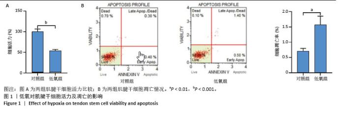
2.1 低氧对肌腱干细胞生物学功能的影响 2.1.1 细胞活力及凋亡指标 CCK-8结果显示,与对照组相比,低氧处理48 h后,肌腱干细胞活力显著下降(图1A)。流式分析结果显示,与对照组相比,低氧处理显著诱导肌腱干细胞凋亡(图1B)。"
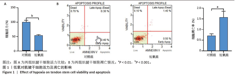
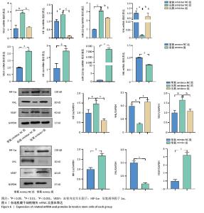
2.1.2 低氧环境下对肌腱干细胞中相关蛋白及mRNA表达的影响 RT-qPCR结果显示,与对照组相比,低氧条件下肌腱干细胞中miR-223-3p、低氧诱导因子1α、血管内皮生长因子的mRNA表达量显著升高(图2A)。Western blot蛋白定量结果显示,与对照组相比,低氧条件下肌腱干细胞中低氧诱导因子1α和血管内皮生长因子的蛋白表达水平显著升高(图2B)。"

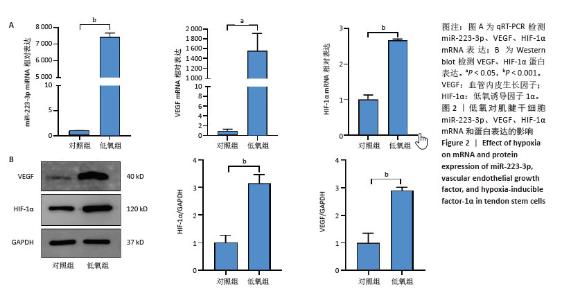
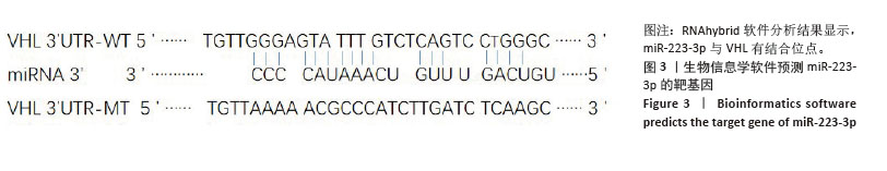
2.2 miR-223-3p与VHL的靶向结合验证 2.2.1 筛选作用于miR-223-3p的靶基因 通过生物信息学方法预测miR-223-3p潜在的靶基因,RNAhybrid 软件分析miR-223-3p和VHL序列,结果显示,miR-223-3p与VHL序列能够很好地互补配对,提示miR-223-3p可能与VHL相互结合(图3)。"
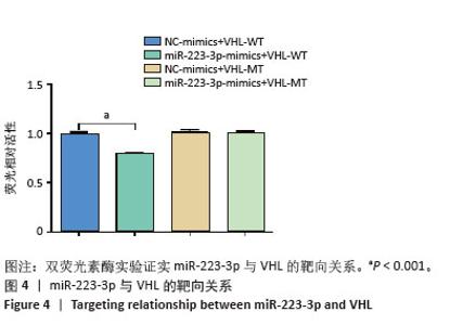
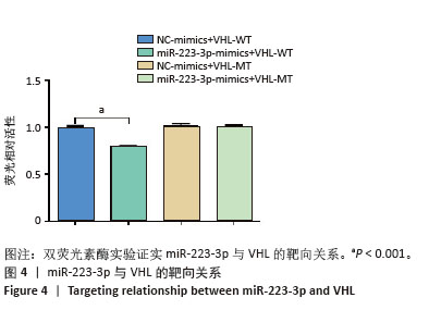
2.2.2 双荧光素酶报告基因验证miR-223-3p靶向调控VHL 为进一步证明上述观点,构建包含VHL基因3’非翻译区野生型(VHL-WT)和突变型(VHL-MT)的报告载体,分别与miR-223-3p mimics或阴性对照(NC mimics)共转染至肌腱干细胞,结果显示,转染miR-223-3p mimics的VHL-WT组荧光素酶活性较阴性对照组显著降低,而VHL-MT组活性无显著变化(图4),这表明miR-223-3p可直接结合VHL的3’非翻译区。"
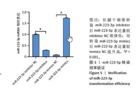
2.3 miR-223-3p对肌腱干细胞功能的调控作用 2.3.1 肌腱干细胞miR-223-3p敲低与过表达效率验证 qRT-PCR结果显示,转染miR-223-3p inhibitor后miR-223-3p表达量较inhibitor NC组降低;而miR-223-3p mimics组miR-223-3p表达量较mimics NC组升高(图5)。"

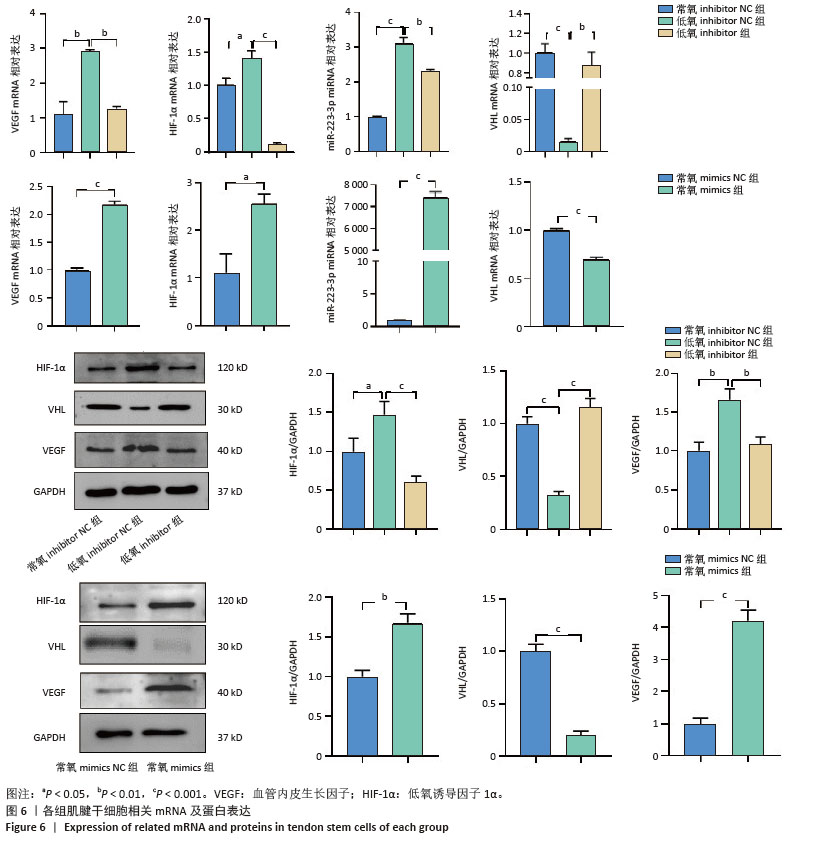
2.3.2 miR-223-3p转染后对肌腱干细胞中相关mRNA及蛋白表达的影响 RT-qPCR检测结果显示,与低氧inhibitor NC组相比,低氧inhibitor组VHL mRNA表达升高,miR-223-3p、低氧诱导因子1α、血管内皮生长因子mRNA表达降低;与常氧inhibitor NC组相比,低氧inhibitor NC组VHL mRNA表达降低,miR-223-3p、低氧诱导因子1α、血管内皮生长因子mRNA表达升高(图6)。与常氧mimics NC组相比,常氧mimics组VHL mRNA表达降低,miR-223-3p、低氧诱导因子1α、血管内皮生长因子mRNA表达升高(图6)。 Western blot检测结果显示,与低氧inhibitor NC组相比,低氧inhibitor组VHL蛋白表达升高,低氧诱导因子1α和血管内皮生长因子蛋白表达降低;与常氧inhibitor NC组相比,低氧inhibitor NC组VHL蛋白表达降低,低氧诱导因子1α和血管内皮生长因子蛋白表达升高(图6)。与常氧mimics NC组相比,常氧mimics组VHL蛋白表达降低,低氧诱导因子1α和血管内皮生长因子蛋白表达升高(图6),表达趋势与qPCR一致。"
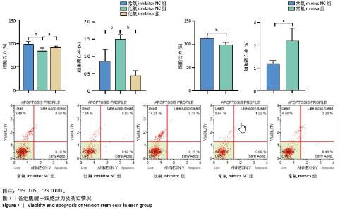
2.3.3 miR-223-3p转染对肌腱干细胞活力与凋亡的影响 CCK-8实验结果显示,与低氧inhibitor NC组相比,低氧inhibitor组肌腱干细胞活力升高;与常氧inhibitor NC组相比,低氧inhibitor NC组肌腱干细胞活力降低;与常氧mimics NC组相比,常氧mimics组肌腱干细胞活力降低(图7)。流式分析结果显示,与低氧inhibitor NC组相比,低氧inhibitor组肌腱干细胞凋亡降低,与常氧inhibitor NC组相比,低氧inhibitor NC组肌腱干细胞凋亡升高;与常氧mimics NC组相比,常氧mimics组肌腱干细胞凋亡升高(图7)。"
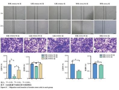
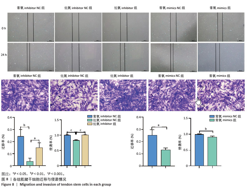
2.3.4 miR-223-3p转染对肌腱干细胞迁移与侵袭能力的影响 划痕实验结果显示,与低氧inhibitor NC组相比,低氧inhibitor组肌腱干细胞迁移率升高,与常氧inhibitor NC组相比,低氧inhibitor NC组肌腱干细胞迁移率降低;与常氧mimics NC组相比,常氧mimics组肌腱干细胞迁移率降低(图8)。Transwell侵袭实验结果显示,与低氧inhibitor NC组相比,低氧inhibitor组肌腱干细胞侵袭增加,与常氧inhibitor NC组相比,低氧inhibitor NC组肌腱干细胞侵袭降低;与常氧mimics NC组相比,常氧mimics组肌腱干细胞侵袭降低(图8)。"
| [1] TANG C, WANG Z, XIE Y, et al. Classification of distinct tendinopathy subtypes for precision therapeutics. Nat Commun. 2024;15(1):9460. [2] PRAKASH N, KIM J, JEON J, et al. Progress and emerging techniques for biomaterial-based derivation of mesenchymal stem cells (MSCs) from pluripotent stem cells (PSCs). Biomater Res. 2023;27(1):31. [3] HE Y, LU S, CHEN W, et al. Exosomes derived from tendon stem/progenitor cells enhance tendon-bone interface healing after rotator cuff repair in a rat model. Bioact Mater. 2024;40:484-502. [4] 逯静薇,吕可馨,蒋莉,等.影响肌腱干细胞分化的因素[J].中国组织工程研究,2024,28(13):2098-2104. [5] CAI Z, XIN Z, WANG H, et al. Extracellular Vesicle-Contained Thrombospondin 1 Retards Age-Related Degenerative Tendinopathy by Rejuvenating Tendon Stem/Progenitor Cell Senescence. Small. 2024;20(38):e2400598. [6] DIENER C, KELLER A, MEESE E. Emerging concepts of miRNA therapeutics: from cells to clinic. Trends Genet. 2022;38(6):613-626. [7] BAO H, PENG Z, CHENG X, et al. GABA induced by sleep deprivation promotes the proliferation and migration of colon tumors through miR-223-3p endogenous pathway and exosome pathway. J Exp Clin Cancer Res. 2023;42(1):344. [8] LIU X, JIN S, LIU J, et al. MiR-223-3p overexpressed adipose mesenchymal stem cell-derived exosomes promote wound healing via targeting MAPK10. Acta Histochem. 2023;125(8):152102. [9] KRYLOVA SV, FENG D. The Machinery of Exosomes: Biogenesis, Release, and Uptake. Int J Mol Sci. 2023;24(2):1337. [10] ZHANG Z, WANG P, ZHENG Y, et al. Exosomal microRNA-223 from neutrophil-like cells inhibits osteogenic differentiation of PDLSCs through the cGMP-PKG signaling pathway. J Periodontal Res. 2023; 58(6):1315-1325. [11] ZHANG MW, SHEN YJ, SHI J, et al. MiR-223-3p in Cardiovascular Diseases: A Biomarker and Potential Therapeutic Target. Front Cardiovasc Med. 2021;7:610561. [12] BARBAGALLO D, PONTI D, BASSANI B, et al. MiR-223-3p in Cancer Development and Cancer Drug Resistance: Same Coin, Different Faces. Int J Mol Sci. 2024;25(15):8191. [13] GAO H, GAO R, ZHANG L, et al. Esrrb plays important roles in maintaining self-renewal of trophoblast stem cells (TSCs) and reprogramming somatic cells to induced TSCs. J Mol Cell Biol. 2019; 11(6):463-473. [14] PRINGELS L, COOK JL, WITVROUW E, et al. Exploring the role of intratendinous pressure in the pathogenesis of tendon pathology: a narrative review and conceptual framework. Br J Sports Med. 2023;57(16):1042-1048. [15] 陆加霖,高尧,李涵,等.富血小板血浆治疗肌腱病的影响因素与机制[J].中国组织工程研究,2023,27(12):1944-1953. [16] ZENG CY, WANG XF, HUA FZ. HIF-1α in Osteoarthritis: From Pathogenesis to Therapeutic Implications. Front Pharmacol. 2022;13: 927126. [17] CHEN W, WU P, YU F, et al. HIF-1α Regulates Bone Homeostasis and Angiogenesis, Participating in the Occurrence of Bone Metabolic Diseases. Cells. 2022;11(22):3552. [18] YANG W, MA J, ZHOU W, et al. Reciprocal regulations between miRNAs and HIF-1α in human cancers. Cell Mol Life Sci. 2019;76(3):453-471. [19] SONG S, ZHANG G, CHEN X, et al. HIF-1α increases the osteogenic capacity of ADSCs by coupling angiogenesis and osteogenesis via the HIF-1α/VEGF/AKT/mTOR signaling pathway. J Nanobiotechnology. 2023;21(1):257. [20] LU X, LI L, LIN J, et al. PAARH promotes M2 macrophage polarization and immune evasion of liver cancer cells through VEGF protein. Int J Biol Macromol. 2024;281(Pt 4):136580. [21] YU Y, LIN L, ZHOU Y, et al. Effect of Hypoxia on Self-Renewal Capacity and Differentiation in Human Tendon-Derived Stem Cells. Med Sci Monit. 2017;23:1334-1339. [22] LI D, JIU J, LIU H, et al. Tissue-engineered mesenchymal stem cell constructs alleviate tendinopathy by suppressing vascularization. Bioact Mater. 2024;36:474-489. [23] JIANG L, LIU T, LYU K, et al. Inflammation-related signaling pathways in tendinopathy. Open Life Sci. 2023;18(1):20220729. [24] SHI J, YAO H, CHONG H, et al. Tissue-engineered collagen matrix loaded with rat adipose-derived stem cells/human amniotic mesenchymal stem cells for rotator cuff tendon-bone repair. Int J Biol Macromol. 2024;282(Pt 4):137144. [25] ZHANG M, DAI GC, ZHANG YW, et al. Enhancing osteogenic differentiation of diabetic tendon stem/progenitor cells through hyperoxia: Unveiling ROS/HIF-1α signalling axis. J Cell Mol Med. 2024; 28(20):e70127. [26] ZHANG W, YAO C, WEI Z, et al. miR-128 promoted adipogenic differentiation and inhibited osteogenic differentiation of human mesenchymal stem cells by suppression of VEGF pathway. J Recept Signal Transduct Res. 2017;37(3):217-223. [27] LI X, YANG N. Exosome miR-223-3p in the bone marrow-derived mesenchymal stem cells alleviates the inflammation and airway remodeling through NLRP3-induced ASC/Caspase-1/GSDMD signaling pathway. Int Immunopharmacol. 2023;123:110746. [28] LONG C, CEN S, ZHONG Z, et al. FOXO3 is targeted by miR-223-3p and promotes osteogenic differentiation of bone marrow mesenchymal stem cells by enhancing autophagy. Hum Cell. 2021;34(1):14-27. [29] YFANTIS A, MYLONIS I, CHACHAMI G, et al. Transcriptional Response to Hypoxia: The Role of HIF-1-Associated Co-Regulators. Cells. 2023; 12(5):798. [30] LI X, LONG J, ZONG L, et al. ZNF561-AS1 Regulates Cell Proliferation and Apoptosis in Myocardial Infarction Through miR-223-3p/NLRP3 Axis. Cell Transplant. 2022;31:9636897221077928. [31] ZHI Y, ZHANG W, WU Z, et al. miR-223-3p Targets KIF4A and Promotes the Oxidative Stress-Mediated Apoptosis of Breast Cancer Cells. Cancer Biother Radiopharm. 2025. doi: 10.1089/cbr.2024.0102. [32] DIEHL CJ, CIULLI A. Discovery of small molecule ligands for the von Hippel-Lindau (VHL) E3 ligase and their use as inhibitors and PROTAC degraders. Chem Soc Rev. 2022;51(19):8216-8257. [33] SEMENZA GL. Involvement of oxygen-sensing pathways in physiologic and pathologic erythropoiesis. Blood. 2009;114(10):2015-2019. [34] PÉREZ-GUTIÉRREZ L, FERRARA N. Biology and therapeutic targeting of vascular endothelial growth factor A. Nat Rev Mol Cell Biol. 2023; 24(11):816-834. [35] APTE RS, CHEN DS, FERRARA N. VEGF in Signaling and Disease: Beyond Discovery and Development. Cell. 2019;176(6):1248-1264. [36] CÉBE SUAREZ S, PIEREN M, CARIOLATO L, et al. A VEGF-A splice variant defective for heparan sulfate and neuropilin-1 binding shows attenuated signaling through VEGFR-2. Cell Mol Life Sci. 2006;63(17): 2067-2077. [37] CHEN C, SONG C, LIU B, et al. Activation of BMP4/SMAD pathway by HIF-1α in hypoxic environment promotes osteogenic differentiation of BMSCs and leads to ectopic bone formation. Tissue Cell. 2024;88: 102376. [38] CHENG X, YUN X, WEI Y, et al. Hypoxia-Mimicking Microenvironment Scaffold for Enhanced Tendon Regeneration. ACS Appl Mater Interfaces. 2025;17(6):8937-8948. [39] MABETA P, STEENKAMP V. The VEGF/VEGFR Axis Revisited: Implications for Cancer Therapy. Int J Mol Sci. 2022;23(24):15585. [40] ASAKIYA C, ZHU L, YUHAN J, et al. Current progress of miRNA-derivative nucleotide drugs: modifications, delivery systems, applications. Expert Opin Drug Deliv. 2022;19(4):435-450. |
| [1] | Haonan Yang, Zhengwei Yuan, Junpeng Xu, Zhiqi Mao, Jianning Zhang. Preliminary study on the mechanisms and efficacy of deep brain stimulation in treating depression [J]. Chinese Journal of Tissue Engineering Research, 2026, 30(在线): 1-9. |
| [2] | Jiang Xinghai, Song Yulin, Li Dejin, Shao Jianmin, Xu Junzhi, Liu Huakai, Wu Yingguo, Shen Yuehui, Feng Sicheng. Vascular endothelial growth factor 165 genes transfected into bone marrow mesenchymal stem cells to construct a vascularized amphiphilic peptide gel module [J]. Chinese Journal of Tissue Engineering Research, 2026, 30(8): 1903-1911. |
| [3] | Guo Jiachen, Gao Jun, Dai Wenhao, Liao Huayuan, Jiang You, Zhang Xi . Effect of compressive stress microenvironment on cytokines during fracture healing [J]. Chinese Journal of Tissue Engineering Research, 2026, 30(4): 908-916. |
| [4] | Wang Zhengye, Liu Wanlin, Zhao Zhenqun. Mechanism by which vascular endothelial growth factor A targets regulation of angiogenesis in the treatment of steroid-induced osteonecrosis of the femoral head [J]. Chinese Journal of Tissue Engineering Research, 2026, 30(3): 671-679. |
| [5] | Zhou Shibo, Yu Xing, Chen Hailong, Xiong Yang. Nanocrystalline collagen-based bone combined with Bushen Zhuangjin Decoction repairs bone defects in osteoporotic rats [J]. Chinese Journal of Tissue Engineering Research, 2026, 30(2): 354-361. |
| [6] | Wang Zhengye, Liu Wanlin, Zhao Zhenqun. Multidimensional target regulation of vascular endothelial growth factor A in articular cartilage development [J]. Chinese Journal of Tissue Engineering Research, 2026, 30(16): 4193-4203. |
| [7] | Wumiti·Taxi, Wang Lining, Li Muzhe, Sun Jie, Chen Shuangliu, Zhu Yihua, Zhou Shijie, Ma Yong, Guo Yang. Wenshen Tongluo Zhitong decoction regulates the bone fat differentiation balance of bone marrow mesenchymal stem cells through exosomal miR-342-3p [J]. Chinese Journal of Tissue Engineering Research, 2026, 30(13): 3258-3269. |
| [8] | Zhang Shilei, Qin Chuanhong, Wang Jianxu, Sun Shui. Osteogenic-adipogenic differentiation imbalance of bone marrow mesenchymal stem cells and osteonecrosis of the femoral head: from molecular mechanisms to therapeutic strategies [J]. Chinese Journal of Tissue Engineering Research, 2026, 30(13): 3350-3358. |
| [9] | Liu Ziwei, Nijati·Tursun, Yin Rui, Li Shuhui, Zhou Jing. Effect of cannabinoid type I receptors on neuronal differentiation of human apical papilla stem cells [J]. Chinese Journal of Tissue Engineering Research, 2026, 30(1): 93-100. |
| [10] | Wu Zhijing, Li Jiali, Zhang Jiaxin, Wang Tangrong, Zheng Yuzhou, Sun Zixuan. Alpha-ketoglutarate engineered small extracellular vesicles delay skin aging [J]. Chinese Journal of Tissue Engineering Research, 2026, 30(1): 120-129. |
| [11] | Xue Hui, Li Dongnan, Zhao Yadi, Chen Chao, Xie Zongyuan. Relationship between BCR/ABL gene expression and recurrence before and after allogeneic transplantation in Ph chromosome positive acute lymphoblastic leukemia [J]. Chinese Journal of Tissue Engineering Research, 2026, 30(1): 139-144. |
| [12] | Lyu Ruyue, Gu Lulu, Liu Qian, Zhou Siyi, Li Beibei, Xue Letian, Sun Peng. Regulatory mechanisms of exosome secretion and its application prospects in biomedicine [J]. Chinese Journal of Tissue Engineering Research, 2026, 30(1): 184-193. |
| [13] | Luo Wenbin, Li Ruoyun, Pan Chaofan, Luo Changjiang. Engineered exosomes for repairing tissue damage: application potential, excellent biological stability, and targeting specificity [J]. Chinese Journal of Tissue Engineering Research, 2026, 30(1): 204-217. |
| [14] | Sun Huiwen, Guo Qiangqiang, Wang Wei, Wu Jie, Xi Kun, Gu Yong. Engineered stem cell bionic periosteum coordinates immune inflammation and vascularization to promote bone regeneration [J]. Chinese Journal of Tissue Engineering Research, 2026, 30(1): 21-33. |
| [15] | Xu Hao, Ding Lu, Li Xiao. Investigating the effect of the mechanical wear on abutment screw in Morse taper connection implant implant system by using finite element analysis [J]. Chinese Journal of Tissue Engineering Research, 2025, 29(在线): 1-9. |
| Viewed | ||||||
|
Full text |
|
|||||
|
Abstract |
|
|||||