Chinese Journal of Tissue Engineering Research ›› 2026, Vol. 30 ›› Issue (14): 3586-3596.doi: 10.12307/2026.302
Previous Articles Next Articles
Nanofibers with gradient deposition of endothelial cell derivatives modulate behavior of Schwann cells
Yao Lijie1, 2, Yan Yuying1, 2, Chen Siyu1, 2, Wang Yuanfei2, Wu Tong2
- 1School of Basic Medicine, Qingdao University, Qingdao 266071, Shandong Province, China; 2Medical Research Center, Affiliated Hospital of Qingdao University, Qingdao 266000, Shandong Province, China
-
Received:2025-04-08Accepted:2025-06-13Online:2026-05-18Published:2025-09-10 -
Contact:Wu Tong, PhD, Professor, Doctoral/Master’s supervisor, Medical Research Center, Affiliated Hospital of Qingdao University, Qingdao 266000, Shandong Province, China -
About author:Yao Lijie, MS, School of Basic Medicine, Qingdao University, Qingdao 266071, Shandong Province, China; Medical Research Center, Affiliated Hospital of Qingdao University, Qingdao 266000, Shandong Province, China -
Supported by:Shandong Natural Science Foundation Project, No. ZR2021YQ17 (to WT)
CLC Number:
Cite this article
Yao Lijie, Yan Yuying, Chen Siyu, Wang Yuanfei, Wu Tong. Nanofibers with gradient deposition of endothelial cell derivatives modulate behavior of Schwann cells[J]. Chinese Journal of Tissue Engineering Research, 2026, 30(14): 3586-3596.
share this article
Add to citation manager EndNote|Reference Manager|ProCite|BibTeX|RefWorks

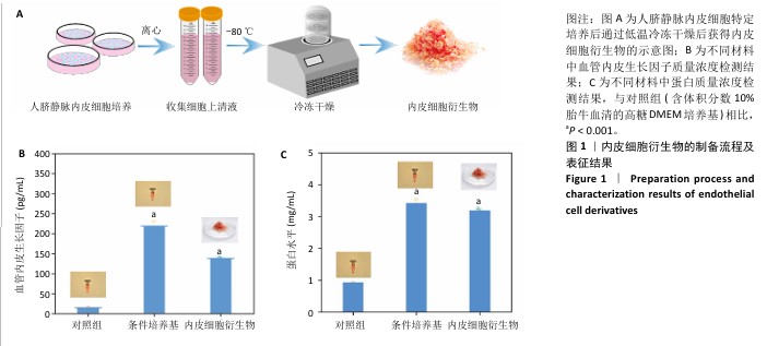
2.1 内皮细胞衍生物的制备及成分测定 内皮细胞衍生物的制备流程见图1A。与对照组中血管内皮生长因子质量浓度(15.77±0.25) pg/mL相比,人脐静脉内皮细胞条件培养基和内皮细胞衍生物中血管内皮生长因子质量浓度均显著增加,分别为(218.85±11.90) pg/mL和(138.89±3.78) pg/mL(图1B)。与对照组中蛋白质量浓度(0.92±0.01) mg/mL相比,人脐静脉内皮细胞条件培养基和内皮细胞衍生物中的蛋白质量浓度均明显增加,分别为(3.41±0.11) mg/mL和(3.16±0.11) mg/mL(图1C)。"
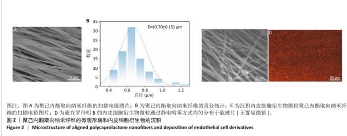
2.2 聚己内酯取向纳米纤维的表征结果 如图2A所示,聚己内酯纳米纤维具有良好的取向性,能够为细胞增殖、迁移提供良好的地形引导。聚己内酯取向纤维的直径达到纳米级别,为(700±150) nm(图2B)。 2.3 内皮细胞衍生物在聚己内酯取向纳米纤维膜上的均一沉积与表征结果 如图2C所示,聚己内酯取向纳米纤维膜上的内皮细胞衍生物以微粒形式沉积,并且纤维保持良好的取向性,表明沉积内皮细胞衍生物微粒的聚己内酯取向纳米纤维能够为细胞提供地形线索和生物信号。如图2D所示,罗丹明B标记的内皮细胞衍生物能够以微粒方式均匀分布于聚己内酯取向纳米纤维膜上。 "

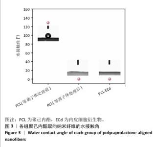
2.4 聚己内酯取向纳米纤维的亲水性表征结果 如图3所示,未经等离子体处理的聚己内酯取向纳米纤维的水接触角为(128.78±2.78)°,具有疏水性;经等离子体处理后,未沉积与沉积内皮细胞衍生物微粒的聚己内酯取向纳米纤维均具有超亲水性,接触角为0°。"

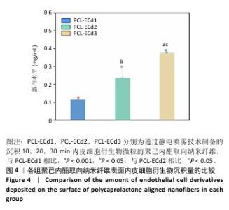
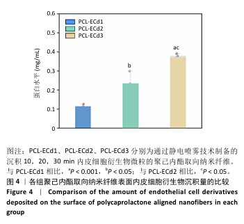
2.5 内皮细胞衍生物最佳沉积时间筛选结果 2.5.1 纳米纤维表面内皮细胞衍生物的沉积量 与纳米纤维PCL-ECd1中蛋白质量浓度(0.11±0.01) mg/mL相比,纳米纤维PCL-ECd2、PCL-ECd3中蛋白质量浓度均显著增加(P < 0.05,P < 0.001),分别为(0.23± 0.07),(0.37±0.02) mg/mL;并且纳米纤维PCL-ECd3中的蛋白质量浓度显著高于PCL-ECd2(P < 0.05),见图4。表明随着沉积时间的增加,内皮细胞衍生物在聚己内酯取向纳米纤维表面的沉积量增加。"

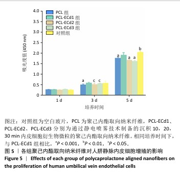
2.5.2 人脐静脉内皮细胞增殖情况 培养第1,3天,与对照组相比,第1天和第3天PCL-ECd1组人脐静脉内皮细胞增殖与对照组比较差异无显著性意义(P > 0.05);培养第3天,PCL-ECd1组人脐静脉内皮细胞增殖多于PCL组、PCL-ECd2组、PCL-ECd3组(P < 0.001或P < 0.05);培养第5天,PCL-ECd1组人脐静脉内皮细胞增殖少于对照组(P < 0.01),多于PCL组、PCL-ECd2组、PCL-ECd3组(P < 0.001),见图5。"

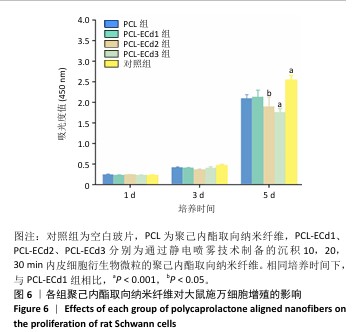
2.5.3 大鼠施万细胞增殖情况 如图6所示,培养第5天时,PCL-ECd1组大鼠施万细胞增殖少于对照组(P < 0.001),多于PCL-ECd2组、PCL-ECd3组(P < 0.05,P < 0.001)。"
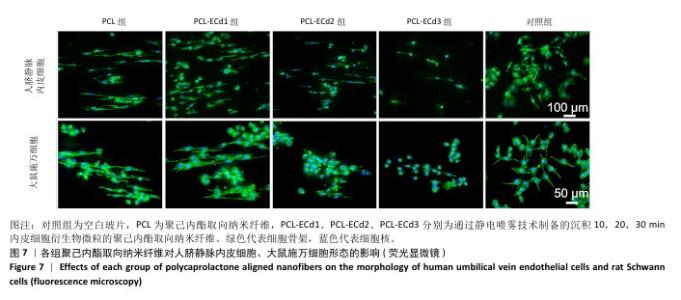
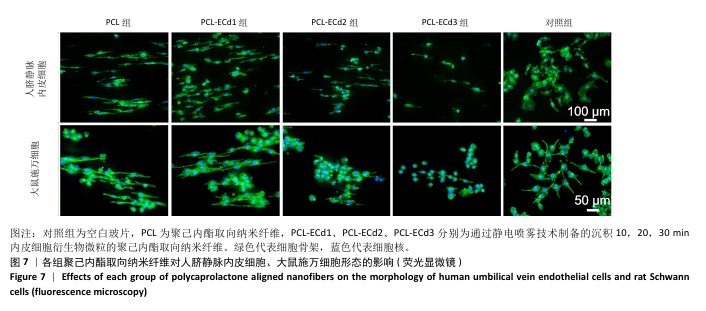
2.5.4 人脐静脉内皮细胞和施万细胞的形态 如图7所示,与对照组相比,PCL组、PCL-ECd1组、PCL-ECd2组、PCL-ECd3组纳米纤维膜上的人脐静脉内皮细胞(或大鼠施万细胞)沿着纤维方向生长。 根据人脐静脉内皮细胞、大鼠施万细胞增殖结果和形态观察结果,最终选取内皮细胞衍生物微粒的沉积时间为10 min,用于后续实验。"

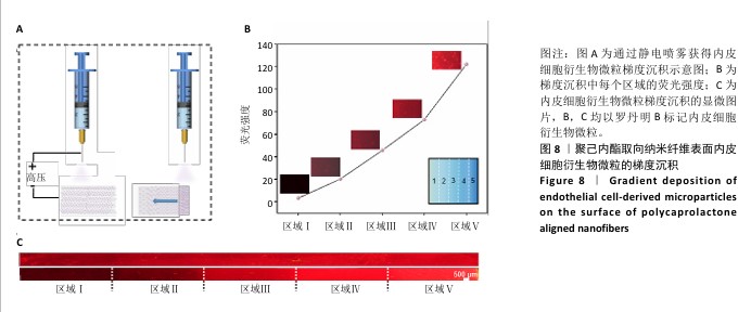
2.6 内皮细胞衍生物微粒的单向线性梯度沉积结果 选择10 min沉积时间,通过改变载玻片的遮挡范围,即载玻片的遮挡范围依次为4个区域、3个区域、2个区域、1个区域和无载玻片遮挡区域,使每个区域内皮细胞衍生物微粒的沉积量逐渐减少,最终获得内皮细胞衍生物微粒单向线性梯度沉积,制备流程见图8A。在罗丹明B标记内皮细胞衍生物微粒单向梯度涂层的聚己内酯取向纳米纤维膜上,荧光强度逐渐增加,并且最高侧的荧光强度约为最低侧的40倍(图8B),表明在聚己内酯取向纳米纤维上成功地沉积了内皮细胞衍生物微粒线性梯度。如图8C所示,荧光显微照片显示以罗丹明B标记的内皮细胞衍生物微粒在聚己内酯取向纳米纤维膜上的单向线性梯度沉积。 "
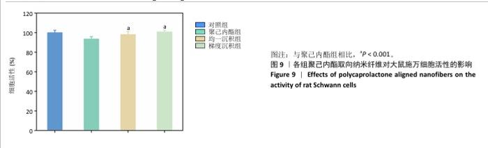
2.7 单向线性梯度沉积内皮细胞衍生物微粒聚己内酯取向纳米纤维对大鼠施万细胞的调控 2.7.1 施万细胞活性检测结果 CCK-8检测结果显示,与对照组相比,均一沉积组与梯度沉积组大鼠施万细胞活性无显著降低(P > 0.05),聚己内酯组大鼠施万细胞活性显著降低(P < 0.001),但各组细胞活性均大于标准化细胞活性(70%)[20](图9)。"


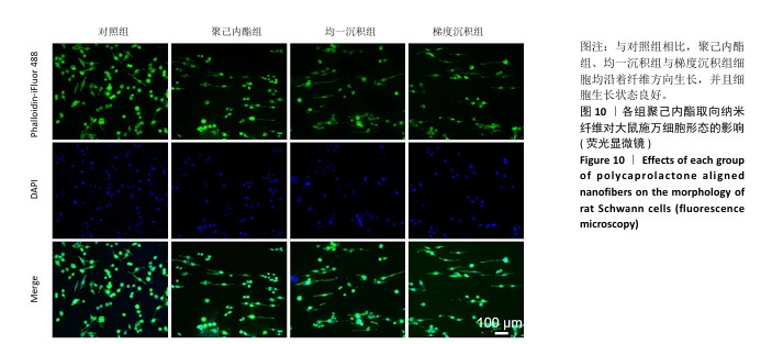
2.7.2 大鼠施万细胞形态观察 荧光显微观察结果显示,与对照组相比,聚己内酯组、均一沉积组与梯度沉积组细胞均沿着纤维方向生长,并且细胞生长状态良好,见图10。"
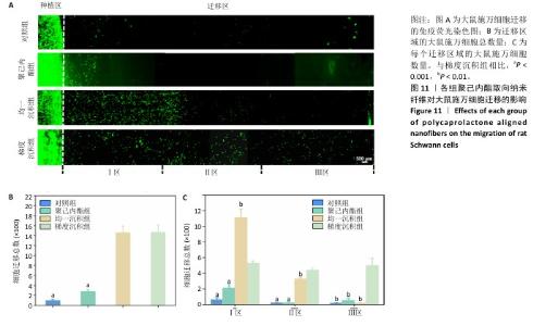
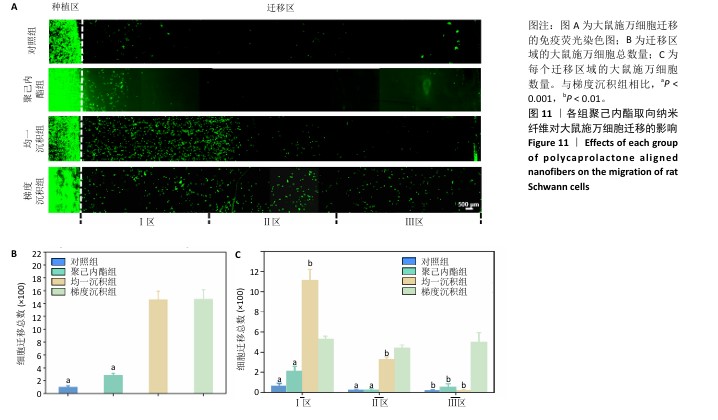
2.7.3 大鼠施万细胞迁移结果 如图11A所示,免疫荧光染色结果显示,与对照组相比,均一沉积组和梯度沉积组大鼠施万细胞均有不同程度的迁移,并且梯度沉积组大鼠施万细胞能够迁移至最远的位置。量化统计分析结果显示,均一沉积组和梯度沉积组大鼠施万细胞迁移总数多于对照组、聚己内酯组(P < 0.01),见图11B。将迁移区平分为3个区域,在迁移Ⅰ区,梯度沉积组大鼠施万细胞迁移数量少于均一沉积组(P < 0.01),多于对照组和聚己内酯组(P < 0.001);在迁移Ⅱ、Ⅲ区,梯度沉积组大鼠施万细胞迁移数量多于对照组、聚己内酯组与均一沉积组(P < 0.001,P < 0.01),见图11C。因此,内皮细胞衍生物的单向线性梯度可以驱使大鼠施万细胞迁移更远,这种效应可能归因于梯度形成的物理和化学信号,蛋白质的均一沉积会使生化信号在微环境中相对均匀分布,细胞接收到的信息较为一致,可能引起细胞整体上向某一特定方向迁移;而梯度沉积会造成生化信号在空间上的不均匀分布,细胞通过在不同位置获取不同信号以满足行为需求,从而影响细胞迁移的方向和速度[21-22]。"
| [1] SHARIFI M, KAMALABADI-FARAHANI M, SALEHI M, et al. Recent advances in enhances peripheral nerve orientation: the synergy of micro or nano patterns with therapeutic tactics. J Nanobiotechnology. 2024;22:194. [2] WANG X, CHEN S, CHEN X, et al. Biomimetic multi-channel nerve conduits with micro/nanostructures for rapid nerve repair. Bioact Mater. 2024;41:577-596. [3] XUE J, WU T, LI J, et al. Promoting the Outgrowth of Neurites on Electrospun Microfibers by Functionalization with Electrosprayed Microparticles of Fatty Acids. Angew Chem Int Ed Engl. 2019;58:3948-3951. [4] HAN Q, GUAN W, SUN S, et al. Anisotropic topological scaffolds synergizing non-invasive wireless magnetic stimulation for accelerating long-distance peripheral nerve regeneration. Chem Eng J. 2024;496:153809. [5] JIN B, YU Y, LOU C, et al. Combining a Density Gradient of Biomacromolecular Nanoparticles with Biological Effectors in an Electrospun Fiber-Based Nerve Guidance Conduit to Promote Peripheral Nerve Repair. Adv Sci (Weinh). 2023;10:e2203296. [6] MOTTA CMM, ENDRES KJ, WESDEMIOTIS C, et al. Enhancing Schwann cell migration using concentration gradients of laminin-derived peptides. Biomaterials. 2019;218:119335. [7] WU P, TONG Z, LUO L, et al. Comprehensive strategy of conduit guidance combined with VEGF producing Schwann cells accelerates peripheral nerve repair. Bioact Mater. 2021;6:3515-3527. [8] CERQUEIRA SR, LEE YS, CORNELISON RC, et al. Decellularized peripheral nerve supports Schwann cell transplants and axon growth following spinal cord injury. Biomaterials. 2018;177:176-185. [9] HUANG Y, YE K, HE A, et al. Dual-layer conduit containing VEGF-A - Transfected Schwann cells promotes peripheral nerve regeneration via angiogenesis. Acta Biomater. 2024;180:323-336. [10] MA T, HAO Y, LI S, et al. Sequential oxygen supply system promotes peripheral nerve regeneration by enhancing Schwann cells survival and angiogenesis. Biomaterials. 2022;289:121755. [11] TANG J, WU C, CHEN S, et al. Combining Electrospinning and Electrospraying to Prepare a Biomimetic Neural Scaffold with Synergistic Cues of Topography and Electrotransduction. ACS Appl Bio Mater. 2020; 3:5148-5159. [12] XUE J, PISIGNANO D, XIA Y. Maneuvering the Migration and Differentiation of Stem Cells with Electrospun Nanofibers. Adv Sci (Weinh). 2020;7:2000735. [13] JI E, SONG YH, LEE JK, et al. Bioadhesive levan-based coaxial nanofibrous membranes with enhanced cell adhesion and mesenchymal stem cell differentiation. Carbohydr Polym. 2025;354:123337. [14] QIAN J, LIN Z, LIU Y, et al. Functionalization strategies of electrospun nanofibrous scaffolds for nerve tissue engineering. Smart Mater Med. 2021;2:260-279. [15] LI Y, GE Z, LIU Z, et al. Integrating electrospun aligned fiber scaffolds with bovine serum albumin-basic fibroblast growth factor nanoparticles to promote tendon regeneration. J Nanobiotechnology. 2024;22:799. [16] WANG Q, ZHANG S, JIANG J, et al. Electrospun radially oriented berberine-PHBV nanofiber dressing patches for accelerating diabetic wound healing. Regen Biomater. 2024;11:rbae063. [17] ZHOU Z, LIN Y, LIU N, et al. Gradient coating of extracellular matrix derived from endothelial cells on aligned PCL nanofibers for rapid endothelialization. Front Bioeng Biotechnol. 2024;12:1527046. [18] ZHANG X, GUO M, GUO Q, et al. Modulating axonal growth and neural stem cell migration with the use of uniaxially aligned nanofiber yarns welded with NGF-loaded microparticles. Mater Today Adv. 2023;17: 100343. [19] XUE J, WU T, QIU J, et al. Accelerating Cell Migration along Radially Aligned Nanofibers through the Addition of Electrosprayed Nanoparticles in a Radial Density Gradient. Part Part Syst Charact. 2022;39(4):2100280. [20] BLACK BJ, ECKER M, STILLER A, et al. In vitro compatibility testing of thiol-ene/acrylate-based shape memory polymers for use in implantable neural interfaces. J Biomed Mater Res A. 2018;106:2891-2898. [21] XUE J, WU T, QIU J, et al. Promoting Cell Migration and Neurite Extension along Uniaxially Aligned Nanofibers with Biomacromolecular Particles in a Density Gradient. Adv Funct Mater. 2020;30(40):2002031. [22] WU T, XUE J, LI H, et al. General Method for Generating Circular Gradients of Active Proteins on Nanofiber Scaffolds Sought for Wound Closure and Related Applications. ACS Appl Mater Interfaces. 2018;10:8536-8545. [23] DAI Y, LU T, LI L, et al. Electrospun Composite PLLA-PPSB Nanofiber Nerve Conduits for Peripheral Nerve Defects Repair and Regeneration. Adv Healthc Mater. 2024;13:e2303539. [24] PATEL DK, WON SY, JUNG E, et al. Recent progress in biopolymer-based electrospun nanofibers and their potential biomedical applications: A review. Int J Biol Macromol. 2025;293:139426. [25] ROBLES KN, ZAHRA FT, MU R, et al. Advances in Electrospun Poly(ε-caprolactone)-Based Nanofibrous Scaffolds for Tissue Engineering. Polymers (Basel). 2024;16(20):2853. [26] SNYDER Y, TODD M, JANA S. Substrates with Tunable Hydrophobicity for Optimal Cell Adhesion. Macromol Biosci. 2024;24:e2400196. [27] KIM K, YANG J, LI C, et al. Anisotropic structure of nanofiber hydrogel accelerates diabetic wound healing via triadic synergy of immune-angiogenic-neurogenic microenvironments. Bioact Mater. 2025;47:64-82. [28] LIU H, PUIGGALÍ-JOU A, CHANSORIA P, et al. Filamented hydrogels as tunable conduits for guiding neurite outgrowth. Mater Today Bio. 2025;31:101471. [29] LIU S, SIMIŃSKA-STANNY J, YAN L, et al. Bioactive ECM-mimicking nerve guidance conduit for enhancing peripheral nerve repair. Mater Today Bio. 2024;29:101324. [30] GRASMAN JM, KAPLAN DL. Human endothelial cells secrete neurotropic factors to direct axonal growth of peripheral nerves. Sci Rep. 2017;7: 4092. [31] ZHOU Z, LIU N, ZHANG X, et al. Manipulating electrostatic field to control the distribution of bioactive proteins or polymeric microparticles on planar surfaces for guiding cell migration. Colloids Surf B. 2022;209: 112185. [32] ZHENG T, WU L, SUN S, et al. Co-culture of Schwann cells and endothelial cells for synergistically regulating dorsal root ganglion behavior on chitosan-based anisotropic topology for peripheral nerve regeneration. Burns Trauma. 2022;10:tkac030. [33] YAO X, XUE T, CHEN B, et al. Advances in biomaterial-based tissue engineering for peripheral nerve injury repair. Bioact Mater. 2025;46: 150-172. [34] HUANG J, LI J, LI S, et al. Netrin-1-engineered endothelial cell exosomes induce the formation of pre-regenerative niche to accelerate peripheral nerve repair. Sci Adv. 2024;10:eadm8454. [35] 宋凯凯,张锴,贾龙.周围神经系统损伤的微环境与修复方式[J]. 中国组织工程研究,2021,25(4):651-656. [36] WEI Y, ZHOU X, LI Z, et al. Genetically Programmed Single-Component Protein Hydrogel for Spinal Cord Injury Repair. Adv Sci (Weinh). 2025; 12(10):e2405054. [37] NING X, WANG R, LIU N, et al. Three-dimensional structured PLCL/ADM bioactive aerogel for rapid repair of full-thickness skin defects. Biomater Sci. 2024;12:6325-6337. [38] XU W, WU Y, LU H, et al. Injectable hydrogel encapsulated with VEGF-mimetic peptide-loaded nanoliposomes promotes peripheral nerve repair in vivo. Acta Biomater. 2023;160:225-238. [39] HUANG J, ZHANG G, LI S, et al. Endothelial cell-derived exosomes boost and maintain repair-related phenotypes of Schwann cells via miR199-5p to promote nerve regeneration. J Nanobiotechnology. 2023;21:10. [40] XIA B, LV Y. Dual-delivery of VEGF and NGF by emulsion electrospun nanofibrous scaffold for peripheral nerve regeneration. Mater Sci Eng C Mater Biol Appl. 2018;82:253-264. [41] SENGUPTA S, PARENT CA, BEAR JE. The principles of directed cell migration. Nat Rev Mol Cell Biol. 2021;22:529-547. [42] IDRISOVA KF, ZEINALOVA AK, MASGUTOVA GA, et al. Application of neurotrophic and proangiogenic factors as therapy after peripheral nervous system injury. Neural Regen Res. 2022;17:1240-1247. [43] MENEZES R, VINCENT R, OSORNO L, et al. Biomaterials and tissue engineering approaches using glycosaminoglycans for tissue repair: Lessons learned from the native extracellular matrix. Acta Biomater. 2023;163:210-227. [44] BARIK D, SHYAMAL S, DAS K, et al. Glycoprotein Injectable Hydrogels Promote Accelerated Bone Regeneration through Angiogenesis and Innervation. Adv Healthc Mater. 2023;12:e2301959. [45] CHU X L, SONG X Z, LI Q, et al. Basic mechanisms of peripheral nerve injury and treatment via electrical stimulation. Neural Regen Res. 2022; 17:2185-2193. |
| [1] | Sun Lei, Zhang Qi, Zhang Yu. Pro-osteoblastic effect of chlorogenic acid protein microsphere/polycaprolactone electrospinning membrane [J]. Chinese Journal of Tissue Engineering Research, 2026, 30(8): 1877-1884. |
| [2] | Wang Qisa, Lu Yuzheng, Han Xiufeng, Zhao Wenling, Shi Haitao, Xu Zhe. Cytocompatibility of 3D printed methyl acrylated hyaluronic acid/decellularized skin hydrogel scaffolds [J]. Chinese Journal of Tissue Engineering Research, 2026, 30(8): 1912-1920. |
| [3] | Wang Ying, Wang Yawen, Xu Yingjie, Wang Yuanfei, Wu Tong. Preparation of polycaprolactone/low molecular weight fucoidan nanofibers by emulsion electrospinning and assessment of their biocompatibility [J]. Chinese Journal of Tissue Engineering Research, 2026, 30(2): 433-442. |
| [4] | Li He, Wang Yu, Xie Zikang, Jiang Tao. Application of electrospinning in artificial dura mater substitutes: diverse biological functions [J]. Chinese Journal of Tissue Engineering Research, 2026, 30(14): 3702-3708. |
| Viewed | ||||||
|
Full text |
|
|||||
|
Abstract |
|
|||||