[1] BENIASH E, STIFLER CA, SUN CY, et al. The hidden structure of human enamel. Nat Commun. 2019;10(1):4383.
[2] GAZZANI F, BELLISARIO D, QUADRINI F, et al. Light-curing process for clear aligners’ attachment reproduction: comparison between two nanocomposites cured by the auxiliary of a new tool. BMC Oral Health. 2022;22(1):376.
[3] SUNDARARAJ D, VENKATACHALAPATHY S, TANDON A, et al. Critical evaluation of incidence and prevalence of white spot lesions during fixed orthodontic appliance treatment: A meta-analysis. J Int Soc Prev Community Dent. 2015;5(6):433-439.
[4] 李爽,胡敏.关于正畸固定矫治中牙釉质脱矿的研究进展[J].口腔医学研究, 2021,37(8):685-688.
[5] HAMDAN AH, SALEAH SA, SEONG D, et al. Three-Dimensional Assessment of Dental Enamel Microcrack Progression After Orthodontic Bracket Debonding Using Optical Coherence Tomography. J Funct Biomater. 2024;16(1):7.
[6] INCHINGOLO F, INCHINGOLO AM, RICCALDO L, et al. Structural and Color Alterations of Teeth following Orthodontic Debonding: A Systematic Review. J Funct Biomater. 2024;15:123.
[7] PENG S, SANG T, WANG H, et al. Bioinspired Anti-demineralization Enamel Coating for Orthodontics. J Dent Res. 2022;101(13):1620-1627.
[8] JANISZEWSKA-OLSZOWSKA J, TANDECKA K, SZATKIEWICZ T, et al. Three-dimensional analysis of enamel surface alteration resulting from orthodontic clean-up -comparison of three different tools. BMC Oral Health. 2015;15(1):146.
[9] MICHALAK M, KIRYK S, KOTELA A, et al. Orthodontic Ceramic Bracket Removal Using Lasers: A Systematic Review. J Funct Biomater. 2025;16:123.
[10] KHALIL AS, TAMISH NM, ELKALZA AR, et al. Assessment of chemical, ultrasonic, diode laser, and Er:YAG laser application on debonding of ceramic brackets. BMC Oral Health. 2022;22(1):79.
[11] GRAZIOLI G, HARDAN L, BOURGI R, et al. Residual Adhesive Removal Methods for Rebonding of Debonded Orthodontic Metal Brackets: Systematic Review and Meta-Analysis. Materials (Basel). 2021;14:6120.
[12] OSORIO R, TOLEDANO M, GARCÍA-GODOY F, et al. Enamel surface morphology after bracket debonding. ASDC J Dent Child. 1998;65(5):313-317,354.
[13] ULUSOY C. Comparison of finishing and polishing systems for residual resin removal after debonding. J Appl Oral Sci. 2009;17(3):209-215.
[14] WEBB BJ, KOCH J, HAGAN JL, et al. Enamel surface roughness of preferred debonding and polishing protocols. J Orthod. 2016;43(1):39-46.
[15] SHAH P, SHARMA P, GOJE SK, et al. Comparative evaluation of enamel surface roughness after debonding using four finishing and polishing systems for residual resin removal-an in vitro study. Prog Orthod. 2019;20(1):18.
[16] JANISZEWSKA-OLSZOWSKA J, SZATKIEWICZ T, TOMKOWSKI R, et al. Effect of orthodontic debonding and adhesive removal on the enamel - current knowledge and future perspectives - a systematic review. Med Sci Monit. 2014;20: 1991-2001.
[17] ELIADES T, KOLETSI D. Minimizing the aerosol-generating procedures in orthodontics in the era of a pandemic: Current evidence on the reduction of hazardous effects for the treatment team and patients. Am J Orthod Dentofacial Orthop. 2020;158(3):330-342.
[18] AL SHAMSI AH, CUNNINGHAM JL, LAMEY PJ, et al. Three-dimensional measurement of residual adhesive and enamel loss on teeth after debonding of orthodontic brackets: an in-vitro study. Am J Orthod Dentofacial Orthop. 2007;131(3):301.e309-315.
[19] CHOUR RG, MODA A, ARORA A, et al. Comparative evaluation of effect of different polishing systems on surface roughness of composite resin: An in vitro study. J Int Soc Prev Community Dent. 2016;6(Suppl 2):S166-170.
[20] GÖMLEKSIZ S, GÖMLEKSIZ O. The effect of contemporary finishing and polishing systems on the surface roughness of bulk fill resin composite and nanocomposites. J Esthet Restor Dent. 2022;34(6):915-923.
[21] GHALEB L, AL-WORAFI NA, THAWABA A, et al. Evaluation of enamel surface integrity after orthodontic bracket debonding: comparison of three different system. BMC Oral Health. 2024;24(1):358.
[22] BOLLEN CM, LAMBRECHTS P, QUIRYNEN M, et al. Comparison of surface roughness of oral hard materials to the threshold surface roughness for bacterial plaque retention: a review of the literature. Dent Mater. 1997;13(4):258-269.
[23] TURKOGLU H, ATIK E. 3-Dimensional topographic enamel surface changes after different debonding techniques for aligner attachments: a micro-CT study. Clin Oral Investig. 2025;29(1):89.
[24] RYF S, FLURY S, PALANIAPPAN S, et al. Enamel loss and adhesive remnants following bracket removal and various clean-up procedures in vitro. Eur J Orthod. 2012;34(1):25-32.
[25] 杨佳佳,罗瑞,许晓,等.不同车针去除正畸粘接剂对牙表面粗糙度的影响[J].口腔材料器械杂志,2017,26(3):124-128.
[26] 刘光俊,马锦锦,陶珂金,等.不同粘接剂去除方式对釉质微观结构及托槽抗剪切粘接强度的影响[J].口腔医学,2022,42(9):785-789.
[27] 钮晔,安贝尔,敬怡,等.5种不同去牙面正畸残余粘接剂方法比较[J].上海口腔医学,2019,28(2):136-140.
[28] PINZAN-VERCELINO CRM, SOUZA COSTA AC, GURGEL JA, et al. Comparison of enamel surface roughness and color alteration after bracket debonding and polishing with 2 systems: A split-mouth clinical trial. Am J Orthod Dentofacial Orthop. 2021;160(5):686-694.
[29] KŘIVKOVÁ T, TICHÝ A, TYCOVÁ H, et al. The Influence of Various Adhesive Systems and Polishing Methods on Enamel Surface Roughness after Debonding of Orthodontic Brackets: A Three-Dimensional In Vitro Evaluation. Materials (Basel). 2023;16:5107.
[30] 徐春华.固定正畸治疗粘接托槽后局部抛光对牙菌斑控制的分析[J].全科口腔医学电子杂志,2018,5(21):74-75.
[31] 范晓川,陈莉,黄晓峰.等.不同方法去除不同材质正畸托槽及粘接剂的对比实验研究[J].北京口腔医学,2017,25(6):337-342.
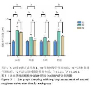
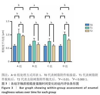
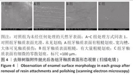
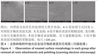
|