[1] BAI Y, WANG Z, HE X, et al. Application of Bioactive Materials for Osteogenic Function in Bone Tissue Engineering. Small Methods. 2024;8:e2301283.
[2] WANG J, LIU M, YANG C, et al. Biomaterials for bone defect repair: Types, mechanisms and effects. Int J Artif Organs. 2024;47:75-84.
[3] KIM K, SU Y, KUCINE AJ, et al. Guided Bone Regeneration Using Barrier Membrane in Dental Applications. ACS Biomater Sci Eng. 2023;9(10): 5457-5478.
[4] ELGALI I, OMAR O, DAHLIN C, et al. Guided bone regeneration: materials and biological mechanisms revisited. Eur J Oral Sci. 2017;125(5):315-337.
[5] ALQAHTANI AM. Guided Tissue and Bone Regeneration Membranes: A Review of Biomaterials and Techniques for Periodontal Treatments. Polymers (Basel). 2023;15(16):3355.
[6] YANG Z, WU C, SHI H, et al. Advances in Barrier Membranes for Guided Bone Regeneration Techniques. Front Bioeng Biotechnol. 2022;10:921576.
[7] ALI M, MOHD NOOR SNF, MOHAMAD H, et al. Advances in guided bone regeneration membranes: a comprehensive review of materials and techniques. Biomed Phys Eng Express. 2024;10(3):32003.
[8] BEE SL, HAMID ZAA. Asymmetric resorbable-based dental barrier membrane for periodontal guided tissue regeneration and guided bone regeneration: A review. J Biomed Mater Res B Appl Biomater. 2022;110(9):2157-2182.
[9] MATEO-SIDRON ANTON MC, PEREZ-GONZALEZ F, MENIZ-GARCIA C. Titanium mesh for guided bone regeneration: a systematic review. Br J Oral Maxillofac Surg. 2024;62:433-440.
[10] VROOM MG, GRUNDEMANN LJ, GALLO P. Clinical Classification of Healing Complications and Management in Guided Bone Regeneration Procedures with a Nonresorbable d-PTFE Membrane. Int J Periodontics Restorative Dent. 2022;42:419-427.
[11] REN Y, FAN L, ALKILDANI S, et al. Barrier Membranes for Guided Bone Regeneration (GBR): A Focus on Recent Advances in Collagen Membranes. Int J Mol Sci. 2022;23(23):14987.
[12] ABTAHI S, CHEN X, SHAHABI S, et al. Resorbable Membranes for Guided Bone Regeneration: Critical Features, Potentials, and Limitations. ACS Mater Au. 2023;3(5):394-417.
[13] ZHOU Z, YUN J, LI J, et al. Comparison of the efficacy of different biodegradable membranes in guided bone/tissue regeneration: a systematic review and network meta-analysis. Biomed Mater. 2023;18:032003.
[14] MATHEW A, VAQUETTE C, HASHIMI S, et al. Antimicrobial and Immunomodulatory Surface-Functionalized Electrospun Membranes for Bone Regeneration. Adv Healthc Mater. 2017;6(10):1601345.
[15] XUE J, HE M, LIU H, et al. Drug loaded homogeneous electrospun PCL/gelatin hybrid nanofiber structures for anti-infective tissue regeneration membranes. Biomaterials. 2014;35:9395-9405.
[16] HAN F, ZHANG P, SUN Y, et al. Hydroxyapatite-doped polycaprolactone nanofiber membrane improves tendon-bone interface healing for anterior cruciate ligament reconstruction. Int J Nanomedicine. 2015; 10:7333-7343.
[17] KARFELD-SULZER LS, GHAYOR C, SIEGENTHALER B, et al. Comparative study of NMP-preloaded and dip-loaded membranes for guided bone regeneration of rabbit cranial defects. J Tissue Eng Regen Med. 2017;11:425-433.
[18] BENITO-GARZON L, GUADILLA Y, DIAZ-GUEMES I, et al. Nanostructured Zn-Substituted Monetite Based Material Induces Higher Bone Regeneration Than Anorganic Bovine Bone and beta-Tricalcium Phosphate in Vertical Augmentation Model in Rabbit Calvaria. Nanomaterials (Basel). 2021; 12:143.
[19] PARK WB, CRASTO GJ, HAN JY, et al. Bone Regenerative Potential of Cross-Linked Collagen Membrane in Peri-Implant Osseous Defect: Case Series with Histologic/Micro-Computed Tomographic Findings. Medicina (Kaunas). 2023;59:176.
[20] ZHANG C, DU T, MU G, et al. Evaluation and ultrastructural changes of amniotic membrane fragility after UVA/riboflavin cross-linking and its effects on biodegradation. Medicine (Baltimore). 2020;99:e20091.
[21] CIARDELLI G, GENTILE P, CHIONO V, et al. Enzymatically crosslinked porous composite matrices for bone tissue regeneration. J Biomed Mater Res A. 2010;92(1):137-151.
[22] TERZI A, STORELLI E, BETTINI S, et al. Effects of processing on structural, mechanical and biological properties of collagen-based substrates for regenerative medicine. Sci Rep. 2018;8:1429.
[23] ZHANG JY, LIU K, LIU RX, et al. Safety and Efficacy of Midface Augmentation Using Bio-Oss Bone Powder and Bio-Gide Collagen Membrane in Asians. J Clin Med. 2023;12(3):959.
[24] APAZA ALCCAYHUAMAN KA, HEIMEL P, TANGL S, et al. Active and Passive Mineralization of Bio-Gide((R)) Membranes in Rat Calvaria Defects. J Funct Biomater. 2024;15(3):54.
[25] 孙晓嘉,马宁.不同口腔修复膜材料在牙种植中的引导骨再生效果分析[J].中国现代药物应用,2024,18(15):51-54.
[26] 尤鹏越,刘玉华,王新知,等.脱细胞猪心包膜生物相容性及成骨性能的体内外评价[J].北京大学学报(医学版),2021,53(4):776-784.
[27] 林季萩,莫安春.牛心包膜在美学区水平骨增量手术中的应用效果分析[J].口腔疾病防治,2025,33(3):203-210.
[28] 高丹妮,郭星,谢言,等.GTR联合根管治疗左上前牙牙周牙髓联合病变1例[J].牙体牙髓牙周病学杂志,2024,29(12):719-722.
[29] MEYER M. Processing of collagen based biomaterials and the resulting materials properties. Biomed Eng Online. 2019;18(1):24.
[30] 王莉莉,佳严,李东升,等.两种新型胶原膜引导骨组织再生的体内外性能研究[J].口腔医学,2019,39(6):481.
[31] BARBECK M, LORENZ J, KUBESCH A, et al. Porcine Dermis-Derived Collagen Membranes Induce Implantation Bed Vascularization Via Multinucleated Giant Cells: A Physiological Reaction? J Oral Implantol. 2015;41:e238-251.
[32] NOBLE C, MORSE D, LERMAN A, et al. Evaluation of Pericardial Tissues from Assorted Species as a Tissue-Engineered Heart Valve Material. Med Biol Eng Comput. 2022;60:393-406.
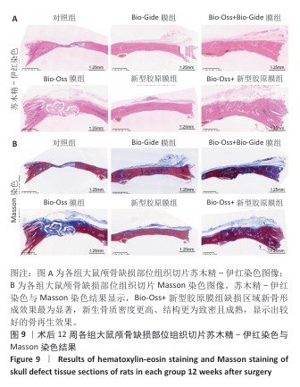
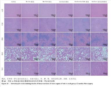
|