Chinese Journal of Tissue Engineering Research ›› 2025, Vol. 29 ›› Issue (20): 4249-4257.doi: 10.12307/2025.695
Previous Articles Next Articles
Pueraria lobata decoction intervenes in neuroinflammatory response and apoptosis in rats with cervical spondylotic myelopathy
Wu Diyou1, Huang Jiajun1, Tao Guangyi1, Zhao Yu1, Huang Junqing2, Yang Bin2, Xue Yun1
- 1Henan University of Chinese Medicine, Zhengzhou 450000, Henan Province, China; 2Henan Province Hospital of Traditional Chinese Medicine, Second Affiliated Hospital of Henan University of Chinese Medicine, Zhengzhou 450000, Henan Province, China
-
Received:2024-07-22Accepted:2024-08-31Online:2025-07-18Published:2024-12-20 -
Contact:Huang Junqing, MS, Chief physician, Henan Province Hospital of Traditional Chinese Medicine, Second Affiliated Hospital of Henan University of Chinese Medicine, Zhengzhou 450000, Henan Province, China -
About author:Wu Diyou, Master’s candidate, Henan University of Chinese Medicine, Zhengzhou 450000, Henan Province, China -
Supported by:Special Projects for Scientific Research on Traditional Chinese Medicine in Henan Province, Nos. 2019JDZX095 (to HJQ) and 2024ZY2066 (to YB)
CLC Number:
Cite this article
Wu Diyou, Huang Jiajun, Tao Guangyi, Zhao Yu, Huang Junqing, Yang Bin, Xue Yun. Pueraria lobata decoction intervenes in neuroinflammatory response and apoptosis in rats with cervical spondylotic myelopathy[J]. Chinese Journal of Tissue Engineering Research, 2025, 29(20): 4249-4257.
share this article
Add to citation manager EndNote|Reference Manager|ProCite|BibTeX|RefWorks
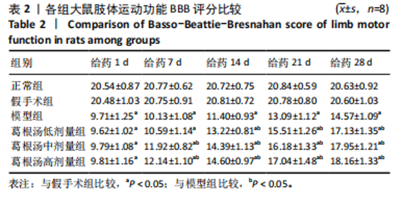
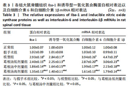
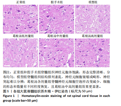
2.1 实验动物数量分析 参与实验60只SD大鼠,在手术过程中模型组和葛根汤低剂量组各有1只大鼠出现死亡。术后1 d,葛根汤中剂量组有1只大鼠出现死亡,可能与出血、窒息等因素有关。最终57只大鼠用于结果分析。 2.2 葛根汤对脊髓型颈椎病大鼠BBB评分的影响 给药后不同时间点,正常组大鼠BBB评分与假手术组比较无显著差异(P > 0.05);模型组、葛根汤低、中、高剂量组大鼠BBB评分与正常组和假手术组比较显著降低(P < 0.05);给药1 d,葛根汤低、中、高剂量组大鼠BBB评分与模型组比较无显著差异(P > 0.05);给药7 d,葛根汤中、高剂量组大鼠BBB评分高于模型组(P < 0.05);给药14,21,28 d,葛根汤低、中、高剂量组大鼠BBB评分高于模型组(P < 0.05),见表2。 2.3 葛根汤对脊髓型颈椎病大鼠脊髓组织病理形态的影响 正常组和假手术组大鼠脊髓组织可见神经元胞体饱满,分布均匀且形态完整清晰;模型组大鼠脊髓组织可见结构排列紊乱,神经元细胞皱缩或畸形,神经突起难以分辨;葛根汤低、中、高剂量组大鼠脊髓神经元细胞空泡样改变减少,细胞的形态和数量有不同程度的恢复。葛根汤中、高剂量组效果较葛根汤低剂量组更显著。见图1。"
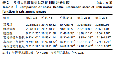
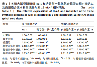
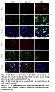
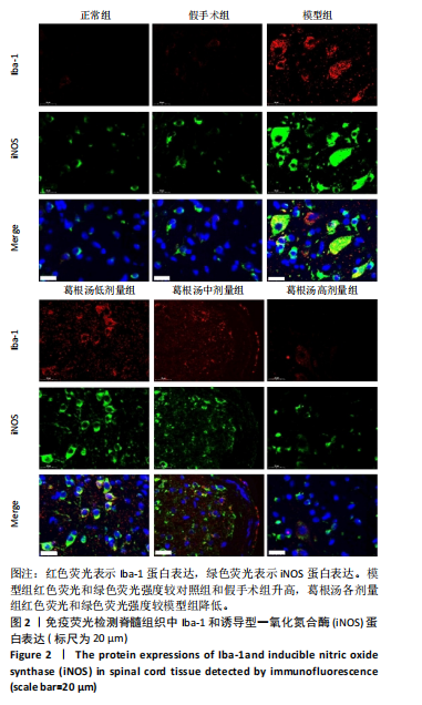
2.4 葛根汤对脊髓型颈椎病大鼠脊髓组织小胶质细胞极化的影响 Iba-1是小胶质细胞的标志蛋白。免疫荧光染色结果显示,与正常组和假手术组比较,模型组脊髓组织Iba-1蛋白表达明显升高(P < 0.05);与模型组比较,葛根汤低、中、高剂量组脊髓组织Iba-1蛋白表达降低(P < 0.05)。此外,诱导型一氧化氮合酶是小胶质细胞M1型活化的特异性指标。免疫荧光结果显示,与正常组和假手术组比较,模型组脊髓组织诱导型一氧化氮合酶蛋白表达升高;与模型组比较,葛根汤低、中、高剂量组脊髓组织诱导型一氧化氮合酶蛋白表达降低(P < 0.05)。小胶质细胞M1型活化能够导致白细胞介素6和白细胞介素1β等促炎因子的表达,进一步加剧神经系统损伤。荧光定量PCR结果显示,与正常组和假手术组比较,模型组脊髓组织白细胞介素6和白细胞介素1β mRNA表达升高(P < 0.05);与模型组比较,葛根汤低、中、高剂量组脊髓组织白细胞介素6和白细胞介素1β mRNA表达降低(P < 0.05)。与葛根汤低、中剂量组比较,葛根汤高剂量组脊髓组织中Iba-1、诱导型一氧化氮合酶蛋白表达以及白细胞介素6和白细胞介素1β mRNA表达降低(P < 0.05)。见图2和表3。"

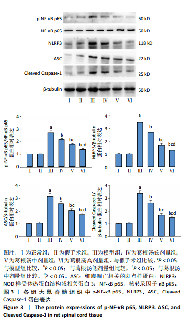
2.5 葛根汤对大鼠脊髓组织p-NF-κB p65、NLRP3、ASC、Cleaved Caspase-1蛋白表达的影响 NF-κB和NLRP3炎症小体的活化与脊髓受压部位神经炎症反应有着密切关系。与正常组和假手术组比较,模型组大鼠脊髓组织中p-NF-κB "
"

p65、NLRP3、ASC、Cleaved Caspase-1蛋白表达升高(P < 0.05);与模型组比较,葛根汤低、中、高剂量组大鼠脊髓组织中p-NF-κB p65、NLRP3、ASC、Cleaved Caspase-1蛋白表达降低( P < 0.05)。与葛根汤低、中剂量组比较,葛根汤高剂量组大鼠脊髓组织中p-NF-κB p65、NLRP3、ASC、Cleaved Caspase-1蛋白表达降低(P < 0.05),见图3。"
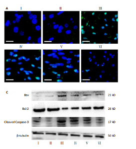
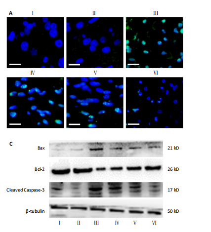
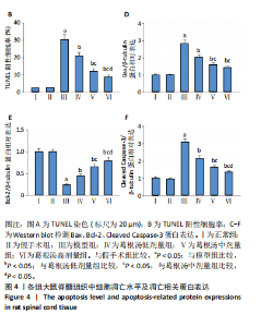
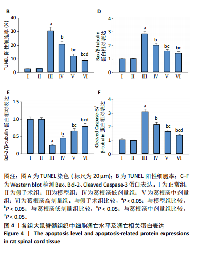
2.6 葛根汤对大鼠脊髓组织细胞凋亡以及Bax、Bcl-2和Cleaved Caspase-3蛋白表达的影响 与正常组和假手术组比较,模型组大鼠脊髓组织中TUNEL阳性细胞率、Bax和Cleaved Caspase-3蛋白表达升高,Bcl-2蛋白表达降低,差异有显著性意义(P < 0.05);与模型组比较,葛根汤低、中、高剂量组大鼠脊髓组织中TUNEL阳性细胞率、Bax和Cleaved Caspase-3蛋白表达降低,Bcl-2蛋白表达升高,差异有显著性意义(P < 0.05)。与葛根汤低剂量组比较,葛根汤中、高剂量组大鼠脊髓组织中TUNEL阳性细胞率、Bax和Cleaved Caspase-3蛋白表达降低(P < 0.05),Bcl-2蛋白表达升高(P < 0.05)。与葛根汤中剂量组比较,葛根汤高剂量组大鼠脊髓组织中TUNEL阳性细胞率和Cleaved Caspase-3蛋白表达降低(P < 0.05),Bcl-2蛋白表达升高(P < 0.05),见图4。"
"
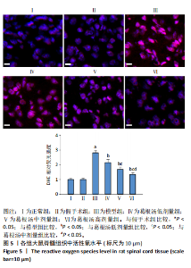
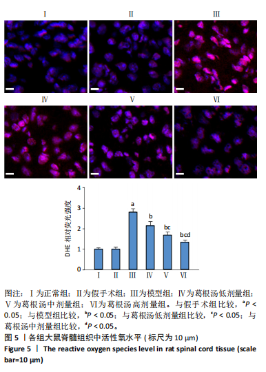
2.7 葛根汤对大鼠脊髓组织活性氧水平的影响 与正常组和假手术组比较,模型组大鼠脊髓组织中活性氧水平升高(P < 0.05);与模型组比较,葛根汤低、中、高剂量组大鼠脊髓组织中活性氧水平降低,差异有显著性意义(P < 0.05)。与葛根汤低剂量组比较,葛根汤中、高剂量组大鼠脊髓组织中活性氧水平降低(P < 0.05)。与葛根汤中剂量组比较,葛根汤高剂量组大鼠脊髓组织中活性氧水平降低(P < 0.05),见图5。"
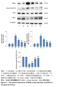
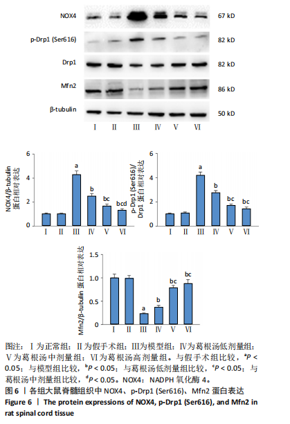
2.8 葛根汤对大鼠脊髓组织NOX4、p-Drp1和Mfn2蛋白表达的影响 与正常组和假手术组比较,模型组大鼠脊髓组织中NOX4和p-Drp1 (Ser616)蛋白表达升高,Mfn2蛋白表达降低,差异有显著性意义(P < 0.05);与模型组比较,葛根汤低、中、高剂量组大鼠脊髓组织中NOX4和p-Drp1 (Ser616)蛋白表达降低,Mfn2蛋白表达升高 (P < 0.05)。与葛根汤低剂量组比较,葛根汤中、高剂量组大鼠脊髓组织中NOX4和p-Drp1 (Ser616)蛋白表达降低,Mfn2蛋白表达升高 ,差异有显著性意义(P < 0.05),见图6。"
| [1] MCCORMICK JR, SAMA AJ, SCHILLER NC, et al. Cervical Spondylotic Myelopathy: A Guide to Diagnosis and Management. J Am Board Fam Med. 2020;33(2):303-313. [2] 刘正蓬,王雅辉,张义龙,等.3D打印椎间融合器置入治疗脊髓型颈椎病:颈椎曲度及椎间高度恢复的半年随访[J].中国组织工程研究,2021,25(6):849-853. [3] KHAN AF, HAYNES G, MOHAMMADI E, et al. Utility of MRI in Quantifying Tissue Injury in Cervical Spondylotic Myelopathy. J Clin Med. 2023;12(9):3337. [4] BONOSI L, MUSSO S, CUSIMANO LM, et al. The role of neuronal plasticity in cervical spondylotic myelopathy surgery: functional assessment and prognostic implication. Neurosurg Rev. 2023;46(1):149. [5] TOCI GR, CANSECO JA, KARAMIAN BA, et al. The impact of preoperative neurological symptom severity on postoperative outcomes in cervical spondylotic myelopathy. J Craniovertebr Junction Spine. 2022;13(1): 94-100. [6] 邱丽英,王利仁.基于“阳化气,阴成形”及“温养督脉”理论探讨脊髓型颈椎病治法[J].陕西中医,2024,45(7):951-954. [7] 睦顺姬,叶秀兰,姚敏,等.中医药治疗脊髓型颈椎病的机理研究概况[J].世界中医药,2017,12(1):222-224+228. [8] BOOGAARTS HD, BARTELS RH. Prevalence of cervical spondylotic myelopathy. Eur Spine J. 2015;24 Suppl 2:139-141. [9] 刘志伟,银河,陈忻,等.益肾养髓方治疗轻中度脊髓型颈椎病的疗效研究[J].中国中医骨伤科杂志,2022,30(8):36-40+45. [10] 莫文,袁文.脊髓型颈椎病中西医结合诊疗指南(2023)[J].中国骨伤,2024,37(1):103-110. [11] 卞正航,许金海,叶洁,等.中药治疗脊髓型颈椎病作用机制的研究进展[J].中医正骨,2023,35(12):36-41. [12] 陈强,逯艳婷,王洪海.《伤寒论》葛根汤古今应用探析[J].辽宁中医药大学学报,2024,26(10):165-169. [13] 阴继爱,戴岳,安树庞.葛根汤的药理和临床研究概况[J].中华中医药学刊,2007(6):1275-1278. [14] 王献宇.葛根汤加味防治脊髓型颈椎病颈椎前路术后发生轴性症状的疗效观察[D].福州:福建中医药大学,2018. [15] 马梦全.补阳还五汤、身痛逐瘀汤、葛根汤复方加减治疗脊髓型颈椎病临床运用效果分析[J].世界最新医学信息文摘,2017, 17(67):145. [16] 张典,银河,朱立国,等.益肾养髓方对脊髓型颈椎病大鼠脊髓中细胞凋亡的影响[J].中国中医骨伤科杂志,2024,32(1):1-6. [17] KARADIMAS SK, GATZOUNIS G, FEHLINGS MG. Pathobiology of cervical spondylotic myelopathy. Eur Spine J. 2015;24 Suppl 2:132-138. [18] KALSI-RYAN S, KARADIMAS SK, FEHLINGS MG. Cervical spondylotic myelopathy: the clinical phenomenon and the current pathobiology of an increasingly prevalent and devastating disorder. Neuroscientist. 2013;19(4):409-421. [19] 刘恩旭,段嘉豪,杨雷,等.杨少锋教授基于六经辨证治疗颈椎病的经验[J].湖南中医药大学学报,2023,43(11):2013-2017. [20] 林少平,伦婷婷,王世雄,等.葛根汤联合高能量体外冲击波对神经根型颈椎病患者血清炎性因子的影响[J/OL].中华中医药学刊,1-11[2024-08-24].http://kns.cnki.net/kcms/detail/21.1546.R.20240306.1737.002.html. [21] 杨博文,朱立国,陈忻,等.益肾养髓方对脊髓型颈椎病大鼠脊髓炎症的影响[J].时珍国医国药,2023,34(5):1056-1059. [22] 李淦.参芪麝蓉丸调控内质网应激抑制神经元凋亡治疗脊髓型颈椎病的机制研究[D].上海:上海中医药大学,2021. [23] 邓文龙.动物中人体剂量换算遵循的原则[J].中药药理与临床, 2016,32(3):196-197. [24] BASSO DM, BEATTIE MS, BRESNAHAN JC. A sensitive and reliable locomotor rating scale for open field testing in rats. J Neurotrauma. 1995;12(1):1-21. [25] IYER A, AZAD TD, THARIN S. Cervical Spondylotic Myelopathy. Clin Spine Surg. 2016;29(10):408-414. [26] 李梁,赵晓玲,詹常森.治疗脊髓型颈椎病的中药组方及临床评价研究进展[J].中成药,2023,45(3):861-865. [27] 卜献忠,钟远鸣,卜保献,等.脊髓伤方促进脊髓型颈椎病(气虚血瘀证)术后恢复的效果研究[J].时珍国医国药,2023,34(12): 2951-2955. [28] 王文雅,王会生,陈萌.基于津液理论探讨经方治疗颈椎病的科学内涵[J].环球中医药,2023,16(3):518-521. [29] 黄代富,王庆龙,闵欢.葛根汤加减配合手法,颈部十字操治疗混合型颈椎病的临床研究[J].贵州中医药大学学报,2023,45(5):57-60. [30] YAO M, LI G, PU PM, et al. Neuroinflammation and apoptosis after surgery for a rat model of double-level cervical cord compression. Neurochem Int. 2022;157:105340. [31] YU WR, LIU T, KIEHL TR, et al. Human neuropathological and animal model evidence supporting a role for Fas-mediated apoptosis and inflammation in cervical spondylotic myelopathy. Brain. 2011;134(Pt 5):1277-1292. [32] 劳泽辉,凌国伟,华国花.circRNAs调控NF-κB信号通路在脊髓型颈椎病中的作用及机制研究[J].中国医学工程,2022,30(12):24-28. [33] PENG L, WEN L, SHI QF, et al. Scutellarin ameliorates pulmonary fibrosis through inhibiting NF-κB/NLRP3-mediated epithelial-mesenchymal transition and inflammation. Cell Death Dis. 2020;11(11):978. [34] O’SHEA TM, BURDA JE, SOFRONIEW MV. Cell biology of spinal cord injury and repair. J Clin Invest. 2017;127(9):3259-3270. [35] BRENNAN FH, LI Y, WANG C, et al. Microglia coordinate cellular interactions during spinal cord repair in mice. Nat Commun. 2022; 13(1):4096. [36] 成志红,冯松,王霞,等.米诺环素对脊神经结扎大鼠脊髓内M1/M2型小胶质细胞极化的影响[J].陆军军医大学学报,2024, 46(15):1740-1750. [37] 杨博文,朱立国,陈忻,等.益肾养髓方对脊髓型颈椎病大鼠小胶质细胞极化的影响[J].中国中医骨伤科杂志,2023,31(3):1-5. [38] JIANG D, YANG X, GE M, et al. Zinc defends against Parthanatos and promotes functional recovery after spinal cord injury through SIRT3-mediated anti-oxidative stress and mitophagy. CNS Neurosci Ther. 2023;29(10):2857-2872. [39] Schmidt J, Quintá HR. Mitochondrial dysfunction as a target in spinal cord injury: intimate correlation between pathological processes and therapeutic approaches. Neural Regen Res. 2023;18(10):2161-2166. [40] TILOKANI L, NAGASHIMA S, PAUPE V, et al. Mitochondrial dynamics: overview of molecular mechanisms. Essays Biochem. 2018;62(3): 341-360. [41] 周文洋,廖烨晖,田明昊,等.NLRP3炎性小体在脊髓损伤后小胶质细胞中的作用[J].中国组织工程研究,2025,29(13):2849-2860. [42] PU PM, LI ZY, DAI YX, et al. Analysis of gene expression profiles and experimental validations of a rat chronic cervical cord compression model. Neurochem Int. 2023;168:105564. |
| [1] | Miao Jiahang, Ma Sheng, Li Qupeng, Yu Huilin, Hu Tianyu, Gao Xiao, Feng Hu. Cervical lordosis ratio can be used as a decision-making indicator for selection of posterior surgical approach for multi-level cervical spondylotic myelopathy [J]. Chinese Journal of Tissue Engineering Research, 2025, 29(9): 1796-1802. |
| [2] | Yu Shuai, Liu Jiawei, Zhu Bin, Pan Tan, Li Xinglong, Sun Guangfeng, Yu Haiyang, Ding Ya, Wang Hongliang. Hot issues and application prospects of small molecule drugs in treatment of osteoarthritis [J]. Chinese Journal of Tissue Engineering Research, 2025, 29(9): 1913-1922. |
| [3] | Yin Lu, Jiang Chuanfeng, Chen Junjie, Yi Ming, Wang Zihe, Shi Houyin, Wang Guoyou, Shen Huarui. Effect of Complanatoside A on the apoptosis of articular chondrocytes [J]. Chinese Journal of Tissue Engineering Research, 2025, 29(8): 1541-1547. |
| [4] | Li Kaiying, Wei Xiaoge, Song Fei, Yang Nan, Zhao Zhenning, Wang Yan, Mu Jing, Ma Huisheng. Mechanism of Lijin manipulation regulating scar formation in skeletal muscle injury repair in rabbits [J]. Chinese Journal of Tissue Engineering Research, 2025, 29(8): 1600-1608. |
| [5] | Jin Kai, Tang Ting, Li Meile, Xie Yuan. Effects of conditioned medium and exosomes of human umbilical cord mesenchymal stem cells on proliferation, migration, invasion, and apoptosis of hepatocellular carcinoma cells [J]. Chinese Journal of Tissue Engineering Research, 2025, 29(7): 1350-1355. |
| [6] | He Longcai, Song Wenxue, Ming Jiang, Chen Guangtang, Wang Junhao, Liao Yidong, Cui Junshuan, Xu Kaya. An experimental method for simultaneous extraction and culture of primary cortical neurons and microglial cells from SD rats [J]. Chinese Journal of Tissue Engineering Research, 2025, 29(7): 1395-1400. |
| [7] | Wan Lingling, Wu Mengying, Zhang Yujiao, Luo Qingqing. Inflammatory factor interferon-gamma affects migration and apoptosis of human vascular smooth muscle cells through pyroptosis pathway [J]. Chinese Journal of Tissue Engineering Research, 2025, 29(7): 1422-1428. |
| [8] | Chi Wenxin, Zhang Cunxin, Gao Kai, Lyu Chaoliang, Zhang Kefeng. Mechanism by which nobiletin inhibits inflammatory response of BV2 microglia [J]. Chinese Journal of Tissue Engineering Research, 2025, 29(7): 1321-1327. |
| [9] | Zhao Ruihua, Chen Sixian, Guo Yang, Shi Lei, Wu Chengjie, Wu Mao, Yang Guanglu, Zhang Haoheng, Ma Yong. Wen-Shen-Tong-Du Decoction promoting spinal cord injury repair in mice [J]. Chinese Journal of Tissue Engineering Research, 2025, 29(6): 1118-1126. |
| [10] | Liu Zhezhe, Yu Meiqing, Wang Tingting, Zhang Min, Li Baiyan. Troxerutin modulates nuclear factor-kappaB signaling pathway to inhibit brain injury and neuronal apoptosis in cerebral infarction rats [J]. Chinese Journal of Tissue Engineering Research, 2025, 29(6): 1137-1143. |
| [11] | He Guanghui, Yuan Jie, Ke Yanqin, Qiu Xiaoting, Zhang Xiaoling. Hemin regulates mitochondrial pathway of oxidative stress in mouse chondrocytes [J]. Chinese Journal of Tissue Engineering Research, 2025, 29(6): 1183-1191. |
| [12] | He Bo, Chen Wen, Ma Suilu, He Zhijun, Song Yuan, Li Jinpeng, Liu Tao, Wei Xiaotao, Wang Weiwei, Xie Jing . Pathogenesis and treatment progress of flap ischemia-reperfusion injury [J]. Chinese Journal of Tissue Engineering Research, 2025, 29(6): 1230-1238. |
| [13] | Lan Shuangli, Xiang Feifan, Deng Guanghui, Xiao Yukun, Yang Yunkang, Liang Jie. Naringin inhibits iron deposition and cell apoptosis in bone tissue of osteoporotic rats [J]. Chinese Journal of Tissue Engineering Research, 2025, 29(5): 888-898. |
| [14] | Bai Jing, Zhang Xue, Ren Yan, Li Yuehui, Tian Xiaoyu. Effect of lncRNA-TNFRSF13C on hypoxia-inducible factor 1alpha in periodontal cells by modulation of #br# miR-1246 #br# [J]. Chinese Journal of Tissue Engineering Research, 2025, 29(5): 928-935. |
| [15] | Zhi Fang, Zhu Manhua, Xiong Wei, Lin Xingzhen. Analgesic effect of acupuncture in a rat model of lumbar disc herniation [J]. Chinese Journal of Tissue Engineering Research, 2025, 29(5): 936-941. |
| Viewed | ||||||
|
Full text |
|
|||||
|
Abstract |
|
|||||