[1] IZUNO H, HORI K, SAWADA M, et al. Physical fitness and oral function in community-dwelling older people: a pilot study. Gerodontology. 2016; 33:470-479.
[2] INUI A, TAKAHASHI I, KURAUCHI S, et al. Oral conditions and dysphagia in Japanese, community-dwelling middle- and older- aged adults, independent in daily living. Clin Interv Aging. 2017;12:515-521.
[3] OSAWA A, MAESHIMA S, MATSUDA H, et al. Functional lesions in dysphagia due to acute stroke: discordance between abnormal findings of bedside swallowing assessment and aspiration on videofluorography. Neuroradiology. 2013;55:413-421.
[4] MAESHIMA S, OSAWA A, YAMANE F, et al. Dysphagia following acute thalamic haemorrhage: clinical correlates and outcomes. Eur Neurol. 2014;71:165-172.
[5] MATSUO K, PALMER JB. Anatomy and physiology of feeding and swallowing: normal and abnormal. Phys Med Rehabil Clin N Am. 2008; 19:691-707,vii.
[6] HUMBERT IA, FITZGERALD ME, MCLAREN DG, et al. Neurophysiology of swallowing: effects of age and bolus type. Neuroimage. 2009;44:982-991.
[7] ZHANG Z, YAN L, XING X, et al. Brain Activation Site of Laryngeal Elevation During Swallowing: An fMRI Study. Dysphagia. 2022. doi: 10.1007/s00455-022-10464-y.
[8] TOOGOOD JA, SMITH RC, STEVENS TK, et al. Swallowing Preparation and Execution: Insights from a Delayed-Response Functional Magnetic Resonance Imaging (fMRI) Study. Dysphagia. 2017;32:526-541.
[9] LIMA MS, MANGILLI LD, SASSI FC, et al. Functional magnetic resonance and swallowing: critical literature review. Braz J Otorhinolaryngol. 2015; 81: 671-680.
[10] KOBER SE, GROSSINGER D, WOOD G. Effects of Motor Imagery and Visual Neurofeedback on Activation in the Swallowing Network: A Real-Time fMRI Study. Dysphagia. 2019;34:879-895.
[11] BU L, HUO C, QIN Y, et al. Effective Connectivity in Subjects With Mild Cognitive Impairment as Assessed Using Functional Near-Infrared Spectroscopy. Am J Phys Med Rehabil. 2019;98:438-445.
[12] KHAN MNA, GHAFOOR U, YOO HR, et al. Acupuncture enhances brain function in patients with mild cognitive impairment: evidence from a functional-near infrared spectroscopy study. Neural Regen Res. 2022; 17(8):1850-1856.
[13] BU L, WU Y, YAN Q, et al. Effects of physical training on brain functional connectivity of methamphetamine dependencies as assessed using functional near-infrared spectroscopy. Neurosci Lett. 2020;715:134605.
[14] SCHOLKMANN F, KLEISER S, METZ AJ, et al. A review on continuous wave functional near-infrared spectroscopy and imaging instrumentation and methodology. Neuroimage. 2014;85 Pt 1:6-27.
[15] SASAI S, HOMAE F, WATANABE H, et al. A NIRS- fMRI study of resting state network. Neuroimage. 2012;63:179-193.
[16] MATSUMOTO T, SAITO K, NAKAMURA A, et al. Dried-bonito aroma components enhance salivary hemodynamic responses to broth tastes detected by near-infrared spectroscopy. J Agric Food Chem. 2012;60: 805-811.
[17] SATO H, OBATA AN, MODA I, et al. Application of near-infrared spectroscopy to measurement of hemodynamic signals accompanying stimulated saliva secretion. J Biomed Opt. 2011;16:047002.
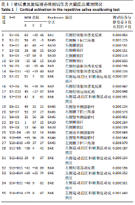
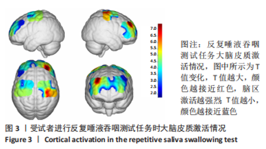
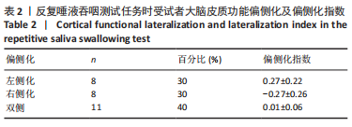
[18] 邰佳慧,李浩正,王婷玮,等.基于功能性近红外光谱技术的吞咽相关任务大脑皮质功能偏侧化研究[J].中国康复医学杂志,2022, 37(5):594-599.
[19] JIA G, LIU G, NIU H. Hemispheric Lateralization of Visuospatial Attention Is Independent of Language Production on Right-Handers: Evidence From Functional Near-Infrared Spectroscopy. Front Neurol. 2021;12: 784821.
[20] VERNOOIJ MW, SMITS M, WIELOPOLSKI PA, et al. Fiber density asymmetry of the arcuate fasciculus in relation to functional hemispheric language lateralization in both right- and left-handed healthy subjects: a combined fMRI and DTI study. Neuroimage. 2007; 35:1064-1076.
[21] KNOLLHOFF SM, HANCOCK AS, BARRETT TS, et al. Cortical Activation of Swallowing Using fNIRS: A Proof of Concept Study with Healthy Adults. Dysphagia 2022. doi: 10.1007/s00455-021-10403-3.
[22] WILMSKOETTER J, BONILHA L, MARTIN-HARRIS B, et al. Mapping acute lesion locations to physiological swallow impairments after stroke. Neuroimage Clin. 2019;22:101685.
[23] TAE WS, LEE S, CHOI S, et al. Effects of aging on brain networks during swallowing: general linear model and independent component analyses. Sci Rep. 2021;11:1069.
[24] SÖRÖS P, INAMOTO Y, MARTIN RE. Functional brain imaging of swallowing: an activation likelihood estimation meta-analysis. Hum Brain Mapp. 2009;30:2426-2439.
[25] GRIFFIN L, KAMARUNAS E, KUO C, et al. Comparing amplitudes of transcranial direct current stimulation (tDCS) to the sensorimotor cortex during swallowing. Exp Brain Res. 2022;240:1811-1822.
[26] KAWAI T, WATANABE Y, TONOGI M, et al. Visual and auditory stimuli associated with swallowing: an fMRI study. Bull Tokyo Dent Coll. 2009; 50:169-181.
[27] DUVERNE S, KOECHLIN E. Rewards and Cognitive Control in the Human Prefrontal Cortex. Cereb Cortex. 2017;27:5024-5039.
[28] LEE J, YAMATE C, TAIRA M, et al. Prefrontal cortex activity during swallowing in dysphagia patients. J Oral Sci. 2018;60:329-335.
[29] KOBER SE, WOOD G. Hemodynamic signal changes during saliva and water swallowing: a near-infrared spectroscopy study. J Biomed Opt. 2018;23:1-7.
[30] KOBER SE, HINTERLEITNER V, BAUERNFEIND G, et al. Trainability of hemodynamic parameters: A near-infrared spectroscopy based neurofeedback study. Biol Psychol. 2018;136:168-180.
[31] KOBER SE, GRESSENBERGER B, KURZMANN J, et al. Voluntary Modulation of Hemodynamic Responses in Swallowing Related Motor Areas: A Near-Infrared Spectroscopy-Based Neurofeedback Study. PLoS One. 2015;10:e0143314.
[32] KOBER SE, BAUERNFEIND G, WOLLER C, et al. Hemodynamic Signal Changes Accompanying Execution and Imagery of Swallowing in Patients with Dysphagia: A Multiple Single-Case Near-Infrared Spectroscopy Study. Front Neurol. 2015;6:151.
[33] KOBER SE, WOOD G. Changes in hemodynamic signals accompanying motor imagery and motor execution of swallowing: a near-infrared spectroscopy study. Neuroimage.2014;93 Pt 1:1-10.
[34] KOGANEMARU S, MIZUNO F, TAKAHASHI T, et al. Event-Related Desynchronization and Corticomuscular Coherence Observed During Volitional Swallow by Electroencephalography Recordings in Humans. Front Hum Neurosci. 2021;15:643454.
[35] MALANDRAKI GA, SUTTON BP, PERLMAN AL, et al. Age-related differences in laterality of cortical activations in swallowing. Dysphagia. 2010;25:238-249.
[36] TEISMANN IK, STEINSTRAETER O, SCHWINDT W, et al. Age-related changes in cortical swallowing processing. Neurobiol Aging. 2010;31: 1044-1050.
[37] DZIEWAS R, SÖRÖS P, ISHII R, et al. Neuroimaging evidence for cortical involvement in the preparation and in the act of swallowing. Neuroimage. 2003;20:135-144.
[38] WILMSKOETTER J, MARTIN-HARRIS B, PEARSON WG JR, et al. Differences in swallow physiology in patients with left and right hemispheric strokes. Physiol Behav. 2018;194:144-152.
[39] MOON HI, YOON SY, YI TI, et al. Lesions Responsible for Delayed Oral Transit Time in Post-stroke Dysphagia. Dysphagia. 2018; 33: 321-328.
[40] KOBER SE, WOOD G, KURZMANN J, et al. Near-infrared spectroscopy based neurofeedback training increases specific motor imagery related cortical activation compared to sham feedback. Biol Psychol. 2014;95: 21-30.
|