[1] 王国民,陈伟.泌尿系统肿瘤治疗的进展与展望[J].肿瘤防治研究, 2014,41(2):97-101.
[2] 纪建鹏,黎丽华,杨荣骞,等.基于分水岭算法的交互式三维分割方法[J].中国组织工程研究,2011,15(39):7351-7354.
[3] 吕晓琪,郭金鸽,赵宇红,等.基于图像分割的Tamura纹理特征算法的研究与实现[J].中国组织工程研究,2012,16(17):3160-3163.
[4] 杜雪婷,杨洋,黄文华,等.基于医学影像技术的3D打印临床应用与突破[J].中国组织工程研究,2021,25(18):2887-2894.
[5] 杨谊,喻德旷.基于改进水平集图像分割方法的乳腺超声病灶提取[J].计算机应用与软件,2014,31(11):217-221.
[6] 秦进,付茂臣,任泽民.结合扩展LoB滤波的距离保持水平集方法[J].遵义师范学院学报,2012,14(6):85-88.
[7] CHAN TF, VESE LA. Active Contours without Edges. IEEE Trans Image Process. 2001;10(2):266-277.
[8] 李社蕾,黄梦醒.改进几何活动轮廓模型的水下图像分割算法研究[J].小型微型计算机系统,2019,40(3):671-675.
[9] 赵辉,芮修业,岳有军,等.复杂背景下基于AD-GAC模型和最大熵阈值法的叶片病斑分割[J].江苏农业科学,2019,47(18):136-140.
[10] 刘晨,李丙春,王文龙,等.基于偏微分方程的GAC水平集图像分割模型[J].安徽大学学报(自然科学版),2020,44(4):45-51.
[11] 孟颖慧,潘杨,朱磊,等.使用测地线活动轮廓模型的合成孔径雷达图像分割方法[J].科学技术与工程,2020,20(20):8310-8315.
[12] 李渊强.病理图像中细胞核自动分割算法研究[D].南京:南京理工大学,2019.
[13] 潘改.偏微分方程在图像分割中的应用研究[D].沈阳:东北大学, 2013.
[14] HELLER N, SATHIANATHEN N, KALAPARA A, et al. The KiTS19 challenge data: 300 kidney tumor cases with clinical context, CT semantic segmentations, and surgical outcomes. arXiv. 2019:13.
[15] 张品,梁艳梅,常胜江.基于改进C-V模型的肾脏CT图像分割方法[J].光电子•激光,2013,24(3):602-607.
[16] 钱芸,张英杰.水平集的图像分割方法综述[J].中国图象图形学报, 2008(1):7-13.
[17] CASELLES V, KIMMEL R, SAPIRO G. Geodesic Active Contours. Int J Comput Vis. 1997;22(1):694-699.
[18] 伍艺萌. 基于变分偏微分方程的医学影像分割方法[D].南京:邮电大学,2020.
[19] 朱泽华,闫士举,阮渊,等.基于改进DRLSE模型的前列腺磁共振图像分割[J].波谱学杂志,2020,37(4):447-455.
[20] 房巾莉,吕毅斌,王樱子,等.基于水平集的医学图像分割算法[J].电子科技,2021,34(2):12-20.
[21] LI C, XU C, GUI C, et al. Distance regularized level set evolution and its application to image segmentation. IEEE Trans Image Process. 2010; 19(12):3243-3254.
[22] 陈皓,李广,刘洋,等.一种在MR图像中进行脑胶质瘤检测和病灶分割的方法[J].电子与信息学报,2021,43(4):992-1002.
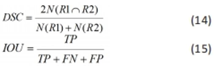
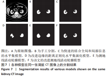
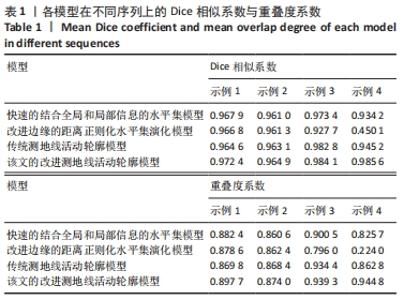
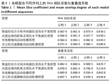
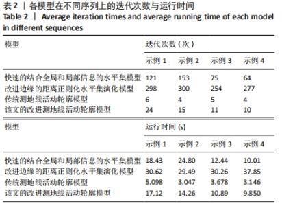
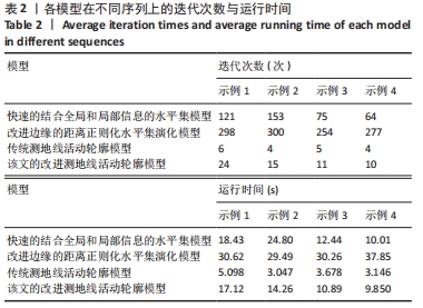
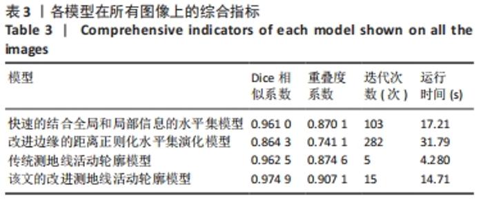
|