Chinese Journal of Tissue Engineering Research ›› 2021, Vol. 25 ›› Issue (26): 4168-4174.doi: 10.12307/2021.116
Previous Articles Next Articles
Extracellular matrix promotes fibroblast proliferation and post-operation knee arthrofibrosis through ERK signaling pathway
Hou Jingzhao1, Zhang Zhen2
- 1Jingjiang People’s Hospital, Taizhou 214500, Jiangsu Province, China; 2Dalian Medical University, Dalian 116044, Liaoning Province, China
-
Received:2020-07-27Revised:2020-07-29Accepted:2020-08-19Online:2021-09-18Published:2021-05-10 -
Contact:Zhang Zhen, Physician, Dalian Medical University, Dalian 116044, Liaoning Province, China E-mail:1395637072@qq.com -
About author:Hou Jingzhao, Master, Associate chief physician, Jingjiang People’s Hospital, Taizhou 214500, Jiangsu Province, China
CLC Number:
Cite this article
Hou Jingzhao, Zhang Zhen. Extracellular matrix promotes fibroblast proliferation and post-operation knee arthrofibrosis through ERK signaling pathway[J]. Chinese Journal of Tissue Engineering Research, 2021, 25(26): 4168-4174.
share this article
Add to citation manager EndNote|Reference Manager|ProCite|BibTeX|RefWorks
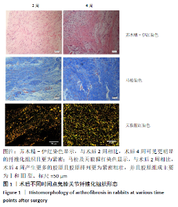
2.1 体内实验结果 2.1.1 实验动物数量分析 所有兔都很好地耐受手术,术后至取材前没有明显的伤口感染、皮肤坏死,整个实验过程中无动物死亡。 2.1.2 组织学分析结果 苏木精-伊红染色显示,与术后2周相比,术后4周可见更明显的纤维化组织且更为紧密;马松及天狼猩红染色显示,与术后2周相比,术后4周产生更多的胶原且胶原排列更为紧密粗壮,并且通过天狼猩红染色中胶原颜色能够看到其胶原组成主要为Ⅰ和 Ⅲ 型,见图1,说明外基质与膝关节纤维化的发展具有相关性。"

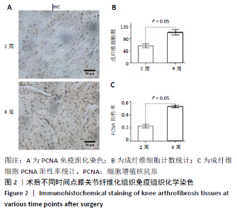
2.1.3 免疫组织化学染色结果 成纤维细胞计数分析显示,术后4周的膝关节纤维化组织中成纤维细胞更多,约为术后2周时的2倍;PCNA免疫组织化学染色结果显示,术后4周可见更多的细胞核聚集,处于增殖期的成纤维细胞约术后2周的2.4倍,见图2。"
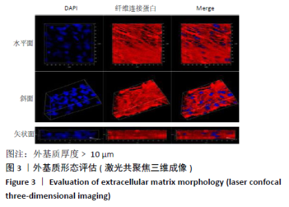
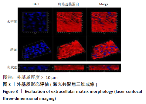
2.2 体外实验结果 2.2.1 外基质构建结果 前期研究发现,细胞分泌外基质厚度 ≥10 μm。激光共聚焦三维显微镜显示,纤维连接蛋白呈现网状结构,构成细胞生存的支架,且细胞核散在分布于网状结构的空隙,横切面显示外基质厚度≥10 μm,见图3。"
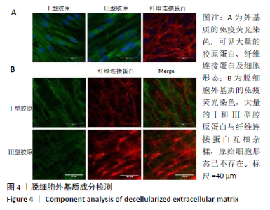
2.2.2 免疫荧光染色结果 未脱细胞外基质可见大量的胶原蛋白、纤维连接蛋白及细胞形态,见图4A。脱细胞后,大量的Ⅰ和 Ⅲ 型胶原蛋白与纤维连接蛋白互相杂糅,构成细胞生存的网络系统,原始细胞形态已不存在,见图4B。结合动物纤维化组织中的胶原构成,表明脱细胞外基质基本能够模拟体内的外基质组成。"
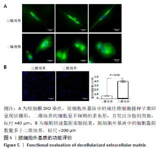
2.2.3 细胞膜染色与细胞黏附结果 细胞膜染色显示,三维培养的细胞呈现纺锤形,二维培养的细胞则呈现不规则图形并出现大量散乱的突触,见图5A,说明脱细胞外基质更利于细胞的早期功能维持。快速黏附实验结果示,脱细胞外基质更利于成纤维细胞的早期黏附,数量较二维培养增加了约5倍,见图5B。"


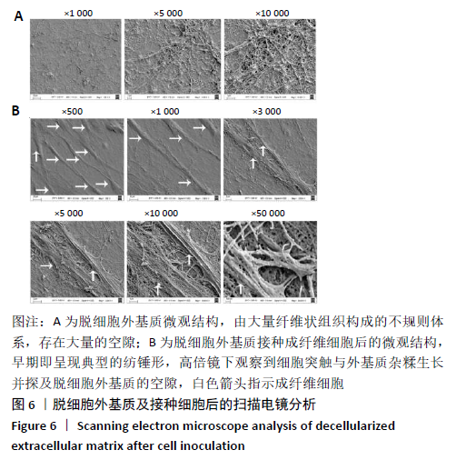
2.2.4 扫描电镜观察结果 扫描电镜显示,脱细胞外基质是由大量纤维状组织构成的不规则体系,存在大量的空隙,见图6A。接种成纤维细胞5 h后,成纤维细胞呈现典型的纺锤形,其突触与脱细胞外基质的纤维状组织交织并可深入到脱细胞外基质的空隙,见图6B,从而促进细胞的黏附、迁移及生存。"

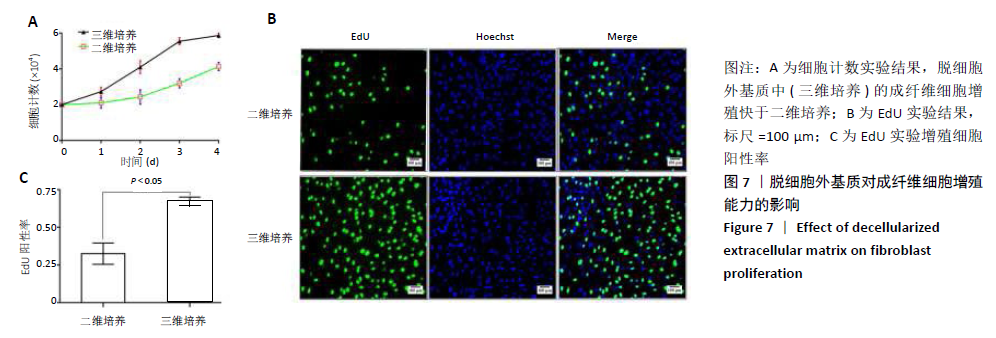
2.2.5 细胞计数与EdU实验结果 细胞计数结果显示,接种于脱细胞外基质中的成纤维细胞能够更快地增殖,至72 h时细胞的增殖逐渐减缓;二维培养的细胞起始增长较慢,至72 h时细胞才处于快速增长阶段,但数量依然低于三维培养,见图7A。EdU实验结果显示,相较于二维培养,脱细胞外基质中处于增殖状态的成纤维细胞数量增加了1倍,见图7B,C。"
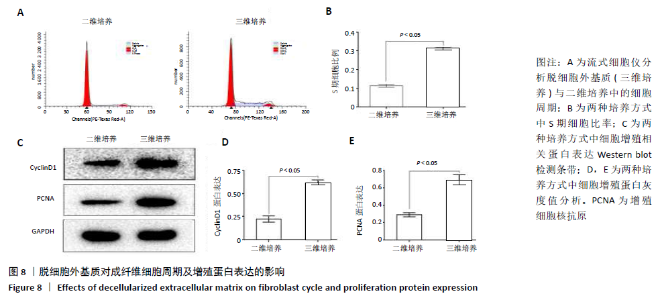
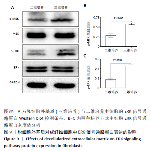
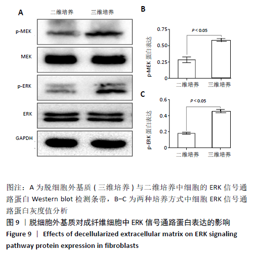
2.2.6 细胞周期分析结果 结果显示,脱细胞外基质中处于S期的成纤维细胞数量约为二维培养的3倍,见图8A,B。 2.2.7 Western blot检测结果 Western blot检测结果示,脱细胞外基质中细胞的增殖蛋白PCNA、Cyclin D1表达水平高于二维培养,见图8C-E,说明脱细胞外基质更利于成纤维细胞的增殖;脱细胞外基质中细胞的p-MEK、p-ERK蛋白表达高于二维培养(P < 0.05),见图9。"
"
| [1] EKHTIARI S, HORNER NS, DE SA D, et al. Arthrofibrosis after ACL reconstruction is best treated in a step-wise approach with early recognition and intervention: a systematic review. Knee Surg Sports Traumatol Arthrosc. 2017;25(12):3929-3937. [2] CHEUY VA, FORAN JRH, PAXTON RJ, et al. Arthrofibrosis Associated With Total Knee Arthroplasty. J Arthroplasty. 2017;32(8):2604-2611. [3] FAUST I, TRAUT P, NOLTING F, et al. Human xylosyltransferases--mediators of arthrofibrosis? New pathomechanistic insights into arthrofibrotic remodeling after knee replacement therapy. Sci Rep. 2015;28(5):512-537. [4] HAMM-FABER TE, GULTUNA I, VAN GORP EJ, et al. High-Dose Spinal Cord Stimulation for Treatment of Chronic Low Back Pain and Leg Pain in Patients With FBSS, 12-Month Results: A Prospective Pilot Study. Neuromodulation. 2020;23(1):118-125. [5] CZAMARA A, KUZNIECOW M, KROLIKOWSKA A. Arthrofibrosis of the Knee Joint - the Current State of Knowledge. Literature Review. Ortop Traumatol Rehabil. 2019;21(2):95-106. [6] 吴海啸,王鹏,张超,等.膝关节粘连:治疗和预防研究新进展[J].中国组织工程研究,2017,21(36):5879-5885. [7] LIU L, SUI T, HONG X, et al. Inhibition of epidural fibrosis after microendoscopic discectomy with topical application of mitomycin C: a randomized, controlled, double-blind trial. J Neurosurg Spine. 2013;18(5):421-427. [8] YILDIZ KH, GEZEN F, IS M, et al. Mitomycin C, 5-fluorouracil, and cyclosporin A prevent epidural fibrosis in an experimental laminectomy model. Eur Spine J. 2007;16(9):1525-1530. [9] MANRIQUE J, GOMEZ MM, PARVIZI J. Stiffness after total knee arthroplasty. J Knee Surg. 2015;28(2):119-126. [10] SU EP, SU SL, DELLA VALLE AG. Stiffness after TKR: how to avoid repeat surgery. Orthopedics. 2010;33(9):658. [11] ECKENRODE BJ. An algorithmic approach to rehabilitation following arthroscopic surgery for arthrofibrosis of the knee. Physiother Theory Pract. 2018;34(1):66-74. [12] BRAM JT, GAMBONE AJ, DEFRANCESCO CJ, et al. Use of Continuous Passive Motion Reduces Rates of Arthrofibrosis After Anterior Cruciate Ligament Reconstruction in a Pediatric Population. Orthopedics. 2019; 42(1):e81-e85. [13] 阳庆军,汪鑫.水下牵伸、关节松动对膝关节僵硬的康复疗效观察[J].中国康复,2020,35(3):147-149. [14] WATSON RS, GOUZE E, LEVINGS PP, et al. Gene delivery of TGF-beta1 induces arthrofibrosis and chondrometaplasia of synovium in vivo. Lab Invest. 2010;90(11):1615-1627. [15] BAYRAM B, LIMBERG AK, SALIB CG, et al. Molecular pathology of human knee arthrofibrosis defined by RNA sequencing. Genomics. 2020;112(4):2703-2712. [16] LI X, CHEN S, YAN L, et al. Prospective application of stem cells to prevent post-operative skeletal fibrosis. J Orthop Res. 2019;37(6): 1236-1245. [17] WAN Q, CHEN H, XIONG G, et al. Artesunate protects against surgery-induced knee arthrofibrosis by activating Beclin-1-mediated autophagy via inhibition of mTOR signaling. Eur J Pharmacol. 2019;85(4):149-158. [18] FRANCO-BARRAZA J, BEACHAM DA, AMATANGELO MD, et al. Preparation of Extracellular Matrices Produced by Cultured and Primary Fibroblasts. Curr Protoc Cell Biol. 2016;71(10):1-34. [19] CHROBOK J, VRBA I, STETKAROVA I. Selection of surgical procedures for treatment of failed back surgery syndrome (FBSS). Chir Narzadow Ruchu Ortop Pol. 2005;70(2):147-153. [20] 陈一鑫,陈小莉,王开龙,等.伸直型膝关节僵硬外治方法研究进展[J].辽宁中医药大学学报,2020,22(2):111-114. [21] RUTHERFORD RW, JENNINGS JM, LEVY DL, et al. Revision Total Knee Arthroplasty for Arthrofibrosis. J Arthroplasty. 2018;33(7S):S177-S181. [22] LI X, CHEN H, WANG S, et al. Tacrolimus induces fibroblasts apoptosis and reduces epidural fibrosis by regulating miR-429 and its target of RhoE. Biochem Biophys Res Commun. 2017;490(4):1197-1204. [23] THOMPSON R, NOVIKOV D, CIZMIC Z, et al. Arthrofibrosis After Total Knee Arthroplasty: Pathophysiology, Diagnosis, and Management. Orthop Clin North Am. 2019;50(3):269-279. [24] HALLER JM, HOLT DC, MCFADDEN ML, et al. Arthrofibrosis of the knee following a fracture of the tibial plateau. Bone Joint J. 2015;97-B(1): 109-114. [25] USHER KM, ZHU S, MAVROPALIAS G, et al. Pathological mechanisms and therapeutic outlooks for arthrofibrosis. Bone Res. 2019;7:9. [26] CUKIERMAN E, PANKOV R, STEVENS DR, et al. Taking cell-matrix adhesions to the third dimension. Science. 2001;294(5547):1708-1712. [27] HERRERA J, HENKE CA, BITTERMAN PB. Extracellular matrix as a driver of progressive fibrosis. J Clin Invest. 2018;128(1):45-53. [28] CHO N, RAZIPOUR SE, MCCAIN ML. Featured Article: TGF-beta1 dominates extracellular matrix rigidity for inducing differentiation of human cardiac fibroblasts to myofibroblasts. Exp Biol Med (Maywood). 2018;243(7):601-612. |
| [1] | Pu Rui, Chen Ziyang, Yuan Lingyan. Characteristics and effects of exosomes from different cell sources in cardioprotection [J]. Chinese Journal of Tissue Engineering Research, 2021, 25(在线): 1-. |
| [2] | An Yang, Liao Yinan, Xie Chengxin, Li Qinglong, Huang Ge, Jin Xin, Yin Dong. Mechanism of Inulae flos in the treatment of osteoporosis: an analysis based on network pharmacology [J]. Chinese Journal of Tissue Engineering Research, 2021, 25(在线): 1-8. |
| [3] | Xu Feng, Kang Hui, Wei Tanjun, Xi Jintao. Biomechanical analysis of different fixation methods of pedicle screws for thoracolumbar fracture [J]. Chinese Journal of Tissue Engineering Research, 2021, 25(9): 1313-1317. |
| [4] | Jiang Yong, Luo Yi, Ding Yongli, Zhou Yong, Min Li, Tang Fan, Zhang Wenli, Duan Hong, Tu Chongqi. Von Mises stress on the influence of pelvic stability by precise sacral resection and clinical validation [J]. Chinese Journal of Tissue Engineering Research, 2021, 25(9): 1318-1323. |
| [5] | Zhang Tongtong, Wang Zhonghua, Wen Jie, Song Yuxin, Liu Lin. Application of three-dimensional printing model in surgical resection and reconstruction of cervical tumor [J]. Chinese Journal of Tissue Engineering Research, 2021, 25(9): 1335-1339. |
| [6] | Zhang Yu, Tian Shaoqi, Zeng Guobo, Hu Chuan. Risk factors for myocardial infarction following primary total joint arthroplasty [J]. Chinese Journal of Tissue Engineering Research, 2021, 25(9): 1340-1345. |
| [7] | Wei Wei, Li Jian, Huang Linhai, Lan Mindong, Lu Xianwei, Huang Shaodong. Factors affecting fall fear in the first movement of elderly patients after total knee or hip arthroplasty [J]. Chinese Journal of Tissue Engineering Research, 2021, 25(9): 1351-1355. |
| [8] | Wang Jinjun, Deng Zengfa, Liu Kang, He Zhiyong, Yu Xinping, Liang Jianji, Li Chen, Guo Zhouyang. Hemostatic effect and safety of intravenous drip of tranexamic acid combined with topical application of cocktail containing tranexamic acid in total knee arthroplasty [J]. Chinese Journal of Tissue Engineering Research, 2021, 25(9): 1356-1361. |
| [9] | Xiao Guoqing, Liu Xuanze, Yan Yuhao, Zhong Xihong. Influencing factors of knee flexion limitation after total knee arthroplasty with posterior stabilized prostheses [J]. Chinese Journal of Tissue Engineering Research, 2021, 25(9): 1362-1367. |
| [10] | Huang Zexiao, Yang Mei, Lin Shiwei, He Heyu. Correlation between the level of serum n-3 polyunsaturated fatty acids and quadriceps weakness in the early stage after total knee arthroplasty [J]. Chinese Journal of Tissue Engineering Research, 2021, 25(9): 1375-1380. |
| [11] | Zhang Chong, Liu Zhiang, Yao Shuaihui, Gao Junsheng, Jiang Yan, Zhang Lu. Safety and effectiveness of topical application of tranexamic acid to reduce drainage of elderly femoral neck fractures after total hip arthroplasty [J]. Chinese Journal of Tissue Engineering Research, 2021, 25(9): 1381-1386. |
| [12] | Wang Haiying, Lü Bing, Li Hui, Wang Shunyi. Posterior lumbar interbody fusion for degenerative lumbar spondylolisthesis: prediction of functional prognosis of patients based on spinopelvic parameters [J]. Chinese Journal of Tissue Engineering Research, 2021, 25(9): 1393-1397. |
| [13] | Lü Zhen, Bai Jinzhu. A prospective study on the application of staged lumbar motion chain rehabilitation based on McKenzie’s technique after lumbar percutaneous transforaminal endoscopic discectomy [J]. Chinese Journal of Tissue Engineering Research, 2021, 25(9): 1398-1403. |
| [14] | Chen Xinmin, Li Wenbiao, Xiong Kaikai, Xiong Xiaoyan, Zheng Liqin, Li Musheng, Zheng Yongze, Lin Ziling. Type A3.3 femoral intertrochanteric fracture with augmented proximal femoral nail anti-rotation in the elderly: finite element analysis of the optimal amount of bone cement [J]. Chinese Journal of Tissue Engineering Research, 2021, 25(9): 1404-1409. |
| [15] | Du Xiupeng, Yang Zhaohui. Effect of degree of initial deformity of impacted femoral neck fractures under 65 years of age on femoral neck shortening [J]. Chinese Journal of Tissue Engineering Research, 2021, 25(9): 1410-1416. |
| Viewed | ||||||
|
Full text |
|
|||||
|
Abstract |
|
|||||