Chinese Journal of Tissue Engineering Research ›› 2021, Vol. 25 ›› Issue (28): 4465-4472.doi: 10.12307/2021.059
Previous Articles Next Articles
Preparation of platelet-derived growth factor loaded three-dimensional bio-printed Meniscus scaffold
李 浩1,2,杨 振1,2,高仓健1,2,付力伟1,2,苑志国2,眭 翔2,刘舒云2,郭全义2
- 1Medical College of Nankai University, Tianjin 300071, China; 2Institute of Orthopedics, the First Medical Center, Chinese PLA General Hospital, Beijing Key Laboratory of Regenerative Medicine in Orthopedics, Key Laboratory of Musculoskeletal Trauma & War Injuries, PLA, Beijing 100853, China
-
Received:2020-06-20Revised:2020-06-30Accepted:2020-08-04Online:2021-10-08Published:2021-05-19 -
Contact:Guo Quanyi, Professor, Institute of Orthopedics, the First Medical Center, Chinese PLA General Hospital, Beijing Key Laboratory of Regenerative Medicine in Orthopedics, Key Laboratory of Musculoskeletal Trauma & War Injuries, PLA, Beijing 100853, China -
About author:Li Hao, Master candidate, Medical College of Nankai University, Tianjin 300071, China; Institute of Orthopedics, the First Medical Center, Chinese PLA General Hospital, Beijing Key Laboratory of Regenerative Medicine in Orthopedics, Key Laboratory of Musculoskeletal Trauma & War Injuries, PLA, Beijing 100853, China -
Supported by:the National Natural Science Foundation of China, No. 81972070 (to GQY)
CLC Number:
Cite this article
李 浩, 杨 振, 高仓健, 付力伟, 苑志国, 眭 翔, 刘舒云, 郭全义. Preparation of platelet-derived growth factor loaded three-dimensional bio-printed Meniscus scaffold[J]. Chinese Journal of Tissue Engineering Research, 2021, 25(28): 4465-4472.
share this article
Add to citation manager EndNote|Reference Manager|ProCite|BibTeX|RefWorks
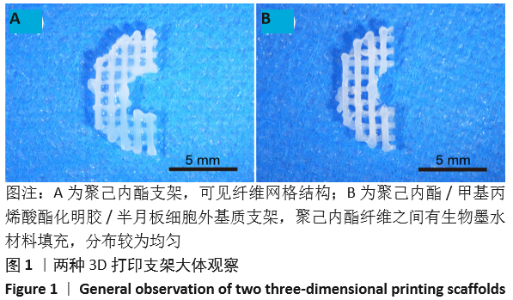
2.1 3D生物打印支架的大体观 所有支架均成功打印,大体观可见两种支架呈半圆环状结构,PCL支架呈白色的十字交叉网格结构,纤维排列均匀整齐,支架内部孔隙互通性较好;3D生物打印支架可见PCL纤维之间有生物墨水材料填充,分布较为均匀,见图1。"

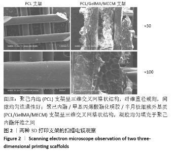
2.2 3D打印支架的扫描电镜观察 扫描电镜下观察发现,PCL支架、PCL/GelMA/MECM支架均呈三维交叉网格状结构,PCL支架纤维直径规则,间隙均匀连通性好;PCL/GelMA/MECM支架可见凝胶均匀填充于PCL纤维之间,冷冻干燥处理后表面较为疏松多孔,见图2。"
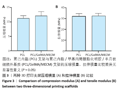
2.3 3D打印支架的力学性能 单纯PCL支架的压缩模量为(11.10±0.95) MPa,3D生物打印PCL/GelMA/MECM支架为(12.136±1.29) MPa,两者之间比较差异无显著性意义(P > 0.05);单纯PCL支架的拉伸模量为(32.08±2.18) MPa,3D生物打印PCL/GelMA/MECM支架为(32.51±2.01) MPa,两者之间比较差异无显著性意义(P > 0.05),说明复合生物墨水打印并未明显影响到支架的力学性能,见图3。"

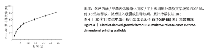
2.4 3D打印支架的缓释特性 负载于GelMA-MECM生物墨水中的PDGF-BB在缓释检测的28 d内,前3 d的短期释放情况表现为突释现象,释放较快,因子累计释放率达30.51%,随后的释放速度逐渐减缓,3-28 d的释放规律类似于零级释放动力学,可以持续缓释因子达28 d,累计释放率达80.42%,并且28 d后仍有缓慢释放的趋势,见图4。"
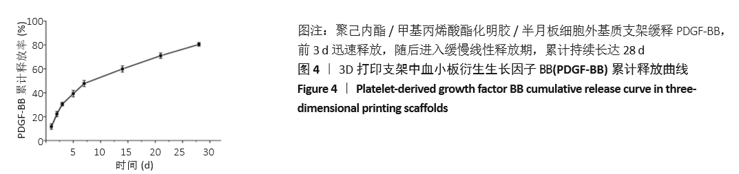
2.5 不同支架对滑膜间充质干细胞迁移的作用 各组细胞迁移及统计分析结果见图5所示, PCL支架组和阴性对照组细胞迁移数最少,且两者比较差异无显著性意义(P > 0.05);与阴性对照组相比,PCL/GelMA/MECM支架组、PCL/GelMA/MECM/PDGF-BB支架组细胞迁移数明显增多(P < 0.001,P < 0.000 1);PCL/GelMA/MECM/PDGF-BB支架组细胞迁移数多于PCL/GelMA/MECM支架组(P < 0.000 1)。"


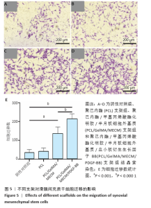
2.6 不同支架对滑膜间充质干细胞增殖的影响 支架浸提液培养细胞1,4,7 d后细胞数量明显增多,见图6。"
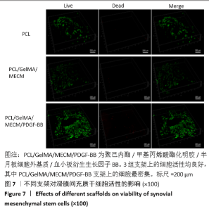
2.7 不同支架上的细胞活性观察 滑膜间充质干细胞在不同支架上培养4 d后的死活染色结果如图7所示,3组支架上的细胞活力均较好,染色成红色的死细胞较少,细胞大多密集生长在接种细胞的支架表面,相对于PCL支架,3D生物打印PCL/GelMA/MECM支架上的细胞数量更多,负载PDGF支架上的细胞生长最为密集。"
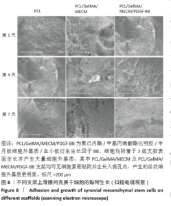
2.8 不同支架上细胞的黏附及生长观察 滑膜间充质干细胞在3D生物打印组织工程半月板支架上的第1,4,7的扫描电镜结果如图8所示,微观角度观察可见细胞均附着于支架表面生长,且形态多变且产生大量细胞外基质,PCL/GelMA/MECM及PCL/GelMA/MECM/PDGF-BB支架均可见细胞紧密贴附并生长入微孔内,产生的丝状细胞外基质相较于对照组更为明显。"

| [1] PATEL M, BRZEZINSKI A, RAOLE DA, et al. Interference screw versus suture endobutton fixation of a fiber-reinforced meniscus replacement device in a human cadaveric knee model. Am J Sports Med. 2018;46(9): 2133-2141. [2] ARNOCZKY SP, WARREN RF. Microvasculature of the human meniscus. Am J Sports Med. 1982;10(2):90-95. [3] JACOB G, SHIMOMURA K, KRYCH AJ, et al. The Meniscus Tear: A Review of Stem Cell Therapies. Cells. 2020;9(1):92. [4] KWON H, BROWN WE, LEE CA, et al. Surgical and tissue engineering strategies for articular cartilage and meniscus repair. Nat Rev Rheumatol. 2019;15(9):550-570. [5] MONK P, GARFJELD ROBERTS P, PALMER A J, et al. The urgent need for evidence in arthroscopic meniscal surgery: a systematic review of the evidence for operative management of meniscal tears. Am J Sports Med. 2017;45(4):965-973. [6] SMITH N, COSTA M, SPALDING T. Meniscal allograft transplantation: rationale for treatment.Bone Joint J. 2015;97(5):590-594. [7] RICHTER W. Mesenchymal stem cells and cartilage in situ regeneration. J Intern Med. 2009;266(4):390-405. [8] QU F, GUILAK F, MAUCK RL. Cell migration: implications for repair and regeneration in joint disease. Nat Rev Rheumatol. 2019;15(3):167-179. [9] LUO Y, WEI X, HUANG P. 3D bioprinting of hydrogel‐based biomimetic microenvironments. J Biomed Mater Res B ApplBiomater. 2019;107(5): 1695-1705. [10] LUO Y, LIN X, HUANG P. 3D Bioprinting of artificial tissues: construction of biomimetic microstructures. MacromolBiosci. 2018;18(6):1800034. [11] BAHCECIOGLU G, BILGEN B, HASIRCI N, et al. Anatomical meniscus construct with zone specific biochemical composition and structural organization. Biomaterials. 2019;218:119361. [12] LEE CH, RODEO SA, FORTIER LA, et al. Protein-releasing polymeric scaffolds induce fibrochondrocytic differentiation of endogenous cells for knee meniscus regeneration in sheep. SciTransl Med. 2014; 6(266):266ra171-266ra171. [13] MONDAL D, GRIFFITH M, VENKATRAMAN SS. Polycaprolactone-based biomaterials for tissue engineering and drug delivery: Current scenario and challenges. Int J Polym Mater. 2016;65(5):255-265. [14] 冯子嫣,樊逸菲,郭玖思,等.组织工程半月板支架材料的研究进展[J].中国修复重建外科杂志,2019,33(8):1019-1028. [15] YUE K, TRUJILLO-DE SANTIAGO G, ALVAREZ MM, et al. Synthesis, properties, and biomedical applications of gelatin methacryloyl (GelMA) hydrogels. Biomaterials. 2015;73:254-271. [16] GAO S, GUO W, CHEN M, et al. Fabrication and characterization of electrospunnanofibers composed of decellularized meniscus extracellular matrix and polycaprolactone for meniscus tissue engineering. J Mater Chem B. 2017;5(12):2273-2285. [17] MISHIMA Y, LOTZ M. Chemotaxis of human articular chondrocytes and mesenchymal stem cells. J Orthop Res. 2008;26(10):1407-1412. [18] LEE KI, OLMER M, BAEK J, et al. Platelet-derived growth factor-coated decellularized meniscus scaffold for integrative healing of meniscus tears.Acta Biomater. 2018;76:126-134. [19] 苑志国,刘舒云,郝春香,等.脱细胞半月板细胞外基质/脱钙骨基质双相半月板支架的制备及其生物相容性的研究[J].中国医药生物技术,2016,11(1):4-12. [20] YUAN Z, LIU S, HAO C, et al. AMECM/DCB scaffold prompts successful total meniscus reconstruction in a rabbit total meniscectomy model. Biomaterials. 2016;11:113-126. [21] BAEK J, LEE E, LOTZ MK, et al. Bioactive proteins delivery through core-shell nanofibers for meniscal tissue regeneration. Nanomedicine. 2020;23:102090. [22] ZHANG Y, LI J, DAVIS ME, et al. Delineation of in vitro chondrogenesis of human synovial stem cells following preconditioning using decellularized matrix. Acta Biomater. 2015;20:39-50. [23] CHEN S, XU Z, SHAO J, et al. MicroRNA-218 promotes early chondrogenesis of mesenchymal stem cells and inhibits later chondrocyte maturation. BMC Biotechnol. 2019;19(1):1-10. [24] QU D, ZHU JP, CHILDS HR, et al. Nanofiber-based transforming growth factor-β3 release induces fibrochondrogenic differentiation of stem cells. Acta Biomater. 2019;93:111-122. [25] KLOTZ BJ, GAWLITTA D, ROSENBERG AJ, et al. Gelatin-methacryloyl hydrogels: towards biofabrication-based tissue repair. Trends Biotechnol. 2016;34(5):394-407. [26] RUPRECHT JC, WAANDERS TD, ROWLAND CR, et al. Meniscus-derived matrix scaffolds promote the integrative repair of meniscal defects. Sci Rep. 2019;9(1):1-13. [27] CHEN M, FENG Z, GUO W, et al. PCL-MECM-Based Hydrogel Hybrid Scaffolds and Meniscal FibrochondrocytesPromote Whole Meniscus Regeneration in a Rabbit Meniscectomy Model. ACS Appl Mater Interfaces. 2019;11(44):41626-41639. [28] 周建,田壮,田沁玉,等.不同交联密度甲基丙烯酸酯明胶/脱细胞半月板细胞外基质复合水凝胶的性能[J].中国组织工程研究, 2020,24(16):2493-2499. [29] VISSER J, MELCHELS FP, JEON JE, et al. Reinforcement of hydrogels using three-dimensionally printed microfibres. Nat Commun. 2015;6(1):1-10. [30] UOMIZU M, MUNETA T, OJIMA M, et al. PDGF-induced proliferation and differentiation of synovial mesenchymal stem cells is mediated by the PI3K-PKB/Akt pathway. J Med Dent Sci. 2018;65(2):73-82. [31] NAZARI M, NI NC, LüDKE A, et al. Mast cells promote proliferation and migration and inhibit differentiation of mesenchymal stem cells through PDGF. J Mol Cell Cardiol. 2016;94:32-42. [32] ANDRAE J, GALLINI R, BETSHOLTZ C. Role of platelet-derived growth factors in physiology and medicine. Genes Dev. 2008;22(10):1276-1312. [33] WANG F, HOU K, CHEN W, et al. Transgenic PDGF-BB/sericin hydrogel supports for cell proliferation and osteogenic differentiation. Biomater Sci. 2020;8(2):657-672. [34] RISAU W, DREXLER H, MIRONOV V, et al. Platelet-derived growth factor is angiogenic in vivo. Growth Factors. 1992;7(4):261-266. [35] PHILLIPS GD, STONE AM. PDGF-BB induced chemotaxis is impaired in aged capillary endothelial cells. Mech Ageing Dev. 1994;73(3):189-196. [36] IBáN MÁR, MELERO NC, MARTINEZ-BOTAS J, et al. Growth factor expression after lesion creation in the avascular zone of the meniscus: A quantitative PCR study in rabbits. Arthroscopy. 2014;30(9):1131-1138. [37] KOU L, XIAO S, SUN R, et al. Biomaterial-engineered intra-articular drug delivery systems for osteoarthritis therapy. Drug Deliv. 2019;26(1): 870-885. [38] PATEL JM, SALEH KS, BURDICK JA, et al. Bioactive factors for cartilage repair and regeneration: improving delivery, retention, and activity. Acta biomater. 2019;93:222-238. [39] LAI T, YU J, TSAI W. Gelatin methacrylate/carboxybetaine methacrylate hydrogels with tunable crosslinking for controlled drug release. J Mater Chem B. 2016;4(13):2304-2313. [40] MODARESIFAR K, HADJIZADEH A, NIKNEJAD H. Design and fabrication of GelMA/chitosan nanoparticles composite hydrogel for angiogenic growth factor delivery.Artif Cells Nanomed Biotechnol. 2018;46(8):1799-1808. [41] JEON O, WOLFSON DW, ALSBERG E. In‐situ formation of growth‐factor‐loaded coacervatemicroparticle‐embedded hydrogels for directing encapsulated stem cell fate. Adv Mater. 2015;27(13):2216-2223. [42] ZHANG Y, CHENG N, MIRON R, et al. Delivery of PDGF-B and BMP-7 by mesoporousbioglass/silk fibrin scaffolds for the repair of osteoporotic defects. Biomaterials. 2012;33(28):6698-6708. [43] AGRAWAL V, BROWN BN, BEATTIE AJ, et al. Evidence of innervation following extracellular matrix scaffold‐mediated remodelling of muscular tissues. J Tissue Eng Regen Med. 2009;3(8):590-600. [44] YIN H, WANG Y, SUN Z, et al. Induction of mesenchymal stem cell chondrogenic differentiation and functional cartilage microtissue formation for in vivo cartilage regeneration by cartilage extracellular matrix-derived particles. Acta Biomater. 2016;33:96-109. |
| [1] | Xu Feng, Kang Hui, Wei Tanjun, Xi Jintao. Biomechanical analysis of different fixation methods of pedicle screws for thoracolumbar fracture [J]. Chinese Journal of Tissue Engineering Research, 2021, 25(9): 1313-1317. |
| [2] | Jiang Yong, Luo Yi, Ding Yongli, Zhou Yong, Min Li, Tang Fan, Zhang Wenli, Duan Hong, Tu Chongqi. Von Mises stress on the influence of pelvic stability by precise sacral resection and clinical validation [J]. Chinese Journal of Tissue Engineering Research, 2021, 25(9): 1318-1323. |
| [3] | Zhang Tongtong, Wang Zhonghua, Wen Jie, Song Yuxin, Liu Lin. Application of three-dimensional printing model in surgical resection and reconstruction of cervical tumor [J]. Chinese Journal of Tissue Engineering Research, 2021, 25(9): 1335-1339. |
| [4] | Zhang Yu, Tian Shaoqi, Zeng Guobo, Hu Chuan. Risk factors for myocardial infarction following primary total joint arthroplasty [J]. Chinese Journal of Tissue Engineering Research, 2021, 25(9): 1340-1345. |
| [5] | Wei Wei, Li Jian, Huang Linhai, Lan Mindong, Lu Xianwei, Huang Shaodong. Factors affecting fall fear in the first movement of elderly patients after total knee or hip arthroplasty [J]. Chinese Journal of Tissue Engineering Research, 2021, 25(9): 1351-1355. |
| [6] | Wang Jinjun, Deng Zengfa, Liu Kang, He Zhiyong, Yu Xinping, Liang Jianji, Li Chen, Guo Zhouyang. Hemostatic effect and safety of intravenous drip of tranexamic acid combined with topical application of cocktail containing tranexamic acid in total knee arthroplasty [J]. Chinese Journal of Tissue Engineering Research, 2021, 25(9): 1356-1361. |
| [7] | Xiao Guoqing, Liu Xuanze, Yan Yuhao, Zhong Xihong. Influencing factors of knee flexion limitation after total knee arthroplasty with posterior stabilized prostheses [J]. Chinese Journal of Tissue Engineering Research, 2021, 25(9): 1362-1367. |
| [8] | Huang Zexiao, Yang Mei, Lin Shiwei, He Heyu. Correlation between the level of serum n-3 polyunsaturated fatty acids and quadriceps weakness in the early stage after total knee arthroplasty [J]. Chinese Journal of Tissue Engineering Research, 2021, 25(9): 1375-1380. |
| [9] | Zhang Chong, Liu Zhiang, Yao Shuaihui, Gao Junsheng, Jiang Yan, Zhang Lu. Safety and effectiveness of topical application of tranexamic acid to reduce drainage of elderly femoral neck fractures after total hip arthroplasty [J]. Chinese Journal of Tissue Engineering Research, 2021, 25(9): 1381-1386. |
| [10] | Wang Haiying, Lü Bing, Li Hui, Wang Shunyi. Posterior lumbar interbody fusion for degenerative lumbar spondylolisthesis: prediction of functional prognosis of patients based on spinopelvic parameters [J]. Chinese Journal of Tissue Engineering Research, 2021, 25(9): 1393-1397. |
| [11] | Lü Zhen, Bai Jinzhu. A prospective study on the application of staged lumbar motion chain rehabilitation based on McKenzie’s technique after lumbar percutaneous transforaminal endoscopic discectomy [J]. Chinese Journal of Tissue Engineering Research, 2021, 25(9): 1398-1403. |
| [12] | Chen Xinmin, Li Wenbiao, Xiong Kaikai, Xiong Xiaoyan, Zheng Liqin, Li Musheng, Zheng Yongze, Lin Ziling. Type A3.3 femoral intertrochanteric fracture with augmented proximal femoral nail anti-rotation in the elderly: finite element analysis of the optimal amount of bone cement [J]. Chinese Journal of Tissue Engineering Research, 2021, 25(9): 1404-1409. |
| [13] | Du Xiupeng, Yang Zhaohui. Effect of degree of initial deformity of impacted femoral neck fractures under 65 years of age on femoral neck shortening [J]. Chinese Journal of Tissue Engineering Research, 2021, 25(9): 1410-1416. |
| [14] | Zhang Shangpu, Ju Xiaodong, Song Hengyi, Dong Zhi, Wang Chen, Sun Guodong. Arthroscopic suture bridge technique with suture anchor in the treatment of acromioclavicular dislocation [J]. Chinese Journal of Tissue Engineering Research, 2021, 25(9): 1417-1422. |
| [15] | Liang Yan, Zhao Yongfei, Xu Shuai, Zhu Zhenqi, Wang Kaifeng, Liu Haiying, Mao Keya. Imaging evaluation of short-segment fixation and fusion for degenerative lumbar scoliosis assisted by highly selective nerve root block [J]. Chinese Journal of Tissue Engineering Research, 2021, 25(9): 1423-1427. |
| Viewed | ||||||
|
Full text |
|
|||||
|
Abstract |
|
|||||