Chinese Journal of Tissue Engineering Research ›› 2013, Vol. 17 ›› Issue (41): 7255-7264.doi: 10.3969/j.issn.2095-4344.2013.41.011
Previous Articles Next Articles
Rapid canine distal movement through distraction osteogenesis of the periodontal ligament
Mai Zhi-hui1, Zhang Jing-lan2, Lu Hong-fei1, Chen Qi1, Liang Huang-you1, Ai Hong1
- 1 Department of Stomatology, the Third Affiliated Hospital of Sun Yat-sen University, Guangzhou 510630, Guangdong Province, China;2 Department of Pediatric Dentistry, Guangdong Provincial Stomatological Hospital, Guangzhou 510280, Guangdong Province, China
-
Received:2013-03-30Revised:2013-05-22Online:2013-10-08Published:2013-11-01 -
Contact:Ai Hong, Master, Professor, Department of Stomatology, the Third Affiliated Hospital of Sun Yat-sen University, Guangzhou 510630, Guangdong Province, China Aih_zssy09@126.com -
About author:Mai Zhi-hui☆, M.D., Attending physician, Department of Stomatology, the Third Affiliated Hospital of Sun Yat-sen University, Guangzhou 510630, Guangdong Province, China maiya2007@126.com Zhang Jing-lan, Master, Department of Pediatric Dentistry, Guangdong Provincial Stomatological Hospital, Guangzhou 510280, Guangdong Province, China Zhang Jing-lan and Mai Zhi-hui contributed equally to this paper. -
Supported by:Natural Science Foundation of Guangdong Province, No. s2011010004629*
CLC Number:
Cite this article
Mai Zhi-hui, Zhang Jing-lan, Lu Hong-fei, Chen Qi, Liang Huang-you, Ai Hong. Rapid canine distal movement through distraction osteogenesis of the periodontal ligament[J]. Chinese Journal of Tissue Engineering Research, 2013, 17(41): 7255-7264.
share this article
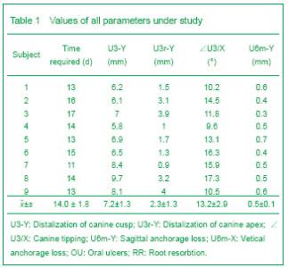
General situation of experimental subjects After 12-16 days, mean (4.0±1.8) days of distraction, the canines moved to the expected position in all cases. The patients only felt mild discomfort during activation of the distraction device, a little tense in the canine area, but the symptoms disappeared after 10-15 minutes. No hot and cold stimulated pain or spontaneous pain happened. Oral ulcers were found in three patients because of the oppression on the mucosa by the distraction device. There were no difficulties in activating the distraction device. No loosening and falling off of distraction device happened. Tooth looseness of canine When the distraction was completed, 4/18 of maxillary canines had looseness of angle class Ⅱ, 14/18 of maxillary canines had looseness of angle classⅠ. One month after placement of fixed appliance, all canines had looseness of angle classⅠ. There was no looseness of maxillary first molar and second premolar. Cephalometric analysis(Table 1) After canine distraction, the amount of canine retraction achieved was an average of 7.18 mm, at a rate of 0.5 mm per day. The distal tipping of canine was (13.24 ±2.87)° at T2, but it reduced to (6.43±1.14)° at T3. The mean sagittal (U6m-Y) and vertical (U6m-X) anchorage loss was 0.5 mm and 0.2 mm at T2, and was 0.8 mm and 0.4 mm at T3, respectively. No signi?cant changes were observed in the other measurements (Table 1). "
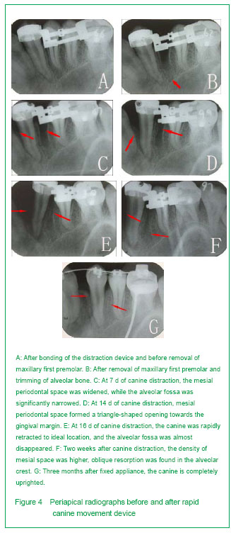
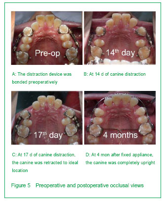
Root resorption Root resorption of the canines was minimal. After distraction, apical root resorption of S1 occurred in 2/18 of the canines, while rdiographic examination showed no evidence of complications, such as root fracture and ankylosis in all teeth (canines, incisors, second premolars, ?rst molars), in any patient. No apical or lateral root resorption (S0) was detected in any subject at T1, T2 and T3. Remodeling of the canine periodontal tissues Newly formed alveolar bone in mesial canines was matured at 3 months after distraction. The periapical radiographs showed a process from empty to solid in the mesial distraction space of canine. One week after canine distraction (Figure 4C), the mesial periodontal space was widened (the space of cervical area was wider than the apical area), showing low density, and the original lamina dura turned indistinct, while the distal lamina dura remained clear. The alveolar fossa was significantly narrowed because of pressure and showed no significant increase of density. No obvious fracture line was found in distal alveolar bone of canine. At the end of the canine distraction (Figure 4E), the mesial periodontal space of canine became obvious, with a triangle-shaped opening towards the gingival margin. The lamina dura was invisible. Low-density was showed in the apical area of canine. Distal periodontal space was widened and the lamina dura disappeared. The alveolar fossa was significantly narrower than before, but the boundary was still clear. The density of alveolar fossa was higher than that in the first week of distraction. Obvious deformation caused by pressure was found in the distal interval alveolar bone of the canine. The boundary of the alveolar crest was indistinct, and the height of it was decreased by about 1 mm. A low-density shadow was found in the lower 1/3 of the interval alveolar bone, which was considered to be a fracture line. One month after placement of fixed appliance (Figure 4F), the mesial space of canine was still empty, but the density was higher than that in the first two weeks. The density around the canine root was significantly increased, but no obvious formation of lamina dura was found. The boundary of alveolar fossa was still indistinct. The density of alveolar fossa was high, but still lower than normal. The periodontal space of canine was still clear. Oblique resorption was found in the alveolar crest, and the height of it was decreased by about 1.5 mm. Three months after placement of fixed appliance (Figure 4G), the width of periodontal space of canine returned to normal, with a clear lamina dura. The height of interval alveolar bone returned to the level before distraction; while the density of neonatal mesial interval alveolar bone was slightly lower (Figure 5)."

"
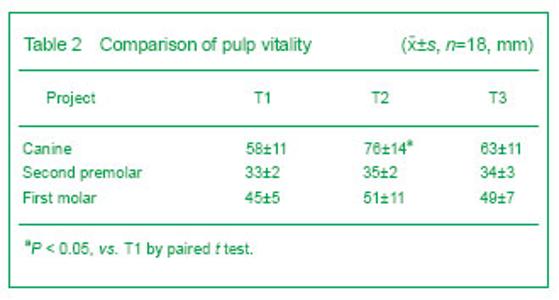
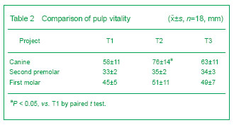
Pulp vitality of maxillary canines, second premolar and first molar At T2, 2/18 of the canines were in a state of shock, decline of pulp vitality appeared in 16/18 of the canines and it had significant differences with that of T1 (P < 0.05). Three months after the fixed appliance treatment, the canine pulp vitality recovered partly and had no significant differences with that of T1 (P > 0.05). The second premolar and first molar had no pulp vitality change (Table 2)."
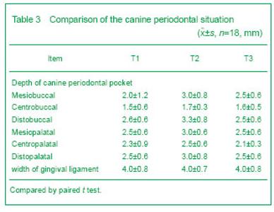
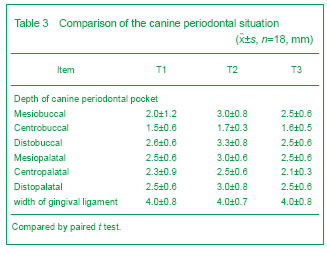
Canine periodontal situation There were significant differences in canine periodontal probing depth and mucogingianl junction width between T1, T2 and T3. During canine distration, the periodontal tissue was healthy without edema and inflammation. Oral ulcers were found in three patients because of the oppression on the mucosa by the distraction device (Table 3)."
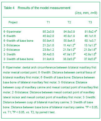
Model measurement After canine distraction, the measurements of 6-6/perimeter, 6-6/width, and 6-6/width of base bone had no significant difference (P > 0.05). There were sigfniciant differences in the measurements of 3-6/distance, 2-6/distance, and 3-3/width before and after canine distraction (P < 0.05). Three months after placement of fixed appliance, there were no significant differences in the measurements of 6-6/width, and 6-6/width of base bone before and after canine distraction (P > 0.05), whereas the measurements of 6-6/perimeter, 3-6/distance, 2-6/distance, and 3-3/width of base bone had significant difference from those before distraction (P < 0.05), and the measurements of 3-3/width had significant difference from those before and after distraction (P < 0.05) (Table 4)."
| [1] Codivilla A. The classic: On the means of lengthening, in the lower limbs, the muscles and tissues which are shortened through deformity. 1905. Clin Orthop Relat Res. 2008;466(12):2903-2909.
[2] Ilizarov GA. The principles of the Ilizarov method. Bull Hosp Jt Dis Orthop Inst. 1988;48(1):1-11.
[3] McCarthy JG. The role of distraction osteogenesis in the reconstruction of the mandible in unilateral craniofacial microsomia. Clin Plast Surg. 1994;21(4):625-631.
[4] Liou EJ, Huang CS. Rapid canine retraction through distraction of the periodontal ligament. Am J Orthod Dentofacial Orthop. 1998;114(4):372-382.
[5] Sharpe W, Reed B, Subtelny JD, et al. Orthodontic relapse, apical root resorption, and crestal alveolar bone levels. Am J Orthod Dentofacial Orthop. 1987;91(3):252-258.
[6] von Böhl M, Maltha JC, Von Den Hoff JW, et al. Focal hyalinization during experimental tooth movement in beagle dogs. Am J Orthod Dentofacial Orthop. 2004;125(5): 615-623.
[7] von Böhl M, Kuijpers-Jagtman AM. Hyalinization during orthodontic tooth movement: a systematic review on tissue reactions. Eur J Orthod. 2009;31(1):30-36.
[8] Kim SJ, Park YG, Kang SG. Effects of Corticision on paradental remodeling in orthodontic tooth movement. Angle Orthod. 2009;79(2):284-291.
[9] Chung KR, Mitsugi M, Lee BS, et al. Speedy surgical orthodontic treatment with skeletal anchorage in adults--sagittal correction and open bite correction. J Oral Maxillofac Surg. 2009;67(10):2130-2148.
[10] Ghoneima AA, Allam ES, Zunt SL, et al. Bisphosphonates treatment and orthodontic considerations. Orthod Craniofac Res. 2010;13(1):1-10.
[11] Ahn JJ, Shin HI. Bone tissue formation in extraction sockets from sites with advanced periodontal disease: a histomorphometric study in humans. Int J Oral Maxillofac Implants. 2008;23(6):1133-1138.
[12] Prabhakar AR, Tauro DP, Shubha AB. Management of an unusual maxillary dentoalveolar fracture: a case report. J Dent Child (Chic). 2006;73(2):112-115.
[13] Brusveen EM, Brudvik P, Bøe OE, et al. Apical root resorption of incisors after orthodontic treatment of impacted maxillary canines: a radiographic study. Am J Orthod Dentofacial Orthop. 2012;141(4):427-435.
[14] Weltman B, Vig KW, Fields HW, et al. Root resorption associated with orthodontic tooth movement: a systematic review. Am J Orthod Dentofacial Orthop. 2010;137(4):462-476; discussion 12A.
[15] Cope JB, Harper RP, Samchukov ML. Experimental tooth movement through regenerate alveolar bone: A pilot study. Am J Orthod Dentofacial Orthop. 1999;116(5):501-505.
[16] Nakamoto N, Nagasaka H, Daimaruya T, et al. Experimental tooth movement through mature and immature bone regenerates after distraction osteogenesis in dogs. Am J Orthod Dentofacial Orthop. 2002;121(4):385-395.
[17] Kharkar VR, Kotrashetti SM. Transport dentoalveolar distraction osteogenesis-assisted rapid orthodontic canine retraction. Oral Surg Oral Med Oral Pathol Oral Radiol Endod. 2010;109(5):687-693.
[18] Kumar PS, Saxena R, Patil S, et al. Clinical investigation of periodontal ligament distraction osteogenesis for rapid orthodontic canine retraction. Aust Orthod J. 2009;25(2):147-152.
[19] Gürgan CA, I?eri H, Ki?ni?ci R. Alterations in gingival dimensions following rapid canine retraction using dentoalveolar distraction osteogenesis. Eur J Orthod. 2005;27(4):324-332.
[20] Ren Y, Maltha JC, Kuijpers-Jagtman AM. Optimum force magnitude for orthodontic tooth movement: a systematic literature review. Angle Orthod. 2003;73(1):86-92.
[21] Alkumru P, Erdem D, Altug-Atac AT. Evaluation of changes in the vertical facial dimension with different anchorage systems in extraction and non-extraction subjects treated by Begg fixed appliances: A retrospective study. Eur J Orthod. 2007;29(5):508-516.
[22] Sayin S, Bengi AO, Gürton AU, et al. Rapid canine distalization using distraction of the periodontal ligament: a preliminary clinical validation of the original technique. Angle Orthod. 2004;74(3):304-315.
[23] Tanne K, Yoshida S, Kawata T, et al. An evaluation of the biomechanical response of the tooth and periodontium to orthodontic forces in adolescent and adult subjects. Br J Orthod. 1998;25(2):109-115.
[24] Yoshihara T, Matsumoto Y, Suzuki J, et al. Effect of serial extraction alone on crowding: spontaneous changes in dentition after serial extraction. Am J Orthod Dentofacial Orthop. 2000;118(6):611-616.
[25] Wei S, Fu MK. Posttreatment stability of four first bicuspid extraction cases treated with the Alexander technique--model analysis. Zhonghua Kou Qiang Yi Xue Za Zhi. 2005;40(4):271-274.
[26] Miyajima K, Nakamura S. Distalization with 'driftodontics'. J Clin Orthod. 1994;28(7):393-394.
[27] Miura H, Hasegawa S, Okada D, et al. The measurement of physiological tooth displacement in function. J Med Dent Sci. 1998;45(2):103-115.
[28] Gritsch K, Laroche N, Morgon L, et al. A systematic review of methods for tissue analysis in animal studies on orthodontic mini-implants. Orthod Craniofac Res. 2012; 15(3):135-147.
[29] Ai H, Xu QF, Lu HF, et al. Rapid tooth movement through distraction osteogenesis of the periodontal ligament in dogs. Chin Med J (Engl). 2008;121(5):455-462.
[30] Kharkar VR, Kotrashetti SM. Transport dentoalveolar distraction osteogenesis-assisted rapid orthodontic canine retraction. Oral Surg Oral Med Oral Pathol Oral Radiol Endod. 2010;109(5):687-693.
[31] Kurt G, I?eri H, Ki?ni?ci R. Rapid tooth movement and orthodontic treatment using dentoalveolar distraction (DAD). Long-term (5 years) follow-up of a Class II case. Angle Orthod. 2010;80(3):597-606.
[32] Ding Y, Liu Y, Cao M, et al. Periodontal tissues changes in tooth-borne distraction osteogenesis: an experimental study of closure of wide alveolar bone defects in dogs. Br J Oral Maxillofac Surg. 2009;47(2):111-115.
[33] Thiruvenkatachari B, Pavithranand A, Rajasigamani K, et al. Comparison and measurement of the amount of anchorage loss of the molars with and without the use of implant anchorage during canine retraction. Am J Orthod Dentofacial Orthop. 2006;129(4):551-554. |
| [1] | Tian Shancan, Bai Rui, Xu Xiaomei, Huang Yue, Zhang Li, Yu Xingyue, Cheng Qian . Three-dimensional finite element analysis of extrusion of the maxillary canine during orthodontic treatment with invisible aligner without brackets [J]. Chinese Journal of Tissue Engineering Research, 2019, 23(10): 1489-1495. |
| [2] | Ma Xiao-zhou, Li Hong-fa, Zhao Yan-hong, Wu Jie, Zhang Ming-can, Zhao Wei. Frictional force of different ligations with NiTi archwires of different sizes [J]. Chinese Journal of Tissue Engineering Research, 2018, 22(22): 3491-3497. |
| [3] |
Mo Si-su, Bao Wei, Shen Qing-yi.
Construction of a three-dimensional finite element model of pulp-exposed maxillary premolar with wedge-shaped defect restored by fiber post
[J]. Chinese Journal of Tissue Engineering Research, 2018, 22(2): 183-188.
|
| [4] | Li Zeng-cai, Liu Xin-qiang. Bilateral condylar morphology in adult patients with unilateral cleft lip and palate [J]. Chinese Journal of Tissue Engineering Research, 2015, 19(24): 3818-3823. |
| [5] | Shen Qing-yi, Wang Dong-mei, Zhong Qun, Chen Dong. Three-dimensional finite element analysis of severe wedge-shaped defective premolar restored with fiber post and composite resin [J]. Chinese Journal of Tissue Engineering Research, 2014, 18(30): 4777-4782. |
| [6] | Shen Qing-yi, Li Guo-qiang, Zhang Qiang, Weng Bei-jun, Weng Jia-wei. Fracture resistance ability of severe wedge-shaped premolar defects restored with fiber reinforced composite post by different methods [J]. Chinese Journal of Tissue Engineering Research, 2014, 18(25): 4004-4008. |
| [7] | Wang Fa-sheng, Li Dong. Clinical comparison of two kinds of orthodontic adhesives bonding buccal tubes of posterior teeth [J]. Chinese Journal of Tissue Engineering Research, 2014, 18(16): 2538-2543. |
| Viewed | ||||||
|
Full text |
|
|||||
|
Abstract |
|
|||||