Chinese Journal of Tissue Engineering Research ›› 2026, Vol. 30 ›› Issue (6): 1384-1389.doi: 10.12307/2026.557
Previous Articles Next Articles
Feasibility of optimizing radiation dose for three-dimensional printing of the maxillofacial bone based on low-dose CT technology
Li Guan1, Wang Haopeng2, Wang Jinbao2, Song Xinhao1, Qin Guochu1, Shao Yang3
- 1Department of Radiology, Nanjing Drum Tower Hospital, Affiliated Hospital of Medical School, Nanjing University, Nanjing 210008, Jiangsu Province, China; 2Department of Radiology, 3Department of Stomatology, General Hospital of Northern Theater Command, Shenyang 110000, Liaoning Province, China
-
Received:2024-09-06Accepted:2025-02-11Online:2026-02-28Published:2025-07-14 -
Contact:Shao Yang, MS, Associate chief physician, Department of Stomatology, General Hospital of Northern Theater Command, Shenyang 110000, Liaoning Province, China -
About author:Li Guan, PhD, Associate chief physician, Department of Radiology, Nanjing Drum Tower Hospital, Affiliated Hospital of Medical School, Nanjing University, Nanjing 210008, Jiangsu Province, China
CLC Number:
Cite this article
Li Guan, Wang Haopeng, Wang Jinbao, Song Xinhao, Qin Guochu, Shao Yang. Feasibility of optimizing radiation dose for three-dimensional printing of the maxillofacial bone based on low-dose CT technology[J]. Chinese Journal of Tissue Engineering Research, 2026, 30(6): 1384-1389.
share this article
Add to citation manager EndNote|Reference Manager|ProCite|BibTeX|RefWorks
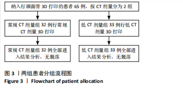
2.1 参与者数量分析 纳入行颌面骨3D打印的患者65例,其中常规CT剂量组32例,低CT剂量组33例,全部进入结果分析,无脱落。两组患者均无辐射以外的不良事件发生。 2.2 试验流程图 见图3。"

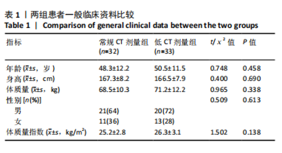
2.3 基线资料比较 两组患者在年龄、身高、体质量、性别及体质量指数上差异均无显著性意义(均 P > 0.05),见表1。"
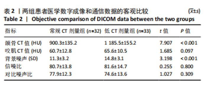
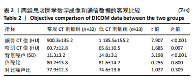
2.4 DICOM数据客观评价 低CT剂量组的颌骨CT值及背景噪声SD值均高于常规CT剂量组(均P < 0.001),而两组患者的咬肌CT值、信噪比、对比噪声比相比差异均无显著性意义(均P > 0.05),见表2。"
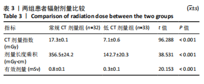
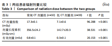
2.5 辐射剂量比较 比较常规CT剂量组和低CT剂量组的辐射剂量,结果发现低CT剂量组的CT剂量指数、剂量长度乘积和有效剂量值较常规CT剂量组分别减少约59.0%,60.0%和62.5%(均P < 0.001),见表3。"
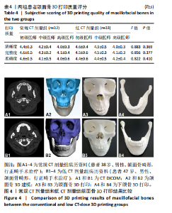
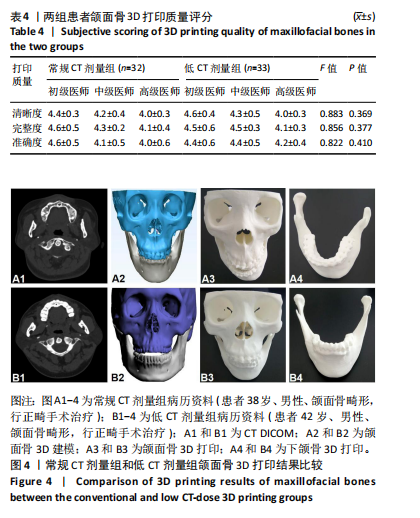
2.6 颌面骨3D打印质量结果比较 6名医师对两组方案完成的颌面骨3D打印质量进行主观打分,结果发现常规CT剂量组和低CT剂量组在清晰度、完整度及准确度指标上未见明显差异(P > 0.05),见表4及图4;且不同年资医师间的主观一致性较好,初级、中级和高级医师Kappa值分别为0.85,0.80和0.76。且常规CT剂量组和低CT剂量组的Bland-Altman图分析结果表明,两组的差值均值为-0.08,差值的95%置信区间为-1.34-1.18,所有测量数据均介于95%置信区间内,两组一致性水平良好。"
| [1] WANG H, CHI Y, HUANG H, et al. Combined use of 3D printing and computer-assisted navigation in the clinical treatment of multiplemaxillofacial fractures. Asian J Surg. 2023;46(6):2284-2292. [2] 何雪锋, 熊爱兵. 3D打印技术在整形外科的研究及应用进展[J]. 中国组织工程研究,2017,21(3):428-432. [3] QIN HL, WEI YM, HAN JY, et al. 3D printed bioceramic scaffolds: Adjusting pore dimension is beneficial for mandibular bone defects repair. J Tissue Eng Regen Med. 2022;16(4):409-421. [4] GOETZE E, ZELLER AN, PABST A. Approaching 3D printing in oral and maxillofacial surgery-suggestions for structured clinical standards. Oral Maxillofac Surg. 2024;28(2):795-802. [5] WANG H, CHI Y, HUANG H, et al. Combined use of 3D printing and computer-assisted navigation in the clinical treatment of multiple maxillofacial fractures. Asian J Surg. 2023;46(6):2284-2292. [6] LIU Z, ZHONG Y, LYU X, et al. Accuracy of the modified tooth-supported 3D printing surgical guides based on CT, CBCT, and intraoral scanning in maxillofacial region: A comparison study. J Stomatol Oral Maxillofac Surg. 2024;125(5S2):101853. [7] MATTEO M, ADRIEN N, GUIDO MM, et al. 3D printed bone models in oral and cranio-maxillofacial surgery: a systematic review. 3D Print Med. 2020;6(1): 30. [8] 郑坤, 许苑晶, 于文强, 等. 3D打印医工结合门诊在数字医学临床实践教学中的应用 [J]. 中国组织工程研究,2022,26(15):2317-2322. [9] HAN JJ, SODNOM-ISH B, EO MY, et al. Accurate mandible reconstruction by mixed reality, 3D printing, and robotic-assisted navigation integration. J Craniofac Surg. 2022;33(6):e701-e706. [10] TAHERI OTAGHSARA SS, JODA T, THIERINGER FM. Accuracy of dental implant placement using static versus dynamic computer-assisted implant surgery: An in vitro study. J Dent. 2023;132:104487. [11] HUANG YH, LEE B, CHUY JA, et al. 3D printing for surgical planning of canine oral and maxillofacial surgeries. 3D Print Med. 2022;8(1):17. [12] GAO L, SUN M, LIU J, et al. Study on mechanical properties of dual-channel cryogenic 3D printing scaffold for mandibular defect repair. Med Biol Eng Comput. 2024;62(8):2435-2448. [13] ANTUNES D, MAYEUR O, MAUPRIVEZ C, et al. 3D-printed model for gingival flap surgery simulation: Development and pilot test. Eur J Dent Educ. 2024;28(2):698-706. [14] JEDRZEJEK M, PESZEK-PRZYBYLA E, JADCZYK T, et al. 3D printing from transesophageal echocardiography for planning mitral paravalvular leak closure - feasibility study. Postepy Kardiol Interwencyjnej. 2023; 19(3):270-276. [15] QUEIROZ-FONTES R, RIBEIRO P, NUNES T, et al. 3D printing and CBCT anatomical reproducibility assessment in forensic scenarios. J Forensic Leg Med. 2024;106:102719. [16] WU W, LIU S, WANG L, et al. Application of 3D printing individualized guide plates in percutaneous needle biopsy of acetabular tumors. Front Genet. 2022;13:955643. [17] PEKER OZTURK H, AYYILDIZ S. Comparison of different 3D printers in terms of dimensional stability by image data of a dry human mandible obtained from CBCT and CT. Int J Artif Organs. 2024;47(1):49-56. [18] WANG L, CHEN XJ, LIANG JH, et al. Preliminary application of three-dimensional printing in congenital uterine anomalies based on three-dimensional transvaginal ultrasonographic data. BMC Womens Health. 2022;22(1):290. [19] HAN MA, KIM JH. Diagnostic X-Ray Exposure and Thyroid Cancer Risk: Systematic Review and Meta-Analysis. Thyroid. 2018;28(2):220-228. [20] ANSELMINO M, MARCANTONI L, AGRESTA A, et al. Interventional cardiology and X-ray exposure of the head: overview of clinical evidence and practical implications. J Cardiovasc Med (Hagerstown). 2022;23(6):353-358. [21] GU Y, WANG J, WANG Y, et al. Association of low-dose ionising radiation wit site-specific solid cancers: Chinese medical X-ray workers cohort study, 1950-1995. Occup Environ Med. 2023;80(12):687-693. [22] PEREZ JA, ROLDAN VS, GORDILLOordillo AK, et al. Mean glandular dose in the mammary gland and dose of radiation in the thyroid gland and lens in women with and without breast implants during different modalities of mammography. Radiologia (Engl Ed). 2022;64 Suppl 1: 11-19. [23] ASMA’A AA, SARA AA, DANIA T, et al. Do Ultra-low multidetector computed tomography doses and iterative reconstruction techniques affect subjective classification of bone type at dental implant sites? Int J Prosthodont. 2018;31(5): 465-470. [24] THONGVIGITMANEE SS, PONGNAPANG N, AOOTAPHAO S, et al. Radiation dose and accuracy analysis of newly developed cone-beam CT for dental and maxillofacial imaging. Annu Int Conf IEEE Eng Med Biol Soc. 2013;2013:2356-2359. [25] LI G, ZHANG L, LIU T, et al. Exploration of thoracoabdominalaortic mixed reality optimisation and its clinical application value in type A aortic dissection. Eur Radiol. 2023;33(6):4313-4322. [26] BHANOT R, HAMEED ZBM, SHAH M, et al. ALARA in Urology: Steps to Minimise Radiation Exposure During All Parts of the Endourological Journey. Curr Urol Rep. 2022;23(10):255-259. [27] LIPS M. ALARA in practice-4 decades of radiological protection at Goesgen NPP. J Radiol Prot. 2021;41(4). doi: 10.1088/1361-6498/ac1a82. [28] HANNA P, MACIAS C, BUCH E. ALARA in Atrial Fibrillation: Redefining Ablation Success. J Am Coll Cardiol. 2024;84(1):75-77. [29] BAKER SI, KAMBOJ S. Applying ALARA Principles in the Design of New Radiological Facilities. Health Phys. 2022;122(3):452-462. [30] YANG S, PU Q, LEI C, et al. Low-dose CT denoising with a high-level feature refinement and dynamic convolution network. Med Phys. 2023;50(6):3597-3611. [31] NODA Y, KAGA T, KAWAI N, et al. Low-dose whole-body CT using deep learning image reconstruction: image quality and lesion detection. Br J Radiol. 2021;94(1121):20201329. [32] GAN G, GONG W, JIA L, et al. Study of peripheral dose from low-dose CT to adaptive radiotherapy of postoperative prostate cancer.. Front Oncol. 2023;13:1227946. [33] ARK S, YOON JH, JOO I, et al. Image quality in liver CT: low-dose deep learning vs standard-dose model-based iterative reconstructions. Eur Radiol. 2022;32(5):2865-2874. [34] ZHANG J, SHANGGUAN Z, GONG W, et al. A novel denoising method for low-dose CT images based on transformer and CNN. Comput Biol Med. 2023;163:107162. [35] LIU Y, WEI C, XU Q. Detector shifting and deep learning based ring artifact correction method for low-dose CT. Med Phys. 2023;50(7): 4308-4324. [36] LI G, DONG J, CAO Z, et al. Application of low-dose CT to the creation of 3D-printed kidney and perinephric tissue models for laparoscopic nephrectomy. Cancer Med. 2021;10(9):3077-3084. [37] LOIZOU L, ALBIIN N, LEIDNER B, et al. Multidetector CT of pancreatic ductal adenocarcinoma: Effect of tube voltage and iodine load on tumour conspicuity and image quality. Eur Radiol. 2016;26(11):4021-4029. [38] LEE S, YOON SW, YOO SM, et al. Comparison of image quality and radiation dose between combined automatic tube current modulation and fixed tube current technique in CT of abdomen and pelvis. Acta Radiol. 2011;52(10):1101-1106. [39] FAEZEH S, CECIL GW, RISHI A, et al. Feasibility of sub-second CT angiography of the abdomen and pelvis with very low volume of contrast media, low tube voltage, and high-pitch technique, on a third-generation dual-source CT scanner. Clin Imaging. 2022;82:15-20. [40] WANG JJ, Chi XT, Wang WW, et al. Analysis of contrast-enhanced spectral chest CT optimal monochromatic imaging combined with ASIR and ASIR-V. Eur Rev Med Pharmacol Sci. 2022;26(6):1930-1938. [41] BIE Y, YANG S, LI X, et al. Impact of deep learning-based image reconstruction on image quality and lesion visibility in renal computed tomography at different doses. Quant Imaging Med Surg. 2023;13(4):2197-2207. |
| [1] | Wang Qisa, Lu Yuzheng, Han Xiufeng, Zhao Wenling, Shi Haitao, Xu Zhe. Cytocompatibility of 3D printed methyl acrylated hyaluronic acid/decellularized skin hydrogel scaffolds [J]. Chinese Journal of Tissue Engineering Research, 2026, 30(8): 1912-1920. |
| [2] | Zhou Hongli, Wang Xiaolong, Guo Rui, Yao Xuanxuan, Guo Ru, Zhou Xiongtao, He Xiangyi. Fabrication and characterization of nanohydroxyapatite/sodium alginate/polycaprolactone/alendronate scaffold [J]. Chinese Journal of Tissue Engineering Research, 2026, 30(8): 1962-1970. |
| [3] | Jia Yingao, Gao Shitao, Wang Fei. Application of 3D printed titanium cage cutting model in anterior cervical vertebrae subtotal decompression and bone graft fusion [J]. Chinese Journal of Tissue Engineering Research, 2026, 30(3): 604-611. |
| [4] | Wang Jiangjing, Zhao Na, Hu Xiaona, Zhao Na . Safety of 3D printed titanium alloy bone trabecular cup prosthesis combined with modified Kidney Tonifying and Blood Activating Decoction for elderly hip arthroplasty [J]. Chinese Journal of Tissue Engineering Research, 2026, 30(3): 612-619. |
| [5] | Yang Fengli, Zhou Chao, Xiong Wei, Zhou Yuxiang, Li Dengshun, Wang Xin, Li Zhanzhen. 3D printed poly-L-lactic acid bone scaffolds in repair of bone defects [J]. Chinese Journal of Tissue Engineering Research, 2026, 30(2): 507-515. |
| [6] | Ma Chi, Wang Ning, Chen Yong, Wei Zhihan, Liu Fengji, Piao Chengzhe. Application of 3D-printing patient-specific instruments combined with customized locking plate in opening wedge high tibial osteotomy [J]. Chinese Journal of Tissue Engineering Research, 2025, 29(9): 1863-1869. |
| [7] | Zhang Yu, Xu Ruian, Fang Lei, Li Longfei, Liu Shuyan, Ding Lingxue, Wang Yuexi, Guo Ziyan, Tian Feng, Xue Jiajia. Gradient artificial bone repair scaffold regulates skeletal system tissue repair and regeneration [J]. Chinese Journal of Tissue Engineering Research, 2025, 29(4): 846-855. |
| [8] | Ge Xiao, Zhao Zhuangzhuang, Guo Shuyu, Xu Rongyao. HOXA10 gene-modified bone marrow mesenchymal stem cells promote bone regeneration [J]. Chinese Journal of Tissue Engineering Research, 2025, 29(36): 7701-7708. |
| [9] | Hu Chaoran, Cen Chaode, Yang Yang, Zhou Cheng, Huang Huaxian, Yuan Honghao, Luo Qin, Cao Yongfei. 3D printing assisted minimal invasive plate osteosynthesis versus intramedullary nail for treatment of AO12-C middle-proximal humeral fractures [J]. Chinese Journal of Tissue Engineering Research, 2025, 29(33): 7116-7122. |
| [10] | Li Guoliang, Zhao Jianyong, Lyu Deliang, Su Juyue, Liu Qilin, Wang Tieqiang, Wang Xin. Improved 3D printed splint for distal radius fracture based on clinical defects: design and rapid grid-free analysis [J]. Chinese Journal of Tissue Engineering Research, 2025, 29(33): 7123-7129. |
| [11] | Wei Zhiheng, Guan Tianmin, Liu Qing, Gong Jue, Xiang Xianxiang. Application of 3D printing accurate osteotomy guide combined with the revision of anterior cruciate ligament with abnormally increased posterior slope of tibial plateau [J]. Chinese Journal of Tissue Engineering Research, 2025, 29(33): 7130-7136. |
| [12] | Zhuang Yan, Wang Xinyu, Cao Yilin, Ding Yuanxin, Wang Jiaqi, Yu Miao, Luan Chunyang, Ding Yuansheng. Three-dimensional finite element analysis of personalized orthodontic devices for 3D printed maxillary single-rooted rotated tooth [J]. Chinese Journal of Tissue Engineering Research, 2025, 29(30): 6409-6415. |
| [13] | Zhao Xiaoxuan, Liu Shuaiyi, Xing Zheng, Li Qingwen, Chu Xiaolei, Li Qi. Research hotspots and trends in application of tissue engineering in peripheral nerve injury [J]. Chinese Journal of Tissue Engineering Research, 2025, 29(30): 6591-6600. |
| [14] | Lian Zeshuang, Xu Qiang, Wang Aoting, Li Ding, Qin Jialin, Wang Junfang. Application of 3D printing technology in traumatic fractures [J]. Chinese Journal of Tissue Engineering Research, 2025, 29(27): 5883-5889. |
| [15] | Jiang Zhixiu, Ji Yuchen, Liu Danyu, Cao Yilin, Jiang Tingting, Song Yihan, Wang Lei, Wang Xinyu. Biomechanical properties of Gyroid structured titanium bionic bone scaffolds for repairing segmental mandibular defects [J]. Chinese Journal of Tissue Engineering Research, 2025, 29(22): 4621-4628. |
| Viewed | ||||||
|
Full text |
|
|||||
|
Abstract |
|
|||||