Chinese Journal of Tissue Engineering Research ›› 2026, Vol. 30 ›› Issue (3): 604-611.doi: 10.12307/2025.985
Previous Articles Next Articles
Application of 3D printed titanium cage cutting model in anterior cervical vertebrae subtotal decompression and bone graft fusion
Jia Yingao, Gao Shitao, Wang Fei
- Department of Spine Surgery, Affiliated Hospital of Yan’an University, Yan’an 716000, Shaanxi Province, China
-
Received:2024-10-23Accepted:2024-12-17Online:2026-01-28Published:2025-07-03 -
Contact:Wang Fei, MD, Chief physician, Department of Spine Surgery, Affiliated Hospital of Yan’an University, Yan’an 716000, Shaanxi Province, China -
About author:Jia Yingao, Master candidate, Department of Spine Surgery, Affiliated Hospital of Yan’an University, Yan’an 716000, Shaanxi Province, China -
Supported by:Yan’an Science and Technology Plan Project, No. SL2019ZCSZ-006 (to WF)
CLC Number:
Cite this article
Jia Yingao, Gao Shitao, Wang Fei. Application of 3D printed titanium cage cutting model in anterior cervical vertebrae subtotal decompression and bone graft fusion[J]. Chinese Journal of Tissue Engineering Research, 2026, 30(3): 604-611.
share this article
Add to citation manager EndNote|Reference Manager|ProCite|BibTeX|RefWorks
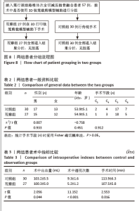
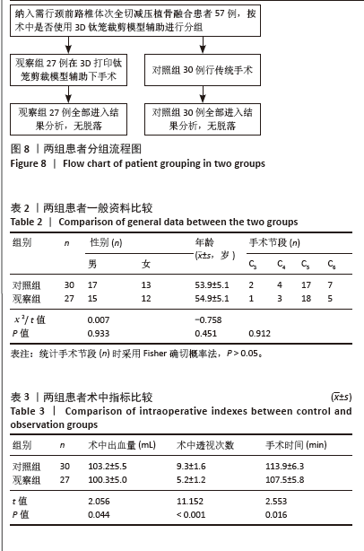
2.1 参与者数量分析 纳入行ACCF术57例患者的病历资料,根据术中是否使用3D打印钛笼裁剪模型分为2组,对照组(传统钛笼植骨)30例,观察组(3D打印钛笼模型)27例。全部进入结果分析,无脱落。 2.2 试验流程图 见图8。 2.3 基线资料比较 两组患者的性别分布、年龄、手术椎体节段比较差异均无显著性意义(P > 0.05),具有可比性,见表2。 2.4 术中数据资料比较 如表3所示,观察组术中出血量、手术时间均少于对照组,差异有显著性意义(P < 0.05)。对照组患者术中C型臂X射线机透视次数平均为(9.3±1.6)次,观察组为(5.2±1.2)次,差异有显著性意义(P < 0.05)。 "
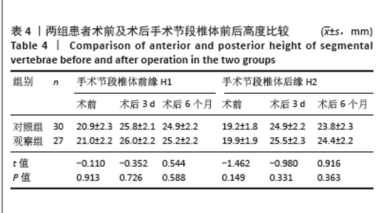
2.5 手术节段钛笼沉降及植骨融合情况 2.5.1 术前、术后手术节段椎体前后高度 两组患者术前、术后3 d及术后6个月H1、H2相比差异均无显著性意义 (P > 0.05),见表4。"
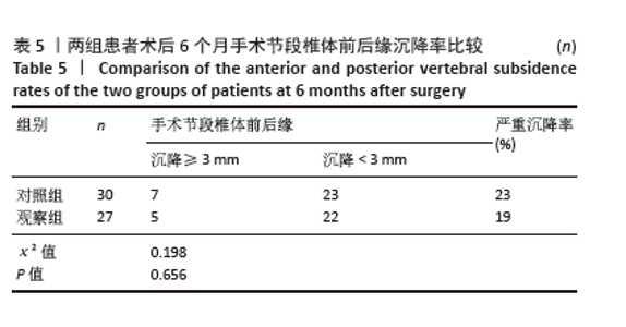
2.5.2 术后6个月手术节段植骨融合情况 在术后6个月末次随访时,观察组及对照组内所有患者手术节段在影像学上均获得骨性融合,融合率为100%,两组在融合率方面差异无显著性意义(P > 0.05)。 2.5.3 术后6个月手术节段椎体前后缘沉降程度 将术后3 d与术后6个月时手术椎体节段椎体H1或H2的下降距离定义为钛笼的下沉距离,根据其他学者的研究成果[16-18],将H1或H2下降≥3 mm定义为内植物(钛笼)严重下沉。对照组患者术后6个月手术节段椎体前后缘发生严重沉降(H1或H2沉降≥3 mm)有7例,严重沉降率为23%;观察组患者术后6个月手术节段椎体前后缘发生严重沉降有5例,严重沉降率为19%。两组钛笼发生严重沉降率相比差异无显著性意义(P > 0.05)。见表5。 "
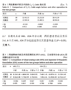
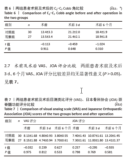
2.6 术前及术后C2-C7 Cobb角比较 两组患者术前及术后3 d、6个月时的C2-C7 Cobb角度比较差异均无显著性意义 (P > 0.05),见表6。 2.8 植入物与宿主的生物相容性 钛网为ACCF术中常用内植物,此次研究旨在使用3D打印技术对钛网实现精准裁剪和置入,钛网生物相容性良好,患者未出现植入物周围感染、过敏反应、免疫反应及排斥反应。"
| [1] IYER A, AZAD TD, THARIN S. Cervical Spondylotic Myelopathy. Clin Spine Surg. 2016;29(10):408-414. [2] MCCORMICK JR, SAMA AJ, SCHILLER NC, et al. Cervical Spondylotic Myelopathy: A Guide to Diagnosis and Management. J Am Board Fam Med. 2020;33(2):303-313. [3] TIAN X, RUDD S, YANG D, et al. Anterior Cervical Hybrid Decompression and Fusion Surgery to Treat Multilevel Cervical Spondylotic Myelopathy. J Vis Exp. 2023;(196). doi: 10.3791/65034. [4] BADHIWALA JH, WILSON JR, WITIW CD, et al. The influence of timing of surgical decompression for acute spinal cord injury: a pooled analysis of individual patient data. Lancet Neurol. 2021;20(2):117-126. [5] MADAN A, THAKUR M, SUD S, et al. Subaxial Cervical Spine Injuries: Outcomes after Anterior Corpectomy and Instrumentation. Asian J Neurosurg. 2019;14(3): 843-847. [6] TATTER C, PERSSON O, BURSTRÖM G, et al. Anterior Cervical Corpectomy and Fusion for Degenerative and Traumatic Spine Disorders, Single-Center Experience of a Case Series of 119 Patients. Oper Neurosurg (Hagerstown). 2020;20(1):8-17. [7] WANG T, WANG H, LIU S, et al. Anterior cervical discectomy and fusion versus anterior cervical corpectomy and fusion in multilevel cervical spondylotic myelopathy: A meta-analysis. Medicine (Baltimore). 2016;95(49):e5437. [8] HEE HT, MAJD ME, HOLT RT, et al. Complications of multilevel cervical corpectomies and reconstruction with titanium cages and anterior plating. J Spinal Disord Tech. 2003;16(1):1-8; discussion 8-9. [9] THALGOTT JS, XIONGSHENG C, GIUFFRE JM. Single stage anterior cervical reconstruction with titanium mesh cages, local bone graft, and anterior plating. Spine J. 2003;3(4):294-300. [10] FANG T, ZHANG M, YAN J, et al. Comparative Analysis of 3D-Printed Artificial Vertebral Body Versus Titanium Mesh Cage in Repairing Bone Defects Following Single-Level Anterior Cervical Corpectomy and Fusion. Med Sci Monit. 2021;27:e928022. [11] 孔金海, 吕国华, 康意军,等. 颈前路钛网植骨融合术后钛网沉陷的原因探讨[J]. 中国脊柱脊髓杂志,2007,17(2):98-102. [12] SHAMJI MF, MASSICOTTE EM, TRAYNELIS VC, et al. Comparison of anterior surgical options for the treatment of multilevel cervical spondylotic myelopathy: a systematic review. Spine (Phila Pa 1976). 2013;38(22 Suppl 1):S195-209. [13] LIANG H, JI T, ZHANG Y, et al. Reconstruction with 3D-printed pelvic endoprostheses after resection of a pelvic tumour. Bone Joint J. 2017; 99-b(2): 267-275. [14] MULFORD JS, BABAZADEH S, MACKAY N. Three-dimensional printing in orthopaedic surgery: review of current and future applications. ANZ J Surg. 2016;86(9):648-653. [15] KABRA DA, GARG DB. Current applications of 3-dimensional printing in spine surgery. J Orthop. 2023;41:28-32. [16] CHENG CC, ORDWAY NR, ZHANG X, et al. Loss of cervical endplate integrity following minimal surface preparation. Spine (Phila Pa 1976). 2007;32(17):1852-1855. [17] LIM TH, KWON H, JEON CH, et al. Effect of endplate conditions and bone mineral density on the compressive strength of the graft-endplate interface in anterior cervical spine fusion. Spine (Phila Pa 1976). 2001;26(8):951-956. [18] YAN D, WANG Z, DENG S, et al. Anterior corpectomy and reconstruction with titanium mesh cage and dynamic cervical plate for cervical spondylotic myelopathy in elderly osteoporosis patients. Arch Orthop Trauma Surg. 2011;131(10):1369-1374. [19] 严广斌. 视觉模拟评分法[J]. 中华关节外科杂志(电子版),2014(2): 273. [20] 姜良海, 谭明生, 杨峰,等. 标杆型3D打印导板辅助颈椎椎弓根置钉的临床应用[J]. 中华骨科杂志,2016,36(5):257-264. [21] ZENG J, DUAN Y, YANG Y, et al. Anterior corpectomy and reconstruction using dynamic cervical plate and titanium mesh cage for cervical spondylotic myelopathy: A minimum 5-year follow-up study. Medicine (Baltimore). 2018;97(5):e9724. [22] NIEDZIELAK TR, PALMER J, MALLOY JPT. Clinical Comparison of Surgical Constructs for Anterior Cervical Corpectomy and Fusion in Patients With Cervical Spondylotic Myelopathy or Ossified Posterior Longitudinal Ligament: A Systematic Review and Meta-Analysis. Clin Spine Surg. 2018;31(6):247-260. [23] BURKETT CJ, BAAJ AA, DAKWAR E, et al. Use of titanium expandable vertebral cages in cervical corpectomy. J Clin Neurosci. 2012;19(3): 402-405. [24] JI C, YU S, YAN N, et al. Risk factors for subsidence of titanium mesh cage following single-level anterior cervical corpectomy and fusion. BMC Musculoskelet Disord. 2020;21(1):32. [25] ZHONG W, LIANG X, TANG K, et al. Nanohydroxyapatite/polyamide 66 strut subsidence after one-level corpectomy: underlying mechanism and effect on cervical neurological function. Sci Rep. 2018;8(1):12098. [26] ARTS M, TORENSMA B, WOLFS J. Porous titanium cervical interbody fusion device in the treatment of degenerative cervical radiculopathy; 1-year results of a prospective controlled trial. Spine J. 2020;20(7):1065-1072. [27] 文睿, 叶飞, 蒲海波, 等. 个体化设计钛网植骨融合内固定恢复颈椎曲度[J]. 中国组织工程研究,2014,18(17):2655-2658. [28] LIU X, CHEN Y, YANG H, et al. The application of a new type of titanium mesh cage in hybrid anterior decompression and fusion technique for the treatment of continuously three-level cervical spondylotic myelopathy. Eur Spine J. 2017;26(1):122-130. [29] CHOU YC, CHEN DC, HSIEH WA, et al. Efficacy of anterior cervical fusion: comparison of titanium cages, polyetheretherketone (PEEK) cages and autogenous bone grafts. J Clin Neurosci. 2008;15(11):1240-1245. [30] WU J, LUO D, YE X, et al. Anatomy-related risk factors for the subsidence of titanium mesh cage in cervical reconstruction after one-level corpectomy. Int J Clin Exp Med. 2015;8(5):7405-7411. [31] ANDALUZ N, ZUCCARELLO M, KUNTZ C. Long-term follow-up of cervical radiographic sagittal spinal alignment after 1- and 2-level cervical corpectomy for the treatment of spondylosis of the subaxial cervical spine causing radiculomyelopathy or myelopathy: a retrospective study. J Neurosurg Spine. 2012;16(1):2-7. [32] LOWE TG, HASHIM S, WILSON LA, et al. A biomechanical study of regional endplate strength and cage morphology as it relates to structural interbody support. Spine (Phila Pa 1976). 2004;29(21): 2389-2394. [33] JANG JW, LEE JK, LEE JH, et al. Effect of posterior subsidence on cervical alignment after anterior cervical corpectomy and reconstruction using titanium mesh cages in degenerative cervical disease. J Clin Neurosci. 2014;21(10):1779-1785. [34] YANG J, CAI H, LV J, et al. In vivo study of a self-stabilizing artificial vertebral body fabricated by electron beam melting. Spine (Phila Pa 1976). 2014;39(8):E486-492. [35] ZHANG Y, QUAN Z, ZHAO Z, et al. Evaluation of anterior cervical reconstruction with titanium mesh cages versus nano-hydroxyapatite/polyamide66 cages after 1- or 2-level corpectomy for multilevel cervical spondylotic myelopathy: a retrospective study of 117 patients. PLoS One. 2014;9(5):e96265. [36] TRUUMEES E, DEMETROPOULOS CK, YANG KH, et al. Effects of disc height and distractive forces on graft compression in an anterior cervical discectomy model. Spine (Phila Pa 1976). 2002;27(22): 2441-2445. [37] 韩树虹, 王建华, 孙贺, 等. 3D打印人工椎体与钛笼在颈椎前路椎体次全切除减压植骨融合术中应用的效果比较[J]. 中国脊柱脊髓杂志,2022,32(5):426-433. [38] CHOY WJ, PARR WCH, PHAN K, et al. 3-dimensional printing for anterior cervical surgery: a review. J Spine Surg. 2018;4(4):757-769. [39] SPETZGER U, FRASCA M, KÖNIG SA. Surgical planning, manufacturing and implantation of an individualized cervical fusion titanium cage using patient-specific data. Eur Spine J. 2016;25(7):2239-2246. [40] LU T, LIU C, YANG B, et al. Single-Level Anterior Cervical Corpectomy and Fusion Using a New 3D-Printed Anatomy-Adaptive Titanium Mesh Cage for Treatment of Cervical Spondylotic Myelopathy and Ossification of the Posterior Longitudinal Ligament: A Retrospective Case Series Study. Med Sci Monit. 2017;23:3105-3114. [41] 王志强, 冯皓宇, 马迅, 等. 3D打印人工椎体及椎间融合器在颈椎前路手术中应用的临床效果[J]. 中国修复重建外科杂志,2021, 35(9):1147-1154. [42] 韩树虹, 王建华, 孙贺, 等. 3D打印人工椎体与钛笼在颈椎前路椎体次全切除减压植骨融合术中应用的效果比较[J]. 中国脊柱脊髓杂志,2022,32(5):426-433. [43] FOGEL G, MARTIN N, WILLIAMS GM, et al. Choice of Spinal Interbody Fusion Cage Material and Design Influences Subsidence and Osseointegration Performance. World Neurosurg. 2022;162: e626-e634. |
| [1] | Li Hanyue, Li Yini, Xiang Linmei, Li Sen. Effects of resistance exercise therapy on pain and function in patients with cervical spondylotic radiculopathy: a meta-analysis [J]. Chinese Journal of Tissue Engineering Research, 2026, 30(4): 987-996. |
| [2] | Abudusalamu·Tuoheti, Xiao Yang, Wang Yixi, Musitapa·Mijiti, Chen Qihao, Maimaitiming·Saiyiti, Guo Hailong, Paerhati·Rexiti. Effects of three internal fixation techniques on biomechanics of adjacent segment degeneration in lumbar interbody fusion [J]. Chinese Journal of Tissue Engineering Research, 2026, 30(3): 586-595. |
| [3] | Wang Yalei, Wang Xuezhi, Zhou Tao, Shen Xinxin, Fang Ding, Chen Hongliang. Effect of sacroiliac joint ankylosis on outcomes of L5/S1 transforminal lumbar interbody fusion and lumbar sagittal parameters [J]. Chinese Journal of Tissue Engineering Research, 2026, 30(3): 634-641. |
| [4] | Wang Zhipeng, Zhang Xiaogang, Zhang Hongwei, Zhao Xiyun, Li Yuanzhen, Guo Chenglong, Qin Daping, Ren Zhen. A systematic review of application value of machine learning to prognostic prediction models for patients with lumbar disc herniation [J]. Chinese Journal of Tissue Engineering Research, 2026, 30(3): 740-748. |
| [5] | Ma Shuangshuang, Gao Dedong, Shan Zhongshu, Xu Wenxu, Lu Zhirui. Finite element analysis and biomechanical validation of revision pedicle screw placement [J]. Chinese Journal of Tissue Engineering Research, 2025, 29(33): 7087-7095. |
| [6] | Yang Yu, Li Yinghao, Duo Zhuangzhi, Zhou Dingrong. Effect of overall functional physical exercise on lumbar biomechanics in patients with lumbar disc herniation after surgery [J]. Chinese Journal of Tissue Engineering Research, 2025, 29(33): 7096-7101. |
| [7] | Li Jing, Lu Guangqi, Zhuang Minghui, Cui Ying, Yu Zhangjingze, Sun Xinyue, Ma Mingming, Zhu Liguo, Yu Jie. Development of a clinical prediction model for cervical instability in young and middle-aged adults based on machine learning [J]. Chinese Journal of Tissue Engineering Research, 2025, 29(33): 7203-7210. |
| [8] | Yan Laijun, Ge Haiya, Wang Zhengming, Yang Zongrui, Niu Lifeng, Zhan Hongsheng. Mechanism by which Tongdu Huoxue Decoction inhibits macrophage inflammation to delay intervertebral disc degeneration in rats [J]. Chinese Journal of Tissue Engineering Research, 2025, 29(32): 6851-6857. |
| [9] | Yu Qinghe, Cai Ziming, Wu Jintao, Ma Pengfei, Zhang Xin, Zhou Longqian, Wang Yakun, Lin Xiaoqin, Lin Wenping. Vanillic acid inhibits inflammatory response and extracellular matrix degradation of endplate chondrocytes [J]. Chinese Journal of Tissue Engineering Research, 2025, 29(30): 6391-9397. |
| [10] | Yu Qinghe, Cai Ziming, Tian He, Li Pian, Ruan Ye, Liang Jinzhu, Lin Shuhui, Lin Wenping. Formononetin inhibits lipopolysaccharide-induced inflammation in nucleus pulposus mesenchymal stem cells [J]. Chinese Journal of Tissue Engineering Research, 2025, 29(25): 5328-5334. |
| [11] | Liu Zhaofeng, Yang Xuejun. Targeted biomarkers of single-cell RNA sequencing in intervertebral disc degeneration [J]. Chinese Journal of Tissue Engineering Research, 2025, 29(20): 4351-4360. |
| [12] | Wang Zikun, Li Shudong, Gao Shuang, Fan Shuhao, Li Cheng, Meng Chunyang. Relationship between intervertebral disc degeneration and 473 gut microbiotas: what can be learned from big data information in the FinnGen database [J]. Chinese Journal of Tissue Engineering Research, 2025, 29(20): 4369-4378. |
| [13] | Wang Qianliang, Chen Jianpeng, Wang Yuanbin, Yan Jun. Mechanism of circ05188 targeting miR-199a-5p involved in nociceptive hypersensitivity in a rat model of lumbar disc herniation [J]. Chinese Journal of Tissue Engineering Research, 2025, 29(20): 4230-4238. |
| [14] | Gao Haoran, Zhang Heling, Jia Fanglin, Guo Di, Jing Li, Shi Yaozhou, Song Hanlin, Gao Xiao, Feng Hu. Risk factors related to intradural lumbar disc herniation analyzed by propensity score matching [J]. Chinese Journal of Tissue Engineering Research, 2025, 29(15): 3199-3205. |
| [15] | Hou Yutong, Huang Chenglan, Yang Yunxiao, Li Ya, Guo Peiwu, Yu Wenqiang, Zhao Yu, Wang Zanbo, Zeng Hong, Ma Zhenjiang, Lu Dezhi, Wang Jinwu. Correlation between lumbar spine and pelvic parameters in Lenke type 5 adolescent idiopathic scoliosis [J]. Chinese Journal of Tissue Engineering Research, 2024, 28(36): 5753-5758. |
| Viewed | ||||||
|
Full text |
|
|||||
|
Abstract |
|
|||||