Chinese Journal of Tissue Engineering Research ›› 2025, Vol. 29 ›› Issue (21): 4486-4491.doi: 10.12307/2025.102
Previous Articles Next Articles
Digital three-dimensional morphological analysis of developmental characteristics of cervical facet joints in adolescents aged 13-18 years
Li Guihua1, He Yujie2, Shi Jun3, Li Kun2, 4, Zhang Shaojie2, 4, Liu Lu2, Li Zhijun2, 4, Wang Xing2, 4
- 1First Hospital of Prison Administration Bureau of Inner Mongolia Autonomous Region, Hohhot 010000, Inner Mongolia Autonomous Region, China; 2Department of Anatomy, 3Department of Physiology, 4Digital Medicine Center, School of Basic Medicine, Inner Mongolia Medical University, Hohhot 010110, Inner Mongolia Autonomous Region, China
-
Received:2023-09-05Accepted:2023-11-30Online:2025-07-28Published:2024-12-05 -
Contact:Li Zhijun, Professor, Doctoral supervisor, Department of Anatomy, and Digital Medicine Center, School of Basic Medicine, Inner Mongolia Medical University, Hohhot 010110, Inner Mongolia Autonomous Region, China Wang Xing, MD, Associate professor, Department of Anatomy, and Digital Medicine Center, School of Basic Medicine, Inner Mongolia Medical University, Hohhot 010110, Inner Mongolia Autonomous Region, China -
About author:Li Guihua, MS, Attending physician, First Hospital of Prison Administration Bureau of Inner Mongolia Autonomous Region, Hohhot 010000, Inner Mongolia Autonomous Region, China He Yujie, Lecturer, Department of Anatomy, School of Basic Medicine, Inner Mongolia Medical University, Hohhot 010110, Inner Mongolia Autonomous Region, China Li Guihua and He Yujie contributed equally to this article. -
Supported by:National Natural Science Foundation of China, No. 81860383 (to LZJ); Natural Science Foundation of Inner Mongolia Autonomous Region, No. 2020LH08021 (to LZJ); National Natural Science Foundation of China, No. 81860382 (to WX); Natural Science Foundation of Inner Mongolia Autonomous Region, No. 2020MS03061 (to WX); Youth Science and Technology Talents Support Program of Higher Education Institutions of Inner Mongolia Autonomous Region, No. NJYT22009 (to WX); Science and Technology Plan Project of Inner Mongolia Autonomous Region, No. 2019GG158 (to WX); Key Research Project of Inner Mongolia Medical University, No. YKD2021ZD011 (to WX); Health Commission Medical Health Technology Project of Inner Mongolia Autonomous Region, No. 202201217 (to WX); Natural Science Foundation of Inner Mongolia Autonomous Region, No. 2019MS08017 (to ZSJ); Youth Fund Project of Inner Mongolia Medical University, No. YKD2021QN011 (to HYJ); Youth Fund Project of Inner Mongolia Medical University, No. YKD2020QNCX055 (to LK)
CLC Number:
Cite this article
Li Guihua, He Yujie, Shi Jun, Li Kun, Zhang Shaojie, Liu Lu, Li Zhijun, Wang Xing. Digital three-dimensional morphological analysis of developmental characteristics of cervical facet joints in adolescents aged 13-18 years[J]. Chinese Journal of Tissue Engineering Research, 2025, 29(21): 4486-4491.
share this article
Add to citation manager EndNote|Reference Manager|ProCite|BibTeX|RefWorks
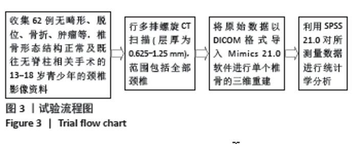
2.1 参与者数据分析 纳入观察对象62例,每一观察指标进行左右侧、上下比较分析,全部进入统计结果分析,无脱落。 2.2 试验流程图 见图3。"
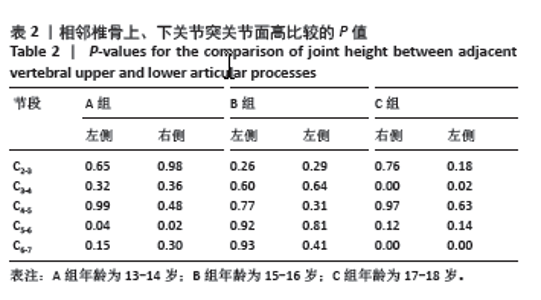
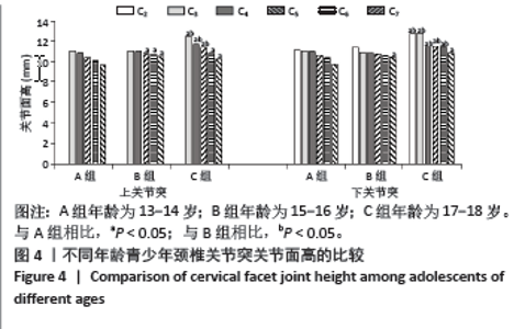
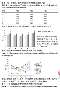
2.3 关节突关节面高 相同年龄段内,上、下关节突关节面高在左右侧别比较中差异均无显著性意义(P > 0.05);在同侧相邻椎骨上、下关节突关节面高比较中,显著性差异(P < 0.05)仅位于A组C5-6与C组C3-4、C6-7。A、B、C组的上、下关节突关节面高均随椎序的增加,整体呈递减趋势,上、下关节突关节面高最小值均位于C7。C组C3-C5的上、下关节突关节面高和C6下关节突关节面高均明显高于A、B组(P < 0.05),A组C6的上关节突关节面高与C7的上、下关节突关节面高明显小于B、C组(P < 0.05)。见表2及图4。"
"
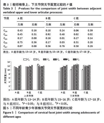
2.4 关节突关节面宽 A、B、C组内,上、下关节突关节面宽在左右侧别比较中差异均无显著性意义(P > 0.05);同侧相邻椎骨上、下关节突关节面高比较,有显著性差异的仅位于A组右侧C4-5、C6-7,B组右侧C4-5、C5-6以及C组C3-4(P < 0.05)。3组的上关节突关节面宽随椎序的增加总体呈“V”字型,最小值均位于C5,下关节突关节面宽随椎序的增加,整体呈递增趋势,最大值均位于C7。上关节突关节面宽中,C组的C3-C5和B组的C5明显大于A组(P < 0.05),下关节突关节面宽中,除C2、C7,其余B组和C组均高于A组(P < 0.05),且C组C3-C5也明显高于B组(P < 0.05)。见表3及图5。"
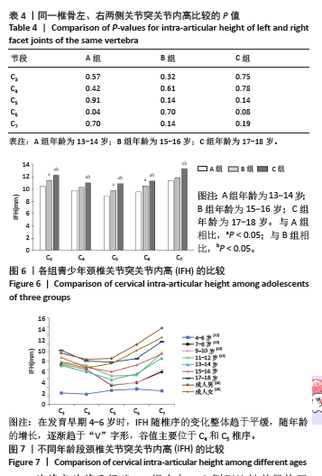
2.5 关节突IFH 同一椎骨左右侧别间除A组C6外,其余差异均无显著性意义(P > 0.5)。3组IFH随椎序的增加整体呈“V”字型,最小值均位于C5。B组的C3、C5和C6明显高于A组(P < 0.05),C组与A、B组相比,差异均有显著性意义(P < 0.05),见表4及图6。在发育早期4-6岁时,IFH随椎序的变化整体趋于平缓,随年龄的增长,逐渐趋于“V”字形,谷值主要位于C4和C5椎序(图7)。 "
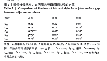
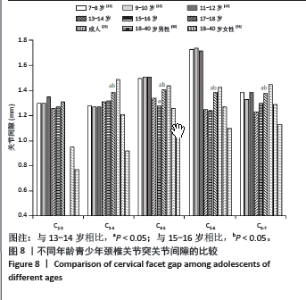
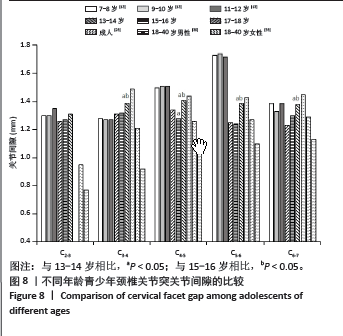
2.6 关节突关节面间隙 3组内左、右侧别比较差异均无显著性意义(P > 0.05),随椎序的增加,走势较为平坦,在不同椎序间比较中,与其余椎序间存在显著性差异的椎序分别位于A组C4-5、B组C3-4和C组C2-3(P < 0.05)。随年龄的增加,关节突关节面间隙呈递减趋势,B组C2-3-C5-6和C组C2-3-C4-5与A组相比差异有显著性意义;与B组相比,C组C2-3关节突关节面间隙较小(P < 0.05)。见表5及图8。"
"
| [1] DAVID D, GIANNINI C, CHIARELLI F, et al. Text Neck Syndrome in Children and Adolescents. Int J Environ Res Public Health. 2021;18(4): 1565-1579. [2] ZIREK E, MUSTAFAOGLU R, YASACI Z, et al. A systematic review of musculoskeletal complaints, symptoms, and pathologies related to mobile phone usage. Musculoskelet Sci Pract. 2020;10(49):102-112. [3] YONGJUN Z, TINGJIE Z, XIAOQIU Y, et al. A survey of chronic pain in China. Libyan J Med. 2020;15(1):1-5. [4] 武宇轩,郑殿芳,云文科,等.“解痉”针法干预颈椎小关节紊乱症100例[J].武警医学,2022,33(11):990-992. [5] 王宝剑,李俊海,黄沪,等.北京市某三甲医院2018-2020年颈椎病门诊患者临床流行病学特征分析[J].中国病案,2022,23(12):40-43. [6] O’LEARY SA, PASCHOS NK, LINK JM, et al. Facet Joints of the Spine: Structure-Function Relationships, Problems and Treatments, and the Potential for Regeneration. Annu Rev Biomed Eng. 2018;4(20):145-170. [7] SHANG Q, WANG D, WANG D, et al. Facet joint degeneration-An initial procedure of the cervical spine degeneration. JOR Spine. 2023;6(1): e1241-1251. [8] TAKESHIMA Y, OKAMOTO A, YOKOYAMA S, et al. Facet Articular Irregularity Is the Most Relevant Risk Factor for Rapidly Progressive Degenerative Cervical Myelopathy. Neurospine. 2023;20(1):365-373. [9] BÜSKEN F, LATASTER A, HERRLER A. The innervation of the cervical facet joints-an anatomical and histological approach. Clin Anat. 2022;35(6):780-788. [10] 李志军,丁韶龙,周强.关节突关节横截面积与退行性颈椎滑脱的关系[J].颈腰痛杂志,2022,43(6):794-797. [11] 徐聪,徐庆平,艾雯,等.颈椎关节突关节矢状位角的不对称性和关节突关节退变关系的影像学研究[J].中国临床解剖学杂志,2021, 39(5):535-545. [12] 和雨洁,张少杰,李志军,等.4-6岁儿童下颈椎关节突关节的三位数字化形态特征[J].中国组织工程研究,2019,23(28):4558-4563. [13] 刘路.7-12岁儿童颈椎关节突关节数字化三维形态研究及其有限元分析[D]. 呼和浩特:内蒙古医科大学,2016. [14] DENIS F. The three column spine and its significance in the classification of acute thoracolumbar spinal injuries.Spine. 1983;8(8):817-831. [15] ELFIKY T, BESSADA B, STIENEN MN, et al. Endplate and Facet Joint Changes in Cervical Spondylotic Myelopathy. World Neurosurg. 2023;7:361-366. [16] BISSON DG, SHENG K, KOCABAS S, et al. Axial rotation and pain are associated with facet joint osteoarthritis in adolescent idiopathic scoliosis. Osteoarthritis Cartilage. 2023;31(8):1101-1110. [17] 田杰,刘慧.颈椎小关节源性疼痛治疗进展及超声应用价值[J].中国疼痛医学杂志,2019,25(8):618-623. [18] 徐庆平.颈椎小关节矢状化与颈椎小关节退变和椎间盘退变的相关性研究[D].南昌:南昌大学,2019. [19] 楚加胜.关节突关节角与退变性下颈椎不稳的相关性分析[D].沈阳:中国医科大学,2020. [20] 文传兵,刘柳,林涛,等.超声横截面扫描探测颈椎关节突关节的可行性及准确性研究[J].中国疼痛医学杂志,2021,27(11):825-828. [21] 张俊麒,王型金,刘浩,等.颈椎关节突关节倾斜角及其不对称的临床意义[J].中国骨与关节杂志,2022,11(7):481-485. [22] 王琪,郭明明,徐森,等.应用影像归档和通信系统测量成人下颈椎关节突关节的临床意义[J].脊柱外科杂志,2017,15(3):171-176. [23] PAL GP, ROUTAL RV, SAGGU SK, et al. The orientation of the articular facets of the zygapophyseal joints at the cervical and upper thoracic region. J Anat. 2001;4(4):431-441. [24] 孟庆兰.颈椎间关节面的形态和面积与颈椎病的关系[J].中国康复理论与实践,2002,8(3):134-135. [25] 卢海波.成人下颈椎关节突关节螺旋CT测量及临床意义[D].汕头:汕头大学,2010. [26] 刘观燚,徐荣明,马维虎,等.下颈椎关节突关节的解剖学测量与经关节螺钉固定的关系[J].中国脊柱脊髓杂志,2007,17(2):140-144. [27] 赵卫东,徐波,张美超,等.颈椎三维运动对关节突关节压力的作用[J].中国组织工程研究与临床康复,2010,14(22):3996-3999. [28] 赵刘军,徐荣明.中下颈椎经关节螺钉的基础与临床研究进展[J].中国骨伤,2007,20(6):430-432. [29] 颜滨,李振宇,闫洪印,等.经关节螺钉固定在下颈椎骨折脱位中的应用[J].中国骨与关节损伤杂志,2008,23(6):447-449. [30] 周雷杰,陆继业,梁彪,等.下颈椎经关节螺钉钉棒系统形式固定的临床研究[J].中国骨伤,2011,24(7):538-540. [31] JAUMARD NV, WELCH WC, WINKELSTEIN BA. Spinal facet joint biomechanics and mechanotransduction in normal, injury and degenerative conditions. J Biomech Eng. 2011;133(7):1-31. [32] KULKARNI AG, SAGANE SS. Cervical facet joint effusion: A sign of instability in cervical degenerative spondylolisthesis. J Craniovertebr Junction Spine. 2022;13(1):38-41. [33] PARK MS, LEE YB, MOON SH, et al. Facet joint degeneration of the cervical spine: a computed tomographic analysis of 320 patients. Spine. 2014;39(12):E713-E718. [34] WANG QA, GUO C, SUN MJ, et al. Three-dimensional spiral CT observation of the facet joints of the lower cervical spine and its clinical significance. Eur Spine J. 2021;30(6):1536-1541. |
| [1] | Wang Yida, Liu Jun, Wang Xiaoling, Wang Liyan, Yang Chengru, Zhang Xuexiao. Effects of wearable electronic device-based interventions on physical activity and sedentary behavior in healthy adolescents: a meta-analysis [J]. Chinese Journal of Tissue Engineering Research, 2025, 29(8): 1693-1704. |
| [2] | Zhao Yuxin, Liang Liang, Jin Feng, Xu Yangyang, Kang Zhijie, Fang Yuan, He Yujie, Wang Xing, Wang Haiyan, Li Xiaohe. Establishment and stress analysis of a finite element model for adolescent cervical disc herniation [J]. Chinese Journal of Tissue Engineering Research, 2025, 29(3): 448-454. |
| [3] | Yi Yuying, Sun Ruifen, Yin Zhaozheng, Li Lei, Zhang Fengzhen, Li Ziyu, Li Kun, Ren Xiaoyan, Wang Xing, Zhang Shaojie. Digital anatomical characteristics of morphological development of neurocentral synchondrosis of cervical vertebra in children [J]. Chinese Journal of Tissue Engineering Research, 2025, 29(15): 3138-3146. |
| [4] | Wang Minglang, Jiang Lin, Song Bin, Zhang Li, Zhang Qiang, Feng Daxiong. Analysis of degeneration degree in the main curvature region of degenerative scoliosis [J]. Chinese Journal of Tissue Engineering Research, 2025, 29(15): 3206-3214. |
| [5] | Sun Feilong, Qiu Haiyang, Ji Yufei, Yang Yipeng, Liu Daming, Wang Longchao, Wang Fei, Lei Wei, Zhang Yang. Finite element analysis of a novel lumbar facet joint fusion device [J]. Chinese Journal of Tissue Engineering Research, 2025, 29(15): 3081-3088. |
| [6] | Wang Menghan, Qi Han, Zhang Yuan, Chen Yanzhi. Three kinds of 3D printed models assisted in treatment of Robinson type II B2 clavicle fracture [J]. Chinese Journal of Tissue Engineering Research, 2024, 28(9): 1403-1408. |
| [7] | Zhang Zhidong, Qi Jialong, Pei Shaobao, Ma Li, Wang Shansong, Liu Yiming. 3D printed guide template technique combined with multiple derotation for severe rigid scoliosis [J]. Chinese Journal of Tissue Engineering Research, 2024, 28(6): 922-926. |
| [8] | Liu Changzhen, Liu Xin, Li Yuefei, Wang Jianye, Feng Zhimeng, Sun Zhaozhong. Imaging landmarks of one-hole split endoscope in the treatment of upper lumbar intervertebral disc herniation under the guidance of three-dimensional reconstruction [J]. Chinese Journal of Tissue Engineering Research, 2024, 28(6): 939-944. |
| [9] | Wang Gang, Gao Xuren, Qiu Shang, Li Gen, Zhang Xichen. Relationship between three-dimensional measurement of acromion coverage and degenerative full-thickness rotator cuff tears [J]. Chinese Journal of Tissue Engineering Research, 2024, 28(36): 5773-5778. |
| [10] | Chen Ji, Zhang Chen, Zhang Bin, Huang Leitao. Quantitative evaluation of lumbar facet arthritis-induced cartilage injury by MR T2* mapping [J]. Chinese Journal of Tissue Engineering Research, 2024, 28(30): 4866-4870. |
| [11] | Zhao Gai, Liu Lingjun, Yin Hao, Ning Rende, Xu Bin. Three-dimensional finite element analysis of medial patellofemoral ligament reconstruction with transosseous and wire anchor fixation [J]. Chinese Journal of Tissue Engineering Research, 2024, 28(24): 3796-3800. |
| [12] | Li Zhongwei, Chu Fuchao, Gao Chunjiu, Yuan Feng. Measurement of three-dimensional parameters of lower cervical facet joints and design of transarticular facet screw guide [J]. Chinese Journal of Tissue Engineering Research, 2024, 28(21): 3339-3343. |
| [13] | Yuan Xinwei, Huang Yixuan, Xi Hongzhong, Guo Mingbin, Mai Jianbin, Sun Guangquan, Liu Xin, Du Bin. Effect of three-dimensional spatial distribution of necrotic and support areas on outcomes of fibular support for hip preservation [J]. Chinese Journal of Tissue Engineering Research, 2024, 28(18): 2789-2794. |
| [14] | Li Peng, Han Xiaosong, Xiang Bingyan, He Yingyi, Huang Kun, Liu Li, Luo Hongjian, Ruan Shiqiang. Application and economic effects of digital three-dimensional reconstruction in hip hemiarthroplasty for intertrochanteric femoral fractures in the elderly [J]. Chinese Journal of Tissue Engineering Research, 2024, 28(18): 2814-2818. |
| [15] | Li Kun, Zhou Zheyuan, Wang Jian, Zhang Yan, Zhao Yan, He Xuetong, Li Ke, Chen Simin, Wu Xingyu, Wang Xing, Zhang Shaojie. Clinical significance of digital measurement of occipital condyle and foramen magnum in children [J]. Chinese Journal of Tissue Engineering Research, 2024, 28(18): 2830-2834. |
| Viewed | ||||||
|
Full text |
|
|||||
|
Abstract |
|
|||||