Chinese Journal of Tissue Engineering Research ›› 2025, Vol. 29 ›› Issue (12): 2475-2483.doi: 10.12307/2025.394
Previous Articles Next Articles
Moxibustion alleviates cartilage lesions in rats with knee osteoarthritis through regulating the circPan3/miR-667-5p/Ghrelin signaling pathway
Liao Qing1, Zeng Jing2, Chen Jun3, Yuan Lixia1, Liu Gang3
- 1School of Traditional Chinese Medicine, Southern Medical University, Guangzhou 510000, Guangdong Province, China; 2Southern University of Science and Technology Hospital, Shenzhen 518000, Guangdong Province, China; 3Department of Rehabilitation, Nanfang Hospital, Southern Medical University, Guangzhou 510000, Guangdong Province, China
-
Received:2024-05-13Accepted:2024-06-21Online:2025-04-28Published:2024-09-09 -
Contact:Liu Gang, MD, Associate professor, Department of Rehabilitation, Nanfang Hospital, Southern Medical University, Guangzhou 510000, Guangdong Province, China -
About author:Liao Qing, MD candidate, School of Traditional Chinese Medicine, Southern Medical University, Guangzhou 510000, Guangdong Province, China -
Supported by:the National Natural Science Foundation of China, Nos. 82247619 (to LG) and 82274400 (to YLX)
CLC Number:
Cite this article
Liao Qing, Zeng Jing, Chen Jun, Yuan Lixia, Liu Gang. Moxibustion alleviates cartilage lesions in rats with knee osteoarthritis through regulating the circPan3/miR-667-5p/Ghrelin signaling pathway[J]. Chinese Journal of Tissue Engineering Research, 2025, 29(12): 2475-2483.
share this article
Add to citation manager EndNote|Reference Manager|ProCite|BibTeX|RefWorks
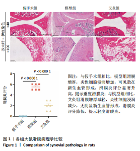
2.1 实验动物数量分析 实验选用30只大鼠,造模过程中无动物死亡,全部进入结果分析。 2.2 各组大鼠滑膜病理学比较 炎症在膝骨关节炎的发生和发展中起重要作用,通过苏木精-伊红染色观察艾灸对膝骨关节炎滑膜的影响。结果显示,与假手术组相比,模型组大鼠滑膜增厚,炎性细胞浸润增加,细胞排列紊乱,可见散在新生血管形成,滑膜炎评分明显升高,提示重度滑膜炎;而相对于模型组,艾灸组大鼠经艾灸治疗后滑膜增厚减轻,细胞排列有序,炎症细胞浸润减少,滑膜炎评分降低,提示轻度滑膜炎,见图1。"

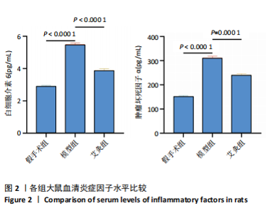
2.3 各组大鼠血清炎症因子水平比较 为了观察艾灸对炎症因子的影响,通过酶联免疫吸附实验检测各组血清中炎症因子白细胞介素6和肿瘤坏死因子α的表达水平,见图 2。结果显示,与假手术组相比,模型组观察到炎症因子白细胞介素6和肿瘤坏死因子α的异常高表达(P < 0.000 1); 而经艾灸治疗后,膝骨关节炎中的白细胞介素6和肿瘤坏死因子α的水平明显降低(P≤0.000 1)。"

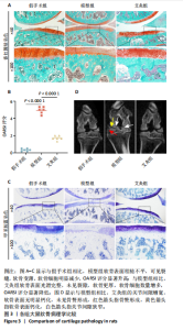
2.4 各组大鼠软骨病理学比较 通过番红固绿染色与甲苯胺蓝染色观察艾灸对膝骨关节炎软骨病变的影响,见图3。图3A,C结果表明,与假手术组相比,模型组的关节软骨表面粗糙不平、破坏显著,可见裂隙,软骨完整性降低,软骨变薄,软骨细胞数量减少且排列混乱;相对于模型组,艾灸组关节软骨表面光滑完整,未见裂隙,软骨更厚,软骨细胞数量增多且排列有序。上述变化也通过OARSI评分来量化软骨损伤的严重程度,结果表明假手术组、艾灸组评分均显著低于模型组(P < 0.000 1),见图 3B。此外,Micro-CT结果显示,与假手术组相比,模型组的关节间隙严重狭窄,软骨表面钙化显著,有骨赘形成;而与模型组相比,艾灸组的关节间隙增宽,软骨表面无明显钙化,未见骨赘形成,见图3D。"

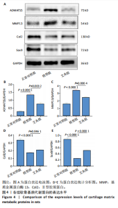
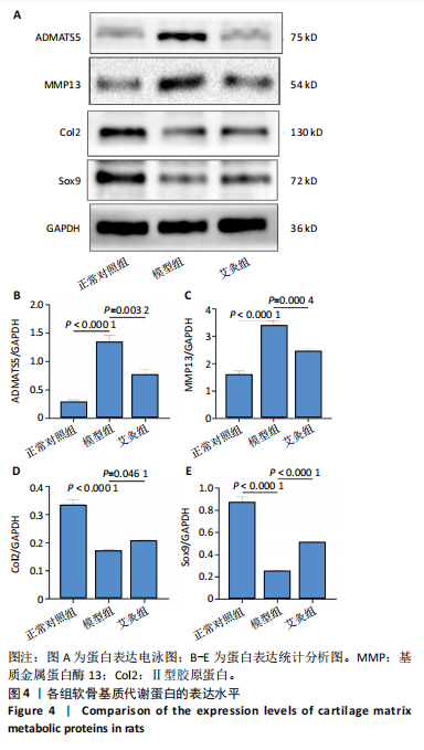
2.5 各组软骨基质代谢蛋白表达水平比较 进一步评估了艾灸对骨关节炎软骨细胞的软骨基质代谢蛋白的影响,见图 4。相对于正常对照组,模型组软骨基质降解蛋白ADAMTS-5和基质金属蛋白酶13的水平显著升高,而软骨基质合成蛋白Ⅱ型胶原蛋白和Sox9的表达明显减少(P < 0.000 1);与模型组相比,艾灸组软骨基质合成分子Ⅱ型胶原蛋白和Sox9蛋白表达增多(P < 0.05,P < 0.000 1),而软骨基质分解蛋白ADAMTS-5和基质金属蛋白酶13的水平显著下降(P < 0.01)。"
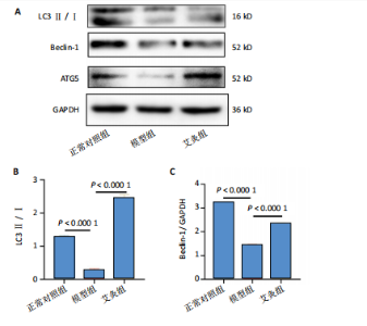
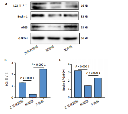
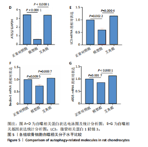
2.6 各组软骨细胞自噬相关分子水平比较 通过Western blot和QPCR观察艾灸对自噬蛋白和基因的影响,见图5。结果表明,与正常对照组相比,自噬标志物ATG5、Beclin-1、微管相关蛋白1轻链3-Ⅱ/Ⅰ蛋白和mRNA水平在模型组中下调(P < 0.01)。而与模型组相比,艾灸治疗后,自噬相关蛋白和基因表达水平显著上升(P < 0.01)。"
"

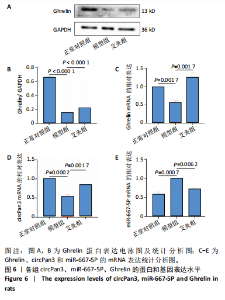
2.7 各组circPan3、miR-667-5P和ghrelin表达水平比较 为了观察艾灸对circPan3/miR-667-5p/Ghrelin信号通路的影响,进行了Western blot和 QPCR 实验,见图6。图 6C-E结果所示,与正常对照组相比,模型组circPan3、ghrelin的mRNA表达减少(P < 0.05,P < 0.01),而miR-667-5p的mRNA表达水平显著升高(P < 0.01);与模型组相比,艾灸组circPan3、ghrelin 的基因表达量显著上调(P < 0.01),miR-667-5p表达明显减少(P < 0.01)。此外western blot结果也表明ghrelin 的蛋白水平和基因水平呈现一致的趋势(P < 0.000 1),见图 6A,B。"


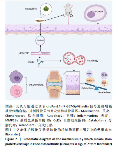
2.8 艾灸对骨关节炎软骨的作用机制 见图7。艾灸可能通过调节circPan3/miR-667-5p/Ghrelin信号通路增强软骨细胞自噬,抑制膝骨关节炎炎症和软骨破坏,对软骨发挥保护作用。艾灸通过调节circPan3/miR-667-5p/Ghrelin信号通路保护膝骨关节炎软骨。"
| [1] FUJII Y, LIU L, YAGASAKI L, et al. Cartilage Homeostasis and Osteoarthritis. Int J Mol Sci. 2022;23(11):6316. [2] HUNTER DJ, MARCH L, CHEW M. Osteoarthritis in 2020 and beyond: a Lancet Commission. Lancet. 2020;396(10264):1711-1712. [3] SHEN X, LAO L, ZHANG Y, et al. Biophysical and clinical research on acupuncture and moxibustion. Evid Based Complement Alternat Med. 2015;2015:518138. [4] 姜泉, 罗成贵, 巩勋, 等. 骨关节炎病证结合诊疗指南[J].中华中医药杂志,2021,36(2):929-933. [5] 张宪基, 汪宗保, 王科文, 等. 艾灸对膝骨关节炎兔软骨Wnt信号通路及血清炎性因子的影响[J]. 江苏中医药,2023,55(12):68-72. [6] HUANG J, CHEN Z, WU Z, et al.Geniposide stimulates autophagy by activating the GLP-1R/AMPK/mTOR signaling in osteoarthritis chondrocytes. Biomed Pharmacother. 2023;167:115595. [7] HE L, XU Z, NIU X, et al. GPRC5B protects osteoarthritis by regulation of autophagy signaling. Acta Pharm Sin B. 2023;13(7):2976-2989. [8] PAN Y, YANG Y, FAN M, et al.Progranulin regulation of autophagy contributes to its chondroprotective effect in osteoarthritis. Genes Dis. 2022;10(4):1582-1595. [9] WANG J, ZHANG Y, CAO J, et al. The role of autophagy in bone metabolism and clinical significance. Autophagy. 2023;19(9): 2409-2427. [10] DEBNATH J, GAMMOH N, RYAN KM. Autophagy and autophagy-related pathways in cancer. Nat Rev Mol Cell Biol. 2023;24(8):560-575. [11] YAMAMOTO H, ZHANG S, MIZUSHIMA N. Autophagy genes in biology and disease. Nat Rev Genet. 2023;24(6):382-400. [12] LIU S, YAO S, YANG H, et al. Autophagy: Regulator of cell death. Cell Death Dis. 2023;14(10):648. [13] XU M, LIU L, SONG C, et al. Ghrelin improves vascular autophagy in rats with vascular calcification. Life Sci. 2017;179:23-29. [14] ZHU K, ZHANG ML, LIU ST, et al. Ghrelin Attenuates Retinal Neuronal Autophagy and Apoptosis in an Experimental Rat Glaucoma Model. Invest Ophthalmol Vis Sci. 2017;58(14):6113-6122. [15] QU R, CHEN X, WANG W, et al. Ghrelin protects against osteoarthritis through interplay with Akt and NF-κB signaling pathways. FASEB J. 2018;32(2):1044-1058. [16] SUN L, ZHANG W. Preconditioning of mesenchymal stem cells with ghrelin exerts superior cardioprotection in aged heart through boosting mitochondrial function and autophagy flux. Eur J Pharmacol. 2021;903:174142. [17] QUIÑONES M, FERNØ J, AL-MASSADI O. Ghrelin and liver disease.Rev Endocr Metab Disord. 2020;21(1):45-56. [18] HAN W, CUI C, ZHANG H, et al. Ghrelin ameliorates diabetes-associated behavioral deficits and NLRP3 inflammasome activation via autophagic flux enhancement. Pharmacol Res. 2022;179:106224. [19] HESHMATI M, SOLTANI A, SANAEI MJ, et al. Ghrelin induces autophagy and CXCR4 expression via the SIRT1/AMPK axis in lymphoblastic leukemia cell lines. Cell Signal. 2020;66:109492. [20] ZENG J, ZHANG Z, LIAO Q, et al. circPan3 Promotes the Ghrelin System and Chondrocyte Autophagy by Sponging miR-667-5p During Rat Osteoarthritis Pathogenesis. Front Cell Dev Biol. 2021;9:719898. [21] XIE W, QI S, DOU L, et al. Achyranthoside D attenuates chondrocyte loss and inflammation in osteoarthritis via targeted regulation of Wnt3a.Phytomedicine. 2023;111:154663. [22] 王成, 袁君, 郭彦玎, 等. 艾灸“足三里”对膝关节骨关节炎与类风湿性关节炎大鼠膝关节滑膜巨噬细胞极化的影响的比较研究[J].针刺研究,2023,48(10):993-1000. [23] 李明静, 张健强, 郭靖, 等.艾灸不同腧穴对KOA大鼠不同组织TNF-α、MMP-13表达的影响[J]. 世界科学技术-中医药现代化, 2023,25(4):1390-1397. [24] 林芯宇, 张思爱, 徐银环, 等.艾灸治疗膝关节骨关节炎的疗效影响因素研究进展[J]. 针刺研究,2024,49(2):185-191. [25] 刘磊, 王敏君, 吴立斌, 等. 不同艾条直径和施灸距离对大鼠穴区皮肤表面温度的影响[J]. 针刺研究,2020,45(5):396-401. [26] FU L, DUAN H, CAI Y, et al. Moxibustion ameliorates osteoarthritis by regulating gut microbiota via impacting cAMP-related signaling pathway. Biomed Pharmacother. 2024;170:116031. [27] KRENN V, MORAWIETZ L, BURMESTER GR, et al. Synovitis score: discrimination between chronic low-grade and high-grade synovitis. Histopathology. 2006;49(4):358-364. [28] GERWIN N, BENDELE AM, GLASSON S, et al. The OARSI histopathology initiative - recommendations for histological assessments of osteoarthritis in the rat. Osteoarthritis Cartilage. 2010;18 Suppl 3:S24-34. [29] ZHU LL, ZHOU JY, LUO L, et al. Comparison of the efficacy between conventional moxibustion and smoke-free moxibustion on knee osteoarthritis: study protocol of a randomized controlled trial. Trials. 2017;18(1):188. [30] CHEN R, CHEN M, XIONG J, et al. Comparative effectiveness of the deqi sensation and non-deqi by moxibustion stimulation: a multicenter prospective cohort study in the treatment of knee osteoarthritis. Evid Based Complement Alternat Med. 2013;2013:906947. [31] 王佩, 王刘玉, 谢舜名, 等. “补肾益髓”法艾灸治疗膝关节骨关节炎的疗效观察[J]. 针刺研究,2024,49(2):171-176. [32] ZHAO L, CHENG K, WANG L, et al. Effectiveness of moxibustion treatment as adjunctive therapy in osteoarthritis of the knee: a randomized, double-blinded, placebo-controlled clinical trial. Arthritis Res Ther. 2014;16(3):R133. [33] MOTTA F, BARONE E, SICA A, et al. Inflammaging and Osteoarthritis. Clin Rev Allergy Immunol. 2023;64(2):222-238. [34] SELLAM J, BERENBAUM F. The role of synovitis in pathophysiology and clinical symptoms of osteoarthritis. Nat Rev Rheumatol. 2010; 6(11):625-635. [35] ZHENG L, ZHANG Z, SHENG P, et al. The role of metabolism in chondrocyte dysfunction and the progression of osteoarthritis. Ageing Res Rev. 2021;66:101249. [36] HOUARD X, GOLDRING MB, BERENBAUM F. Homeostatic mechanisms in articular cartilage and role of inflammation in osteoarthritis. Curr Rheumatol Rep. 2013;15(11):375. [37] 黄宇新, 林勇凯, 卢锦东, 等. 艾灸足三里、肾俞穴对骨性关节炎模型大鼠PGE2、NO及IL-1β的影响[J]. 动物医学进展,2014, 35(11):75-78. [38] JIA YJ, LI TY, HAN P, et al. Effects of different courses of moxibustion treatment on intestinal flora and inflammation of a rat model of knee osteoarthritis. J Integr Med. 2022;20(2):173-181. [39] INUI A. Ghrelin: an orexigenic and somatotrophic signal from the stomach. Nat Rev Neurosci. 2001;2(8):551-560. [40] COLLDÉN G, TSCHÖP MH, MÜLLER TD. Therapeutic Potential of Targeting the Ghrelin Pathway. Int J Mol Sci. 2017;18(4):798. |
| [1] | Lai Pengyu, Liang Ran, Shen Shan. Tissue engineering technology for repairing temporomandibular joint: problems and challenges [J]. Chinese Journal of Tissue Engineering Research, 2025, 29(在线): 1-9. |
| [2] | Miao Jiahang, Ma Sheng, Li Qupeng, Yu Huilin, Hu Tianyu, Gao Xiao, Feng Hu. Cervical lordosis ratio can be used as a decision-making indicator for selection of posterior surgical approach for multi-level cervical spondylotic myelopathy [J]. Chinese Journal of Tissue Engineering Research, 2025, 29(9): 1796-1802. |
| [3] | Yu Shuai, Liu Jiawei, Zhu Bin, Pan Tan, Li Xinglong, Sun Guangfeng, Yu Haiyang, Ding Ya, Wang Hongliang. Hot issues and application prospects of small molecule drugs in treatment of osteoarthritis [J]. Chinese Journal of Tissue Engineering Research, 2025, 29(9): 1913-1922. |
| [4] | Liu Lin, Liu Shixuan, Lu Xinyue, Wang Kan. Metabolomic analysis of urine in a rat model of chronic myofascial trigger points [J]. Chinese Journal of Tissue Engineering Research, 2025, 29(8): 1585-1592. |
| [5] | Su Xiaoyang, Chen Wenting, Fu Yidan, Zhao Yan, Lan Danfeng, Yang Qiuping. Correlation between Mer receptor tyrosine kinase and diabetic peripheral neuropathy in Sprague-Dawley rats [J]. Chinese Journal of Tissue Engineering Research, 2025, 29(8): 1593-1599. |
| [6] | Li Jun, Gong Jingjing, Sun Guobin, Guo Rui, Ding Yang, Qiang Lijuan, Zhang Xiaoli, Fang Zhanhai . miR-27a-3p promotes the proliferation of human hypertrophic scar fibroblasts by regulating mitogen-activated protein kinase signaling pathway [J]. Chinese Journal of Tissue Engineering Research, 2025, 29(8): 1609-1617. |
| [7] | Li Huayuan, Li Chun, Liu Junwei, Wang Ting, Li Long, Wu Yongli. Effect of warm acupuncture on PINK1/Parkin pathway in the skeletal muscle of rats with chronic fatigue syndrome [J]. Chinese Journal of Tissue Engineering Research, 2025, 29(8): 1618-1625. |
| [8] | Jing Ruyi, Chen Yingxin, Cao Lei . Prognosis of deep lamellar keratoplasty versus penetrating keratoplasty in the treatment of stromal corneal dystrophy [J]. Chinese Journal of Tissue Engineering Research, 2025, 29(8): 1626-1633. |
| [9] | Zhou Panpan, Cui Yinglin, Zhang Wentao, Wang Shurui, Chen Jiahui, Yang Tong . Role of cellular autophagy in cerebral ischemic injury and the regulatory mechanism of traditional Chinese medicine [J]. Chinese Journal of Tissue Engineering Research, 2025, 29(8): 1650-1658. |
| [10] | Zhu Hanmin, Wang Song, Xiao Wenlin, Zhang Wenjing, Zhou Xi, He Ye, Li Wei, . Mitophagy regulates bone metabolism [J]. Chinese Journal of Tissue Engineering Research, 2025, 29(8): 1676-1683. |
| [11] | Wang Yida, Liu Jun, Wang Xiaoling, Wang Liyan, Yang Chengru, Zhang Xuexiao. Effects of wearable electronic device-based interventions on physical activity and sedentary behavior in healthy adolescents: a meta-analysis [J]. Chinese Journal of Tissue Engineering Research, 2025, 29(8): 1693-1704. |
| [12] | Zheng Huakun, Yin Mingyue, Liu Qian. Effects of interval and continuous training on the quality of life in physically inactive adults: a meta-analysis [J]. Chinese Journal of Tissue Engineering Research, 2025, 29(8): 1727-1740. |
| [13] | Yin Lu, Jiang Chuanfeng, Chen Junjie, Yi Ming, Wang Zihe, Shi Houyin, Wang Guoyou, Shen Huarui. Effect of Complanatoside A on the apoptosis of articular chondrocytes [J]. Chinese Journal of Tissue Engineering Research, 2025, 29(8): 1541-1547. |
| [14] | Cai Yaohao, Lang Lyu, Li Hong. Assessing the bone mass of the residual alveolar ridge in the first molar for implant placement by cone-beam computed tomography [J]. Chinese Journal of Tissue Engineering Research, 2025, 29(8): 1572-1577. |
| [15] | Hu Taotao, Liu Bing, Chen Cheng, Yin Zongyin, Kan Daohong, Ni Jie, Ye Lingxiao, Zheng Xiangbing, Yan Min, Zou Yong. Human amniotic mesenchymal stem cells overexpressing neuregulin-1 promote skin wound healing in mice [J]. Chinese Journal of Tissue Engineering Research, 2025, 29(7): 1343-1349. |
| Viewed | ||||||
|
Full text |
|
|||||
|
Abstract |
|
|||||