Chinese Journal of Tissue Engineering Research ›› 2025, Vol. 29 ›› Issue (4): 752-760.doi: 10.12307/2024.812
Previous Articles Next Articles
Research frontiers and hotspots of carbon nanomaterials in biomedical field over the past 10 years
Dang Xiaowen, Huang Hailiang, Huang Lei, Wang Yajie
- Rehabilitation Medicine School of Shandong University of Traditional Chinese Medicine, Jinan 250355, Shandong Province, China
-
Received:2023-12-08Accepted:2024-01-16Online:2025-02-08Published:2024-05-30 -
Contact:Huang Hailiang, MD, Professor, Rehabilitation Medicine School of Shandong University of Traditional Chinese Medicine, Jinan 250355, Shandong Province, China -
About author:Dang Xiaowen, Master candidate, Rehabilitation Medicine School of Shandong University of Traditional Chinese Medicine, Jinan 250355, Shandong Province, China -
Supported by:Construction Project of Liu Zhaochun Inheritance Studio for Famous Traditional Chinese Medicine in Shandong Province, No. [2018] 1 (to HHL)
CLC Number:
Cite this article
Dang Xiaowen, Huang Hailiang, Huang Lei, Wang Yajie . Research frontiers and hotspots of carbon nanomaterials in biomedical field over the past 10 years[J]. Chinese Journal of Tissue Engineering Research, 2025, 29(4): 752-760.
share this article
Add to citation manager EndNote|Reference Manager|ProCite|BibTeX|RefWorks
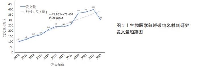
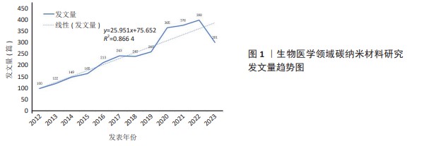
2.1 发文量分析 文献的年发文量是衡量某一领域研究热度和发展现状的重要指标,并在一定程度上可以预测该领域的未来动态[12]。如图1所示,碳纳米材料在医学领域的年发文量整体上呈上升趋势,以上升趋势分为3个阶段。2012-2017年为阶梯式上升期,该时期文献数量缓慢增长,是各国学者将碳纳米材料应用到生物医学领域的初步探索;2017-2019年为稳步发展期,此阶段发文量稍有增长;2019-2022年为快速增长期,这一阶段文献数量增长明显,此时“纳米医学”理论研究热度较高[13-14],对碳纳米材料在生物医学领域的发展产生良性影响。同时线性预测模型表明:生物医学领域碳纳米材料发文量与年份之间有显著相关关系且相关系数较高(R=0.866 4),由此可认为未来此领域会持续受到研究者的关注,发文量将持续增长。"
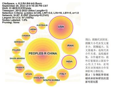
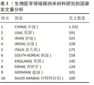
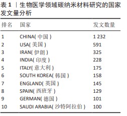
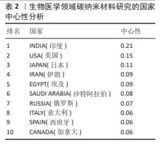
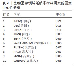
2.2 研究主体 2.2.1 国家分析 图2及表1,2列出了生物医学领域碳纳米材料相关研究的国家共现图谱、文献数量和中心性排名前10位的国家。图2为Citespace软件生成的国家共现网络图谱,文章共纳入发文国家97个,连线数达997条,网络密度为0.182。综合分析文献数量和中心性发现,中国、美国和伊朗在该领域研究成果可观,是碳纳米材料在生物医学领域研究的中坚力量。印度、美国和日本在这一领域与其他国家联系较为密切,在国际上的影响力也较大。中国虽然发文量排名第一,但中心性仅为0.03,表明中国在该领域研究水平还有待提高,应加强与其他国家的合作,增强国际影响力。"

"
"
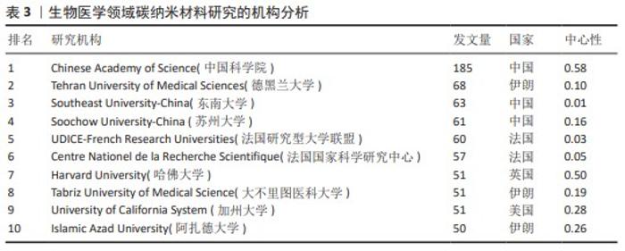
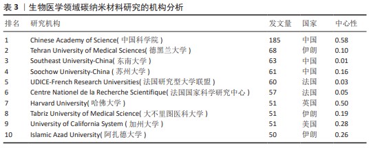
2.2.2 机构分析 表3列出碳纳米材料在生物医学领域研究的前10位机构显示,有3所机构来自中国,总计发文309篇,在此研究领域遥遥领先。图3为该领域核心机构合作共现图谱,文章共提取278个机构,连线数为377条,网络密度仅为0.009 8。最大的合作网络是以中国科学院为中心的合作机构,其中与其直接合作的机构有9所,合作发表185篇文章,主要合作对象为国内研究机构,缺少与国外知名单位的合作,表明中国各研究机构需加强国际间的合作,关注国际发展热点,不断交流学习,弥补自身发展短板,提高中国在生物医学领域碳纳米材料研究的国际影响力。"
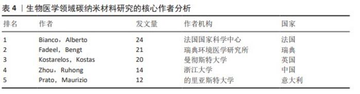
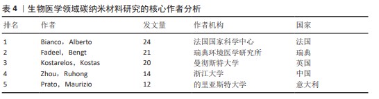
2.2.3 作者分析 对纳入文献的作者及发文量分析,见表4,其中包括546位作者,发文量最多的是来自法国国家科学中心的Bianco,Alberto教授,发表该领域相关文章24篇。根据普莱斯定律通过最高发文量可以得到核心作者的最低发文量N=0.749×ηmax1/2(ηmax为最高产出量作者的发文量)且核心作者将完成该领域一半以上的论文[15]。文章的ηmax=24,由此得出N≈4,即发文量在4篇及以上的作者为核心作者,共66位,总计发文426篇,占总发文量的14.5%,未达到总发文量的1/2,这说明核心作者贡献率低,学术水平有待提高。表4列出了此领域发文量排名前5位的核心作者,其中Bianco,Alberto教授发文24篇位居榜首,其研究方向主要在设计适用于疾病诊断、治疗和成像的多功能碳纳米材料[16]。来自中国的Zhou,Ruhong教授发表文章14篇,位列第4位,其主要从事碳纳米材料与肿瘤药物的研究,并取得一系列突破性进展[17]。"
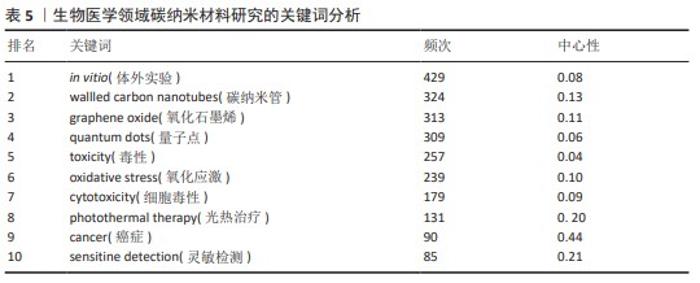
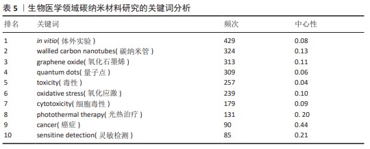
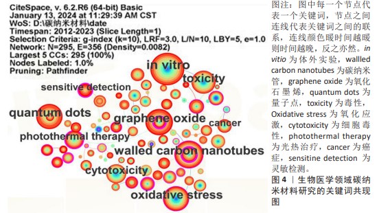
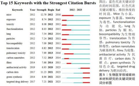
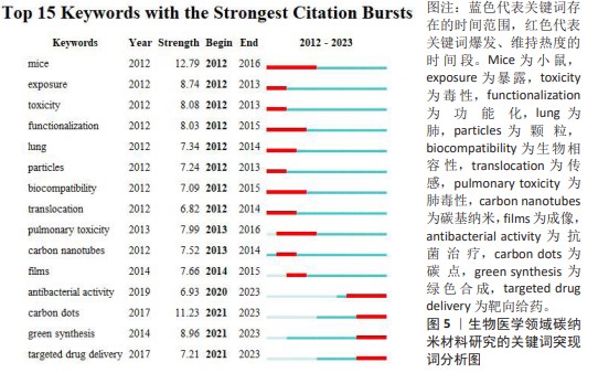
2.3 研究内容 2.3.1 关键词分析 关键词是对文章核心内容的高度凝练,能在一定程度上反映此领域的研究热点[18]。 关键词出现的次数为频次,频次越高则研究越深入;关键词在研究中的位置为中心性,中心性越高则关键词的位置越重要[19]。使用Citespace软件对文献频次及中心性进行统计分析,列出排名前10位的关键词,见表5。其中频次最高的关键词是in vitro (429次),说明了体外实验是碳纳米材料应用于生物医学领域的研究基础。中心性最高的是cancer (0.44),表明癌症医学在此领域的研究较为集中,处于中心位置。通过表5和图4发现,碳纳米管和氧化石墨烯的频次和中心性都较高,说明有关研究相对较多,是此领域探索最深入的碳纳米材料。在研究内容方面,碳纳米材料的毒理学研究在这10年中一直备受关注。此外,关键词的突现词也可以展示该领域的研究热点与前沿,而且还可以呈现出该领域研究热点的时间演变情况[20]。使用Citespace软件对文献关键词进行突现分析见图5。在爆发力最强的前15个突现词中,mice(小鼠)的爆发强度最大,这说明碳纳米材料再生医学领域的研究还处于体外细胞培养和体内动物实验的阶段,实现临床应用需进一步探索。其次carbon dot(碳点)爆发强度位居第二,且其爆发时间开始于2021年,并在2023年持续爆发,说明碳点作为新颖的碳纳米材料将在未来几年持续受到关注,是该领域的研究前沿。碳点因具有低细胞毒性、水溶性和易改性等独特优势在生物传感、生物成像、药物递送、生物抗菌及靶向给药等方面都展现出优越潜力[21-23],其绿色合成的方式近年来也广受关注[24-25]。"

"
"

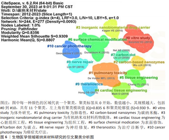
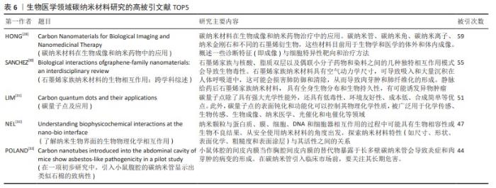
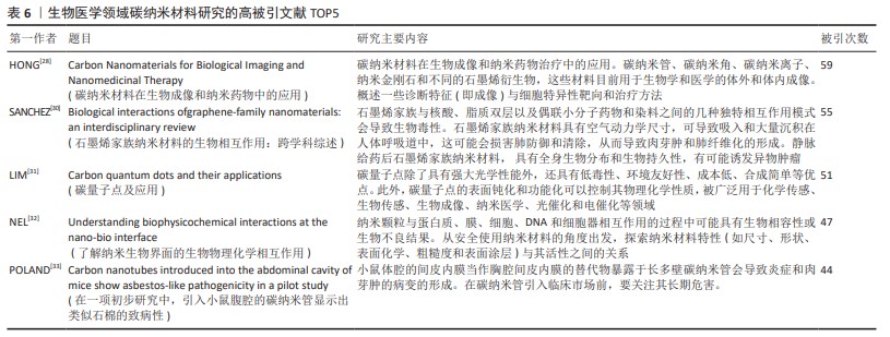
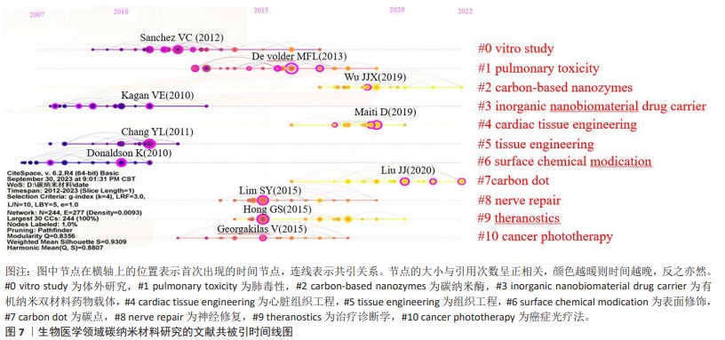
2.3.2 高被引文献分析 高引用研究被认为是该领域最具影响力的研究[26]。共被引分析可用于查找引文中最重要的参考文献,分析这些文章可以阐述该领域最前沿研究背后的演变情况以及对发展趋势和未来研究方向作出合理判断[27]。使用Citespace软件进行文献共被引分析。其中被引次数最多的文献主要介绍了碳纳米材料在生物成像和纳米药物治疗中的应用[28]。其次采用LLR算法进行聚类,运行结果如图6所示。聚类模块值(Q)和聚类轮廓值(S)是评价聚类情况的重要指标,一般Q值大于0.5,S值大于0.7则被认为该聚类结构显著,同质性高,结果令人信服[29],文章列举了共被引频次前5位的文献[28,30-33],见表6。共生成11个聚类,其中Q=0.835 6 > 0.5,S=0.930 9 > 0.7,表明聚类合理,可信度高。聚类标签从0开始,数值越小,其规模越大。对聚类标签进一步分析,可分为3个领域:①围绕碳纳米材料的毒理学研究,包含“#0 vitro study (体外研究)、#1 pulmonary toxicity (肺毒性)、#6 surface chemical modication(表面修饰)”; ②基于碳纳米材料本身材料研究,包含“#2 carbon-based nanozymes(碳纳米酶)、#7 carbon dot(碳点)”; ③碳纳米材料在医学领域的应用究,包含“#3 inorganic nanobiomaterial drug carrier(有机纳米双材料药物载体)、#4 cardiac tissue engineering(心脏组织工程) 、#5 tissue engineering(组织工程)、#8 nerve repair(神经修复)、#9 theranostics (治疗诊断学)、#10 cancer phototherapy(癌症光疗法)。通过以上可以看出研究最广泛的是碳纳米材料在生物医学领域的具体应用,尤其在癌症和组织工程方面。"
"
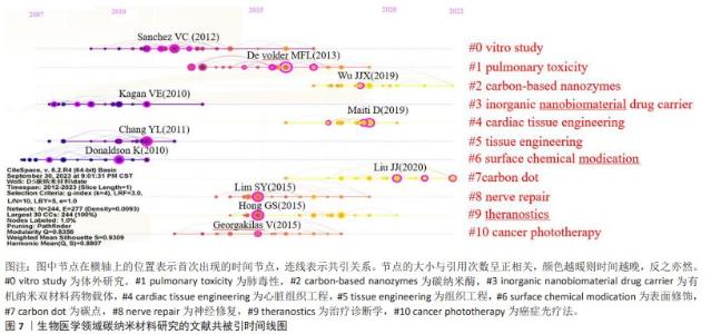
为了进一步揭示生物医学领域碳纳米材料的研究热点和发展趋势,对前11个聚类绘制时间线图,见图7。结合共被引聚类图谱,发现该领域的相关研究中,对碳纳米管和氧化石墨烯的探索较为深入,而碳量子点和碳纳米酶是相对较新的材料。在研究内容方面,对碳纳米材料在药物递送方面研究的时间早,而组织工程领域时间更长,尤其近年来,心脏组织工程、神经组织工程成为新的研究热点。同时体外研究、肺毒性、表面修饰的探索开始时间更早、持续时间更长,虽相关研究的热度随时间变化也稍有降低,但其中出现某些暖黄色节点,说明了生物医学领域碳纳米材料的安全性研究一直以来备受关注,是该领域稳定长期发展的重要基础。"
| [1] PATRICK B, AKHTAR T, KOUSAR R, et al. Carbon nanomaterials: emerging roles in immuno-oncology. Int J Mol Sci. 2023; 24(7):6600. [2] CUI F, LI T, WANG D, et al. Recent advances in carbon-based nanomaterials for combating bacterial biofilm-associated infections. J Hazard Mater. 2022;431: 128597. [3] TORRINHA A, OLIVEIRA TMBF, RIBEIRO FWP, et al. Application of nanostructured carbon-based electrochemical (bio)sensors for screening of emerging pharmaceutical pollutants in waters and aquatic species: a review. Nanomaterials (Basel). 2020;10: 1268. [4] INTISAR A, RAMZAN A, SAWAIRA T, et al. Occurrence, toxic effects, and mitigation of pesticides as emerging environmental pollutants using robust nanomaterials-A review. Chemosphere. 2022;293:133538. [5] FARRE M, KANTIANI L, PETROVIC M, et al. Achievements and future trends in the analysis of emerging organic contaminants in environmental samples by mass spectrometry and bioanalytical techniques. J Chromatogr A. 2012;1259:86-99. [6] TUPONE MG, PANELLA G, D’ANGELO M, et al. An update on graphene-based nanomaterials for neural growth and central nervous system regeneration. Int J Mol Sci. 2021;22:13047. [7] SADEGHI MS, SANGRIZEH FH, JAHANI N, et al. Graphene oxide nanoarchitectures in cancer therapy: drug and gene delivery, phototherapy, immunotherapy, and vaccine development. Environ Res. 2023;237: 117027. [8] LIU Y, LIU H, GUO S, et al. A review of carbon nanomaterials/bacterial cellulose composites for nanomedicine applications. Carbohydr Polym. 2024;323:121445. [9] CUI L, LIANG J, LIU H, et al. Nanomaterials for angiogenesis in skin tissue engineering. Tissue Eng Part B Rev. 2020;26(3):203-216. [10] XU X, SHEN Z, SHAN Y, et al. Application of tissue engineering techniques in tracheal repair: a bibliometric study. Bioengineered. 2023;14:2274150. [11] 陈悦,陈超美,刘则渊,等.CiteSpace知识图谱的方法论功能[J].科学学研究, 2015,33(2):242-253. [12] 贺雅洁,王延博,杨硕.基于CiteSpace对中医药治疗子宫肌瘤的可视化分析[J].世界中西医结合杂志,2022,17(12): 2374-2380. [13] PALMIERIV, DI PIETRO L, PERINI G, et al. Graphene oxide nano-concentrators selectively modulate RNA trapping according to metal cations in solution. Front Bioeng Biotechnol. 2020;8:421. [14] DONG P, RAKESH KP, MANUKUMARH M, et al. Innovative nano-carriers in anticancer drug delivery-a comprehensive review. Bioorg Chem. 2019;85:325-336. [15] 谢恩礼,陶慧敏.血流限制训练在临床康复医学中的应用趋势[J].中国组织工程研究,2024,28(2):258-262. [16] CELLOT G, JACQUEMIN L, REINA G, et al. Bonding of neuropeptide y on graphene oxide for drug delivery applications to the central nervous system. ACS Appl Nano Mater. 2022;5(12):17640-17651. [17] YE R, SONG W, FENG M, et al. Potential interference of graphene nanosheets in immune response via disrupting the recognition of HLA-presented KK10 by TCR: a molecular dynamics simulation study. Nanoscale. 2021;13(45):19255-19263. [18] 李一飞,周开宇,沈建通,等.基于关键词共现分析的我国先心病介入诊疗发展的可视化研究[J]. 临床儿科杂志, 2012,30(7):631-637. [19] 徐鹏,赵京霞,高铸烨,等.2012—2021年国家自然科学基金脑卒中中医内科研究领域申请与资助分析——基于CiteSpace的可视化分析[J].长春中医药大学学报,2022,38(12):1311-1319. [20] LING L, OUYANG Y, HU Y. Research trends on nanomaterials in gastric cancer: a bibliometric analysis from 2004 to 2023. J Nanobiotechnology. 2023;21(1):2481. [21] LI X, VINOTHINI K, RAMESH T, et al. Combined photodynamic-chemotherapy investigation of cancer cells using carbon quantum dot-based drug carrier system. Drug Deliv. 2020;27(1):791-804. [22] SUN L, ZHANG R, ZHANG T, et al. Synthesis, applications and biosafety evaluation of carbon dots derived from herbal medicine. Biomed Mater. 2023. doi:10.1088/1748-605X/acdeb8. [23] KONG B, YANGT, CHENG F, et al. Carbon dots as nanocatalytic medicine for anti-inflammation therapy. J Colloid Interface Sci. 2022;611:545-553. [24] HE JH, CHENG YY, YANG T, et al. Functional preserving carbon dots-based fluorescent probe for mercury (II) ions sensing in herbal medicines via coordination and electron transfer. Anal Chim Acta. 2018;1035:203-210. [25] KANWAL A, BIBI N, HYDER S, et al. Recent advances in green carbon dots (2015-2022): synthesis, metal ion sensing, and biological applications. Beilstein J Nanotechnol. 2022;13:1068-1107. [26] 黄伟,苏晓丽,赵江宁,等.基于CiteSpace的严重创伤患者低体温研究的可视化分析[J].临床医学研究与实践, 2023,8(4):1-4. [27] 尤伟杰,郭青,张楠,等.基于关键词共现和文献共被引的医学期刊微信公众平台热点可视化分析[J].中华医学图书情报杂志,2019,28(2):76-80. [28] HONG G, DIAO S, ANTARIS AL, et al. Carbon nanomaterials for biological imaging and nanomedicinal therapy. Chem Rev. 2015; 115(19SI):10816-10906. [29] 杨丽萍,段培蓓,杨玲,等.肿瘤患者疼痛-疲乏-睡眠障碍症状群的研究现状及热点可视化分析[J].中华全科医学, 2023,21(1):139-143. [30] SANCHEZ VC, JACHAK A, HURT RH, et al. Biological interactions of graphene-family nanomaterials: an interdisciplinary review. Chem Rev. 2012;25(1):15-34. [31] LIM SY, SHEN W, GAO Z. Carbon quantum dots and their applications. Chem Res Toxicol. 2015;44(1):362-381. [32] NEL AE, MAEDLER L, VELEGOL D, et al. Understanding biophysicochemical interactions at the nano-bio interface. Chem Soc Rev. 2009;8(7):543-557. [33] POLAND CA, DUFFIN R, KINLOCH I, et al. Carbon nanotubes introduced into the abdominal cavity of mice show asbestos-like pathogenicity in a pilot study. Nat Mater. 2008;3(7):423-428. [34] 田娇,赵锡丽,冉倩.基于Web of Science数据库的糖尿病饮食研究的可视化分析[J].循证护理,2023,9(21): 3902-3908. [35] 邹婧,楚尧娟,杜秋争,等.酪氨酸激酶抑制剂在HER2阳性乳腺癌中应用的可视化分析[J].中国药房,2023,34(24): 3036-3041. [36] HOSNEDLOVA B, KEPINSKA M, FERNANDEZ C, et al. Carbon nanomaterials for targeted cancer therapy drugs: a critical review. Chem Rec. 2019;19(2-3):502-522. [37] CHEN D, DOUGHERTY CA, ZHU K, et al. Theranostic applications of carbon nanomaterials in cancer: focus on imaging and cargo delivery. J Control Release. 2015;210:230-245. [38] SAJJADI M, NASROLLAHZADEH M, JALEH B, et al. Carbon-based nanomaterials for targeted cancer nanotherapy:recent trends and future prospects. J Drug Target. 2021;29(7):716-741. [39] HAN C, CHEN R, WU X, et al. Fluorescence turn-on immunosensing of HE4 biomarker and ovarian cancer cells based on target-triggered metal-enhanced fluorescence of carbon dots. Anal Chim Acta. 2021;1187: 339160. [40] LIU H, CHEN J, QIAO S, et al. Carbon-based nanomaterials for bone and cartilage regeneration: a review. ACS Biomater Sci Eng. 2021;7(10):4718-4735. [41] LIU C, QIN H, KANG L, et al. Graphitic carbon nitride nanosheets as a multifunctional nanoplatform for photochemical internalization-enhanced photodynamic therapy. J Mater Chem B. 2018;6(47):7908-7915. [42] AHMAD R, FU J, HE N, et al. Advanced gold nanomaterials for photothermal therapy of cancer. J Nanosci Nanotechnol. 2016;16(1):67-80. [43] RADZI M RM, JOHARI NA, ZAWAWI WFAW, et al. In vivo evaluation of oxidized multiwalled-carbon nanotubes-mediated hyperthermia treatment for breast cancer. Biomater Adv. 2022;134:112586. [44] AOKI K, SAITO N. Biocompatibility and carcinogenicity of carbon nanotubes as biomaterials. Nanomaterials (Basel). 2020; 10:2642. [45] 王嘉昀,吴俏兰,高祖,等.基于科学知识图谱的桔梗研究热点与趋势分析[J].中医药导报,2023,29(10):110-118. [46] 张春婷,李波,谢立国,等. 温泉微生物合成的碳纳米材料在生物成像方面的应用[J]. 材料科学与工程学报,2023, 41(4):561-567. [47] SHIN SR, JUNG SM, ZALABANY M, et al. Carbon-nanotube-embedded hydrogel sheets for engineering cardiac constructs and bioactuators. ACS Nano. 2013;7(3):2369-2380. [48] WU Y, SHI X, LI Y, et al. Carbon nanohorns promote maturation of neonatal rat ventricular myocytes and inhibit proliferation of cardiac fibroblasts: a promising scaffold for cardiac tissue engineering. Nanoscale Res Lett. 2016; 11(1):284. [49] MARTINELLI V, CELLOT G, TOMA FM, et al. Carbon nanotubes promote growth and spontaneous electrical activity in cultured cardiac myocytes. Nano Lett. 2012;12(4):1831-1838. [50] FENG Z, WANG T, ZHAO B, et al. Soft graphene nanofibers designed for the acceleration of nerve growth and development. Adv Mater. 2015;27(41): 6462-6468. [51] YAN L, ZHAO B, LIU X, et al. Aligned nanofibers from polypyrrole/graphene as electrodes for regeneration of optic nerve via electrical stimulation. ACS Appl Mater Interfaces. 2016;8(11):6834-6840. [52] JAKUS AE, SECOR EB, RUTZ AL, et al. Three-dimensional printing of high-content graphene scaffolds for electronic and biomedical applications. ACS Nano. 2015;9(4):4636-4648. [53] RAJA IS, KANG MS, HONG SW, et al. State-of-the-art techniques for promoting tissue regeneration: combination of three-dimensional bioprinting and carbon nanomaterials. Int J Bioprint. 2023;9(1): 181-198. [54] ZHANG J, EYISOYLU H, QIN X, et al. 3D bioprinting of graphene oxide-incorporated cell-laden bone mimicking scaffolds for promoting scaffold fidelity, osteogenic differentiation and mineralization. Acta Biomater. 2021;121:637-652. [55] JAKUS AE, SECOR EB, RUTZ AL, et al. Three-dimensional printing of high-content graphene scaffolds for electronic and biomedical applications. ACS Nano. 2015;9(4):4636-4648. [56] OSOULI-BOSTANABAD K, MASALEHDAN T, KAPSA RMI, et al. Traction of 3D and 4D printing in the healthcare industry: from drug delivery and analysis to regenerative medicine. ACS Biomater Sci Eng. 2022;8(7): 2764-2797. [57] POURMASOURMASOUMI P, MOGHADDAM A, MAHAND SN, et al. A review on the recent progress, opportunities, and challenges of 4D printing and bioprinting in regenerative medicine. J Biomater Sci Polym Ed. 2023;34(1):108-146. [58] MIAO S, CUI H, NOWICKI M, et al. Stereolithographic 4D bioprinting of multiresponsive architectures for neural engineering. Adv Biosyst. 2018;2:1800101. [59] QASIM M, CLARKSON ANN, HINKLEY SFR. Green synthesis of carbon nanoparticles (CNPS) from biomass for biomedical applications. Int J Mol Sci. 2023;24:1023. [60] ASHRAFIZADEH M, SAEBAR H, GHOLAMI MH, et al. Doxorubicin-loaded graphene oxide nanocomposites in cancer medicine: stimuli-responsive carriers, co-delivery and suppressing resistance. Expert Opin Drug Deliv. 2022;19(4):355-382. [61] RAJA IS, SONG S, KANG MS, et al. Toxicity of zero- and one-dimensional carbon nanomaterials. Nanomaterials (Basel). 2019;9:1214. [62] JOHNSON AP, GANGADHARAPPA HV, PRAMOD K. Graphene nanoribbons: a promising nanomaterial for biomedical applications. J Control Release. 2020;325: 141-162. [63] RAHMAN MA, ABUL BARKAT H, HARWANSH RK, et al. Carbon-based nanomaterials: carbon nanotubes, graphene, and fullerenes for the control of burn infections and wound healing. Curr Pharm Biotechnol. 2022;23(12):1483-1496. |
| [1] | Israrguli · Maimeti, Jia Sen, Liu Jia. Bone morphogenetic protein 2-loaded hydrogel induces osteogenic differentiation of dental pulp stem cells [J]. Chinese Journal of Tissue Engineering Research, 2025, 29(16): 3301-3310. |
| [2] | Xin Yuan, Wu Xixi, Quan Liang, Zhang Hengtong, Ao Qiang. REG-augmented decellularized porcine cornea/hydroxyethyl methacrylate in situ integrated composite artificial cornea [J]. Chinese Journal of Tissue Engineering Research, 2025, 29(16): 3388-3399. |
| [3] | Wang Renzhi, Chen Yuanfen, Li Jinwei. 3D printed hollow pipe double-crosslinked hydrogel tissue engineering scaffold [J]. Chinese Journal of Tissue Engineering Research, 2025, 29(16): 3432-3439. |
| [4] | Tian Yanping, Li Juan, Liu Xiaobo, Zhang Huiling, Shi Lihong, Jin Rongjiang. Knowledge network mapping of literature regarding platelet-rich plasma in recent 5 years: a visual analysis [J]. Chinese Journal of Tissue Engineering Research, 2021, 25(11): 1745-1752. |
| Viewed | ||||||
|
Full text |
|
|||||
|
Abstract |
|
|||||