Chinese Journal of Tissue Engineering Research ›› 2018, Vol. 22 ›› Issue (35): 5620-5624.doi: 10.3969/j.issn.2095-4344.0364
Previous Articles Next Articles
Best screw placement parameters in S1 pedicle based on three-dimensional reconstruction
Wan Shengyu, Lin Xu, Wu Chao, Deng Jiayan
- Department of Orthopedics, Zigong Fourth People’s Hospital, Zigong 643000, Sichuan Province, China
-
Online:2018-12-18Published:2018-12-18 -
About author:Wan Shengyu, Master, Attending physician, Department of Orthopedics, Zigong Fourth People’s Hospital, Zigong 643000, Sichuan Province, China -
Supported by:the Scientific Research Program of Science and Technology Bureau of Zigong, Sichuan Province, No. 2016ZC11
CLC Number:
Cite this article
Wan Shengyu, Lin Xu, Wu Chao, Deng Jiayan. Best screw placement parameters in S1 pedicle based on three-dimensional reconstruction[J]. Chinese Journal of Tissue Engineering Research, 2018, 22(35): 5620-5624.
share this article
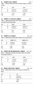
2.1 造模成功情况 3例重建模型因髂骨翼阻挡不能垂直及前内侧置钉,因此105例骨盆三维重建模型进入结果分析,其中男54例,女51例。 2.2 S1椎弓根螺钉置钉的空间参数 采用以S1关节突的外侧缘与下缘交点为置入点,方向朝向骶岬(取得最佳的生物力学稳定性置钉时)参数测量见表1-3。 螺钉置入的左侧内倾角男性与女性比较差异有显著性意义(P=0.013);右侧内倾角男性与女性为比较差异有显著性意义(P=0.003);置入螺钉与水平面的夹角男性左右分别为(19.80±7.15)°,(19.94±7.14)°;女性为(19.35± 6.56)°,(19.37±7.16)°,男女左右侧螺钉与水平面的夹角比较差异无显著性意义(P > 0.05)。 螺钉置入长度男性左右侧分别为(47.90±6.05)mm,(48.67±6.31)mm;女性左右侧分别为(47.48±4.50)mm,(48.54±4.08) mm,男女左右侧置入螺钉的长度比较差异无显著性意义(P > 0.05)。 2.3 骨盆参数 基于S1椎最佳置钉时川南地区男女骨盆参数的解剖学测量见表4,5。 螺帽与髂骨翼的相切距离男性左右侧分别为(2.20±4.33) mm,(2.13±3.93) mm;女性为(8.03±8.81) mm,(7.57±6.70) mm。男、女左右侧螺帽与髂骨翼的相切距离相比较,差异有显著性意义(P=0.000);而男、女螺钉置入平面髂骨之间的距离相比较,差异亦有显著性意义(P =0.000)。 "

| [1] Zindrick MR, Wiltse LL, Widell EH, et al. A biomechanical study of intrapedicularscrew fixation in the lumbosacral spine. Clin Orthop. 1986;203: 99-112.[2] stuik JP. Techniques of internal fixation for degenerative conditions of the spine . Clin Orthop. 1986;203: 2019-2022.[3] Krag MH. Biomechanic of thoraco lumbarspinal fixation: areview. Spine. 1991;16:584-587.[4] Smith SA,Abitbol JJ,Carlson GD,et al.The effects of depth ofpenetration,screw orientation,and bone density on sacral screwfixation. Spine. 1993;18(8):1006-1010.[5] Carlson GD,Abitbol JJ,Anderson DR, et a1.Screw fixation in the human sacrum:an in vitro study of the biomechanics of fixation. Spine. 1992;17(Suppl6):196-203.[6] Fairbank JC, Pynsent PB. The Oswestry Disability Index. Spine(Phila Pa 1976). 2000;25(22):2940-2952.[7] Bernhardt M, Swartz DE,Clothiaux PL,et al.Posterolateral lumbar and lumbosacral fusion with and without pedicle screw internal fixation. ClinOrthop Relat Res.1992;(284):109-115.[8] Horowitch A,Peek RD,Thomas JC Jr, et al.The Wiltse pedicle screw fixation system:Early clinical results.Spine(Phila Pa 1976). 1989;14(4):461-467.[9] Horton WC, Holt RT, Muldowny DS. Controversy. Fusion of L5-S1 in adult scoliosis.Spine(Phila Pa 1976). 1996;21(21):2520-2522.[10] Lehman RA, Kuklo TR,Belmont PJ, et al.Advantage of pedicle screw fixation directed into the apex of the sacral promontory over bicortical fixation: a biomechanicalanalysis.Spine. 2002;27(8):806-811.[11] Zheng YG, Lu WW,Zhu Q, et al. Variation in bone mineral density of the sacrum in young adults and its significance for sacral fixation.Spine. 2000;25(3):353-357.[12] Hadjipavlou AG,Nicodemus CL,al-Hamdan FA,et al. Correlation of bone equivalent mineral density to pull-out resistance of triangulated pedicle screw construct. J Spinal Disord. 1997;10(1):12-19.[13] Pfeiffer M, Hoffman H, Goel VK, et al.In vitro testing of a new transpedicular stabilization technique. Eur Spine J. 1997;6(4):249-255.[14] Matsukawa K, Yato Y, Kato T, et al. Cortical bone trajectory forlumbosacral fixation:penetrating S1 endplate screw technique: technical note.J Neurosurg Spine. 2014;21(2):203-209. [15] 黄宗文,饶书城.脊柱腰骶段经椎弓根固定的应用解剖学研究[J].中国脊柱脊髓杂志,1996,6(3):119-122.[16] 龙源深,梁锦,詹世强,等.改进第1骶椎椎弓根螺钉进入法的解剖学研究与临床应用[J].中华骨科杂志,1999,19(9):537-540.[17] 杨凯,赵海,刘强,等.正常成人骶1椎弓根解剖学测量与临床应用[J].中国矫形外科杂志,1998,5(4):320-321.[18] 严军,郭春,唐天驷,等.经椎弓根椎体间内固定治疗腰椎滑脱症的应用解剖学研究[J].骨与关节损伤杂志, 1996,11(5):278-281[19] Kaptanoglu E, Okutan O, Tekdemir I. Closed posterior superior iliac spine impeding pediculocorporeal S-1 screw insertion.J Neurosurg. 2003;99:229-234.[20] Lehman RA Jr, Kuklo TR, Belmont PJ Jr, et al. Advantage of pedicle screw fixation directed into the apex of the sacral promontory over bicortical fixation: abiomechanical analysis. Spine (Phila Pa 1976). 2002;27(8):806-811.[21] De Peretti F, Argenson C, Bourgeon A, et al. Anatomic and experimental basis for the insertion of a screw at the first sacral vertebra. Surg Radiol Anat. 1991;13(2):133-137.[22] Arman C, Naderi S, Kiray A,et al.The human sacrum and safe approaches for screw placement. J Clin Neurosci. 2009;16(8):1046-1049. [23] Inoue M,Inoue G, Ozawa T,et al.L5 spinal nerve injury caused by misplacement of outwardly-inserted S1 pedicle screws. Eur Spine J. 2013;22(Suppl3):S461-S465.[24] Kim YY ,Ha KY ,Kim SI,et al.A study of sacral anthropometry to determine S1 screw placement for spinal lumbosacral fixation in the Korean population. Eur Spine J. 2015;24 (11):2525-2529.[25] 邓亦奇,许中豹,罗利芳,等.骶骨椎弓根螺钉治疗骶骨骨折的数字解剖学[J].研究中华创伤杂志,2015,31(9):845-849.[26] Krag MH. Biomechanic of transpediclespinal fixation in weinstein JN and Wiesel SW( eds). The lumbar spine. Philadelphia. W B Saunders, 1990: 916-940.[27] Ohlin A, Karlsson M, Duppe H, et al. Complication aftertrans pedicular stabilization of the spine Asurvivorshipanalysis of 163 cases. Spine. 1994;19: 2774- 2778.[28] 王正,沈国平,陈伟兵,等. 椎弓根螺钉内固定稳定性的生物力学测试[J]. 医用生物力学,2002,17(2):80-83[29] Miyawaki S, Tomonari H, Yagi T, et al. Development of a novel spike-like auxiliary skeletal anchorage device to enhance miniscrew stability. Am J Orthod Dentofacial Orthop. 2015;148(2):338-344.[30] Chen Z, Wu B, Zhai X,et al. Basic study for ultrasound-based navigation for pedicle screw insertion using transmission and backscattered methods.PLoS One. 2015;10(4):e0122392.[31] Amaritsakul Y, Chao CK, Lin J. Comparison study of the pullout strength of conventional spinal pedicle screws and a novel design in full and backed-out insertions using mechanical tests. Proc Inst Mech Eng.2014;228(3):250-257. |
| [1] | Shu Qihang, Liao Yijia, Xue Jingbo, Yan Yiguo, Wang Cheng. Three-dimensional finite element analysis of a new three-dimensional printed porous fusion cage for cervical vertebra [J]. Chinese Journal of Tissue Engineering Research, 2021, 25(24): 3810-3815. |
| [2] | Tian Yang, Tang Chao, Liao Yehui, Tang Qiang, Ma Fei, Zhong Dejun. Consistency and repeatability of CT and MRI in measurement of spinal canal area in patients with lumbar spinal stenosis [J]. Chinese Journal of Tissue Engineering Research, 2021, 25(24): 3882-3887. |
| [3] | Yi Meizhi, Luo Guanghua, Xiao Yawen, Hu Rong, Chen Xiaolong, Zhao Heng. MRI findings of anatomical variations of the talus [J]. Chinese Journal of Tissue Engineering Research, 2021, 25(24): 3888-3893. |
| [4] | Zhou Yuanbo, Wang Jindong. Etiology and treatment of femoral trochlear dysplasia: congenital genetic determination or stress stimulation of patella [J]. Chinese Journal of Tissue Engineering Research, 2021, 25(24): 3908-3913. |
| [5] | Cai Qunbin, Yang Lijuan, Li Qiumin, Chen Xinmin, Zheng Liqin, Huang Peizhen, Lin Ziling, Jiang Ziwei . Feasibility of internal fixation removal of intertrochanteric fractures in elderly patients based on fracture mechanics [J]. Chinese Journal of Tissue Engineering Research, 2021, 25(21): 3313-3318. |
| [6] | Wu Yanyu, Zhang Chunlin, Shao Chenglong, Yan Xu, Liu Xiaokang, Wang Yongkui, Li Dongzhe. Quantitative measurement of resorption of cervical herniated disc after cervical microendoscopic laminoplasty by two-dimensional distance method and three-dimensional volume method [J]. Chinese Journal of Tissue Engineering Research, 2021, 25(21): 3390-3394. |
| [7] | Li Yuanyuan, Lu Yingjuan, Ye Yushan, Mustafa M.M Weldali, Chang Shaohai. Constructing finite element models of three maxillary arch forms [J]. Chinese Journal of Tissue Engineering Research, 2021, 25(20): 3125-3129. |
| [8] | Du Xueting, Yang Yang, Huang Wenhua, Chen Wubiao. Clinical application and breakthrough of three-dimensional printing based on medical imaging technology [J]. Chinese Journal of Tissue Engineering Research, 2021, 25(18): 2887-2894. |
| [9] | Chen Xiaolong, Zhao Heng, Hu Rong, Luo Guanghua, Liu Jincai . Correlation of infrapatellar fat pad edema with trochlear and patellofemoral joint morphology: MRI evaluation [J]. Chinese Journal of Tissue Engineering Research, 2021, 25(15): 2410-2415. |
| [10] | Shu Hui, Huang Xiaowei. Different configurations of anterior double-plate fixation in types B and C of sacroiliac joint dislocation: a finite element analysis [J]. Chinese Journal of Tissue Engineering Research, 2021, 25(12): 1810-1814. |
| [11] | Liu Zhengpeng, Wang Yahui, Ming Ying, Sun He. Computer design combined with three-dimensional printing template for spinal orthopedics can improve surgical accuracy and correction effect [J]. Chinese Journal of Tissue Engineering Research, 2021, 25(12): 1826-1830. |
| [12] | He Renjian, Yu Chao, Luo Yuanchao, Liu Xu, Yang Fuguo. Safety dose of bone cement in vertebroplasty assessed by Mimics software [J]. Chinese Journal of Tissue Engineering Research, 2021, 25(10): 1482-1488. |
| [13] | Lu Dezhi, Mei Zhao, Li Xianglei, Wang Caiping, Sun Xin, Wang Xiaowen, Wang Jinwu. Digital design and effect evaluation of three-dimensional printing scoliosis orthosis [J]. Chinese Journal of Tissue Engineering Research, 2021, 25(9): 1329-1334. |
| [14] | Chen Xinmin, Li Wenbiao, Xiong Kaikai, Xiong Xiaoyan, Zheng Liqin, Li Musheng, Zheng Yongze, Lin Ziling. Type A3.3 femoral intertrochanteric fracture with augmented proximal femoral nail anti-rotation in the elderly: finite element analysis of the optimal amount of bone cement [J]. Chinese Journal of Tissue Engineering Research, 2021, 25(9): 1404-1409. |
| [15] | Fan Jiabing, Zhang Junmei. Morphological measurement and analysis of the mandible in adult females with different vertical skeletal types [J]. Chinese Journal of Tissue Engineering Research, 2021, 25(8): 1177-1183. |
| Viewed | ||||||
|
Full text |
|
|||||
|
Abstract |
|
|||||