Chinese Journal of Tissue Engineering Research ›› 2017, Vol. 21 ›› Issue (19): 3031-3037.doi: 10.3969/j.issn.2095-4344.2017.19.013
Previous Articles Next Articles
Biomechanical properties of a novel automatic anti-rotation posterior atlantoaxial internal fixation system: a finite element analysis
Yang Min1, 2, Ma Xiang-yang2, Yang Jin-cheng2, Chen Shu-jin2, Zou Xiao-bao2
- 1Graduate School of Second Military Medical University, Shanghai 200433, China; 2Department of Orthopedics, General Hospital of Guangzhou Command of Chinese PLA, Guangzhou 510010, Guangdong Province, China
-
Online:2017-07-08Published:2017-08-10 -
Contact:Ma Xiang-yang, M.D., Chief physician, Department of Orthopedics, General Hospital of Guangzhou Command of Chinese PLA, Guangzhou 510010, Guangdong Province, China -
About author:Yang Min, Studying for master’s degree, Physician, Graduate School of Second Military Medical University, Shanghai 200433, China; Department of Orthopedics, General Hospital of Guangzhou Command of Chinese PLA, Guangzhou 510010, Guangdong Province, China -
Supported by:the National Natural Science Foundation of China, No. 81672232; the Guangdong Provincial Scientific and Technological Program, No. 2015B020233013
CLC Number:
Cite this article
Yang Min, Ma Xiang-yang, Yang Jin-cheng, Chen Shu-jin, Zou Xiao-bao. Biomechanical properties of a novel automatic anti-rotation posterior atlantoaxial internal fixation system: a finite element analysis [J]. Chinese Journal of Tissue Engineering Research, 2017, 21(19): 3031-3037.
share this article
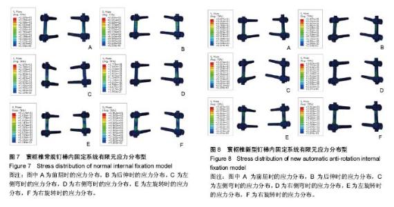
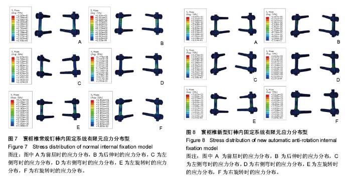
2.1 常规寰枢椎后路钉棒系统有限元模型 常规钉棒系统三维有限元模型包括198 330个节点,964 747个单元,模型外观逼真,几何相似性好,三维有限元模型应力分布云图如图7所示。结果显示不管在哪种工况下,螺钉的主要应力区域为螺钉根部与骨质结合部位,连接棒的主要应力区域为连接棒和螺钉U型槽结合部,应力分布区域符合临床实际,其中连接棒主要应力区域的最大应力值见表2,具体数值见表3。 2.2 自行防旋转钉棒系统有限元模型 自行防旋转钉棒钉棒系统三维有限元模型包括246 788个节点,996 069个单元,三维有限元模型应力分布云图如图8所示。 实验结果显示,各种工况下两种钉棒系统的应力集中部位和一致,其中螺钉的最大应力集中在螺钉根部与骨质结合部位,连接棒的最大应力集中在连接棒和螺钉U型槽结合部,应力分布区域符合临床实际,具体数值见表2,3。"
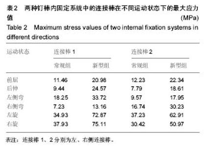
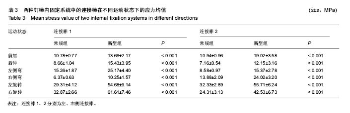
2.3 两种寰枢椎后路钉棒内固定系统的应力差异 常规与新型自行防旋转寰枢椎后路钉棒内固定装置的应力分布区域基本一致,应力分布区域符合临床实际,两种内固定系统均未出现明显的应力集中现象,且主要的应力分布区域均为螺钉根部与骨质结合部,连接棒与螺钉U型槽结合部。分析结果显示新型自行防旋转寰枢椎后路内固定装置的应力均值高于传统内固定装置,两者的应力均值存在明显差异 (P < 0.001),但两者的应力最大值均远远未达到钛合金材料的屈服强度(795-827 MPa)与极限强度(860-896 MPa)[20-21]。"


"
| [1] 谭明生,移平,王文军,等.经寰椎“椎弓根”螺钉内固定技术的临床应用[J].中国脊柱脊髓杂志,2006,16(5): 336-340.[2] 吴增晖,尹庆水,马向阳,等.后路寰枢椎椎弓根钉板固定融合治疗上颈椎不稳[J].中国脊柱脊髓杂志,2004,40(10):591-593.[3] Harms J, Melcher RP. Posterior C1-C2 fusion with polyaxial screw and rod fixation. Spine (Phila Pa 1976). 2001;26(22):2467-2471.[4] Goel A, Laheri V. Plate and screw fixation for atlanto-axial subluxation. Acta Neurochir (Wien). 1994;129(1-2):47-53.[5] Lapsiwala SB, Anderson PA, Oza A, et al. Biomechanical comparison of four C1 to C2 rigid fixative techniques: anterior transarticular, posterior transarticular, C1 to C2 pedicle, and C1 to C2 intralaminar screws. Neurosurgery. 2006;58(3):516-521; discussion 516-521.[6] 马向阳,钟世镇,刘景发,等.寰枢椎后路椎弓根螺钉固定的生物力学评价[J].中国脊柱脊髓杂志,2003,13(12):735-738.[7] 瞿东滨,金大地,欧阳均,等.几种寰枢椎后路内固定术的生物力学评价[J].医用生物力学,1999,14(4):198-201.[8] 马向阳,杨进城,邹小宝,等. 寰枢椎后路专用钉棒固定器械的研制与初步临床应用[J].中国骨科临床与基础研究杂志, 2016,8(5): 261-268.[9] 马向阳,杨进城.一种医用自行防旋转脊柱内固定棒装置:中国, CN201420868545.X[P].2015-06-17.[10] 周风金.后路寰枢椎限制性非融合内固定装置的设计及其可行性研究[D].上海:第二军医大学,2011.[11] Hu Y, Dong WX, Hann S, et al. Construction of Finite Element Model for an Artificial Atlanto-Odontoid Joint Replacement and Analysis of Its Biomechanical Properties. Turk Neurosurg. 2016;26(3):430-436. [12] 郭群峰,陈方经,倪斌,等.带有颅底的全颈椎三维有限元模型的建立及分析[J].中国脊柱脊髓杂志,2014,24(6):550-554.[13] Zhang BC, Liu HB, Cai XH, et al. Biomechanical comparison of a novel transoral atlantoaxial anchored cage with established fixation technique - a finite element analysis. BMC Musculoskelet Disord. 2015;16:261. [14] Faizan A, Goel VK, Garfin SR, et al. Do design variations in the artificial disc influence cervical spine biomechanics? A finite element investigation. Eur Spine J. 2012;21 Suppl 5: S653-662. [15] 关哲,马迅,梁凯恒,等.上颈椎有限元模型的建立及寰椎生物力学有限元分析[J].中国脊柱脊髓杂志,2009,19(7):530-534.[16] Zhang H, Bai J. Development and validation of a finite element model of the occipito-atlantoaxial complex under physiologic loads. Spine (Phila Pa 1976). 2007;32(9): 968-974.[17] Ma X, Peng X, Xiang H, et al. A finite element modeling of posterior atlantoaxial fixation and biomechanical analysis of C2 intralaminar screw fixation. Chin Med J (Engl). 2014; 127(7):1266-1271.[18] Panjabi M, Dvorak J, Crisco JJ 3rd, et al. Effects of alar ligament transection on upper cervical spine rotation. J Orthop Res. 1991;9(4):584-593.[19] Brolin K, Halldin P. Development of a finite element model of the upper cervical spine and a parameter study of ligament characteristics. Spine (Phila Pa 1976). 2004;29(4):376-385.[20] Puttlitz CM, Goel VK, Traynelis VC, et al. A finite element investigation of upper cervical instrumentation. Spine (Phila Pa 1976). 2001;26(22):2449-2455.[21] Cowin SC. Bone Mechanics. Boca Raton, FL: CRC Press, 1989.[22] Gallie WE. Skeletal traction in the treatment of fractures and dislocations of the cervical spine. Ann Surg. 1937;106(4): 770-776.[23] Brooks AL, Jenkins EB. Atlanto-axial arthrodesis by the wedge compression method. J Bone Joint Surg Am. 1978; 60(3):279-284.[24] Hanimoglu H, Hanci L, Kaynar MY, et al. Bilateral C1-C2 claw for atlantoaxial instability. Turk Neurosurg. 2009;19(4): 345-348.[25] Holness RO, Huestis WS, Howes WJ, et al. Posterior stabilization with an interlaminar clamp in cervical injuries: technical note and review of the long term experience with the method. Neurosurgery. 1984;14(3):318-322.[26] Melcher RP, Puttlitz CM, Kleinstueck FS, et al. Biomechanical testing of posterior atlantoaxial fixation techniques. Spine (Phila Pa 1976). 2002;27(22):2435-2440.[27] Richter M, Schmidt R, Claes L, et al. Posterior atlantoaxial fixation: biomechanical in vitro comparison of six different techniques. Spine (Phila Pa 1976). 2002;27(16):1724-1732.[28] Sim HB, Lee JW, Park JT, et al. Biomechanical evaluations of various c1-c2 posterior fixation techniques. Spine (Phila Pa 1976). 2011;36(6):E401-407. [29] Gluf WM, Schmidt MH, Apfelbaum RI. Atlantoaxial transarticular screw fixation: a review of surgical indications, fusion rate, complications, and lessons learned in 191 adult patients. J Neurosurg Spine. 2005;2(2):155-163.[30] Magerl FS. Stable posterior fusion of the atlas and axis by transarticular screw fixation. In: Kehr P, Weidner A, eds. Cervical Spine I. Springer, 1987: 322-327.[31] Mummaneni PV, Haid RW. Atlantoaxial fixation: overview of all techniques. Neurol India. 2005;53(4):408-415.[32] Goel A, Gupta S. Vertebral artery injury with transarticular screws. J Neurosurg. 1999;90(2):376-377.[33] Jeanneret B, Magerl F. Primary posterior fusion C1/2 in odontoid fractures: indications, technique, and results of transarticular screw fixation. J Spinal Disord. 1992;5(4): 464-475.[34] Abou Madawi A, Solanki G, Casey AT, et al. Variation of the groove in the axis vertebra for the vertebral artery. Implications for instrumentation. J Bone Joint Surg Br. 1997;79(5):820-823.[35] Resnick DK, Benzel EC. C1-C2 pedicle screw fixation with rigid cantilever beam construct: case report and technical note. Neurosurgery. 2002;50(2):426-428.[36] Stokes JK, Villavicencio AT, Liu PC, et al. Posterior atlantoaxial stabilization: new alternative to C1-2 transarticular screws. Neurosurg Focus. 2002;12(1):E6.[37] Bhowmick DA, Benzel EC. Posterior atlantoaxial fixation with screw-rod constructs: safety, advantages, and shortcomings. World Neurosurg. 2014;81(2):288-289. [38] Elliott RE, Tanweer O, Boah A, et al. Outcome comparison of atlantoaxial fusion with transarticular screws and screw-rod constructs: meta-analysis and review of literature. J Spinal Disord Tech. 2014;27(1):11-28. [39] Kim JY, Oh CH, Yoon SH, et al. Comparison of outcomes after atlantoaxial fusion with transarticular screws and screw-rod constructs. J Korean Neurosurg Soc. 2014; 55(5):255-260.[40] Meyer B, Kuhlen D. Atlantoaxial fusion: transarticular screws versus screw-rod constructs. World Neurosurg. 2013;80(5): 516-517. [41] Brekelmans WA, Poort HW, Slooff TJ. A new method to analyse the mechanical behaviour of skeletal parts. Acta Orthop Scand. 1972;43(5):301-317.[42] Rajinda P, Towiwat S, Chirappapha P. Comparison of outcomes after atlantoaxial fusion with C1 lateral mass-C2 pedicle screws and C1-C2 transarticular screws. Eur Spine J. 2017;26(4):1064-1072. [43] Teo EC, Ng HW. Evaluation of the role of ligaments, facets and disc nucleus in lower cervical spine under compression and sagittal moments using finite element method. Med Eng Phys. 2001;23(3):155-164.[44] Yoganandan N, Kumaresan S, Voo L, et al. Finite element model of the human lower cervical spine: parametric analysis of the C4-C6 unit. J Biomech Eng. 1997;119(1):87-92.[45] Bozic KJ, Keyak JH, Skinner HB, et al. Three-dimensional finite element modeling of a cervical vertebra: an investigation of burst fracture mechanism. J Spinal Disord. 1994;7(2):102-110.[46] Li Y, Fogel GR, Liao Z, et al. Biomechanical Analysis of Two-level Cervical Disc Replacement with a Stand-alone U-shaped Disc Implant. Spine (Phila Pa 1976). 2017. in press.[47] Fernandes PC, Fernandes PR, Folgado JO, et al. Biomechanical analysis of the anterior cervical fusion. Comput Methods Biomech Biomed Engin. 2012;15(12): 1337-1346.[48] Kang H, Park P, La Marca F, et al. Analysis of load sharing on uncovertebral and facet joints at the C5-6 level with implantation of the Bryan, Prestige LP, or ProDisc-C cervical disc prosthesis: an in vivo image-based finite element study. Neurosurg Focus. 2010;28(6):E9. |
| [1] | Jiang Zi-wei, Huang Feng, Cheng Si-yuan, Zheng Xiao-hui, Sun Shi-dong, Zhao Jing-tao, Cong Hai-chen,Sun Han-qiao, Dong Hang. Design and finite element analysis of digital splint [J]. Chinese Journal of Tissue Engineering Research, 2017, 21(7): 1052-1056. |
| [2] | Wang Fei, Liu Zhi-bin, Tao Hui-ren, Zhang Jian-hua, Li Chang-hong, Cao Qiang, Zheng Jun, Liu Yan-xiong, Qu Xiao-peng. Clinical efficacy of preoperative osteotomy designs using paper-cut technology versus photoshop software for ankylosing spondylitis with kyphosis [J]. Chinese Journal of Tissue Engineering Research, 2017, 21(7): 1057-1063. |
| [3] | Li Hui, Ma Jun-yi, Ma Yuan, Zhu Xu . Establishment of a three-dimensional finite element model of ankylosing spondylitis kyphosis [J]. Chinese Journal of Tissue Engineering Research, 2017, 21(7): 1069-1073. |
| [4] | Ling Guan-han, Ou Zhi-xue, Yao Lan, Wen Li-chun, Wang Guo-xiang, Lin Heng-feng. Establishment of simulating three-dimensional model of China-Japan Friendship Hospital Classification for L type osteonecrosis of the femoral head [J]. Chinese Journal of Tissue Engineering Research, 2017, 21(7): 1074-1079. |
| [5] | Fu Wei-min, Wang Ben-jie. Assessing the degree of necrotic femoral head, and association of blood supply with pathlogical changes: study protocol for a diagnostic animal trial [J]. Chinese Journal of Tissue Engineering Research, 2017, 21(7): 1086-1091. |
| [6] | Zhang Wen-qiang, Ding Qian, Zhang Na. Associations between alpha angle and herniation pit on oblique axial magnetic resonance imaging in asymptomatic hip joints of adults [J]. Chinese Journal of Tissue Engineering Research, 2017, 21(7): 1098-1103. |
| [7] | Sun Xiao-xin1, Zhou Wei2, Zuo Shu-ping3, Liu Hao1, Song Jing-feng1, Liang Chun-yu1. Morphological characteristics for the magnetic resonance imaging assessment of discoid lateral meniscal tears in children [J]. Chinese Journal of Tissue Engineering Research, 2017, 21(7): 1104-1109. |
| [8] | Lin Han-wen, Wen Jun-mao, Huang Chao-yuan, Zhou Chi, Tang Hong-yu. Correlation between the changes in lower limb power line and pain area in the knee osteoarthritis patients: imaging evaluation [J]. Chinese Journal of Tissue Engineering Research, 2017, 21(7): 1110-1114. |
| [9] | Zhang Yun-ge, Song Ke-guan. Periprosthetic osteolysis induced by wear particles: research progress of calcineurin/activated T cell nuclear factor signaling pathway [J]. Chinese Journal of Tissue Engineering Research, 2017, 21(7): 1115-1122. |
| [10] | Liu Wei, Huang Jian. Applied research and progress of three-dimensional printing technology in joint replacement [J]. Chinese Journal of Tissue Engineering Research, 2017, 21(7): 1123-1130. |
| [11] | Liu Jian-kun, Deng Shu-cai. Status and role of three-dimensional printing technology in spine surgery [J]. Chinese Journal of Tissue Engineering Research, 2017, 21(7): 1131-1136. |
| [12] | Xi Li-cheng, Li Hong-yu. Research progress of the influence of alcohol on the local microenvironment of femoral head [J]. Chinese Journal of Tissue Engineering Research, 2017, 21(7): 1137-1142. |
| [13] | Ye Xiang-yang, Sun Xiang, Tang Li-xin, Zhen Ping, Geng Bin, Wang Hua-lei, Zhao Yu-guo. Acetabular liner wear of cross-linked versus conventional polyethylene for total hip arthroplasty: a meta-analysis [J]. Chinese Journal of Tissue Engineering Research, 2017, 21(7): 1143-1148. |
| [14] | Shi Bin, An Jing, Chen Long-gang, Zhang Nan, Tian Ye . Influencing factors for pain after total knee arthroplasty [J]. Chinese Journal of Tissue Engineering Research, 2017, 21(7): 993-997. |
| [15] | Wang Xian-xun. Impact of local compression cryotherapy combined with continuous passive motion on the early functional recovery after total knee arthroplasty [J]. Chinese Journal of Tissue Engineering Research, 2017, 21(7): 998-1003. |
| Viewed | ||||||
|
Full text |
|
|||||
|
Abstract |
|
|||||