Chinese Journal of Tissue Engineering Research ›› 2026, Vol. 30 ›› Issue (26): 6961-6968.doi: 10.12307/2026.399
Hand tendon suturing: optimization of traditional suture techniques and application of novel repair materials
Zhou Ningyu1, Zheng Yuxiang1, Zhang Xiaoyang1, Weng Yuxun1, Yang Yinrui1, Zhou Qijian1, Zheng Jinchen1, Liu Hongying1, Sun Chenchen1, Liu Zheng2
- 1Clinical Medical School of Tianjin Medical University, Tianjin 300270, China; 2Seventh Affiliated Hospital of Sun Yat-sen University, Shenzhen 518107, Guangdong Province, China
-
Accepted:2025-10-17Online:2026-09-18Published:2026-03-16 -
Contact:Liu Zheng, MD, Associate chief physician, Seventh Affiliated Hospital of Sun Yat-sen University, Shenzhen 518107, Guangdong Province, China -
About author:Zhou Ningyu, Clinical Medical School of Tianjin Medical University, Tianjin 300270, China Zheng Yuxiang, Clinical Medical School of Tianjin Medical University, Tianjin 300270, China -
Supported by:Innovation and Entrepreneurship Training Program of National College Students, No. 202413661003 (to ZYX); Shenzhen Municipal Medical and Health “Sanming” Project, No. SZSM202411014 (to LZ)
CLC Number:
Cite this article
Zhou Ningyu, Zheng Yuxiang, Zhang Xiaoyang, Weng Yuxun, Yang Yinrui, Zhou Qijian, Zheng Jinchen, Liu Hongying, Sun Chenchen, Liu Zheng. Hand tendon suturing: optimization of traditional suture techniques and application of novel repair materials[J]. Chinese Journal of Tissue Engineering Research, 2026, 30(26): 6961-6968.
share this article
Add to citation manager EndNote|Reference Manager|ProCite|BibTeX|RefWorks
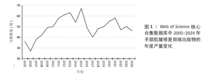
2.1 手部肌腱修复领域文献数量与趋势 2005-2024年间,手部肌腱修复领域相关文献的年度科研产出如图1所示,总体呈现波动增长的趋势。在研究初期阶段(2005-2010年),年度发表文献数量从不足40篇逐步增加至接近50篇,表明该领域逐渐引起研究者的关注。2010-2015年,手部肌腱修复领域文献数量保持稳步增长态势,并在2015年达到峰值。然而,自2016年起,手部肌腱修复领域文献发表数量波动较大,呈现下降趋势,虽然2021年出现了显著回落,但2022,2023年有所回升。"

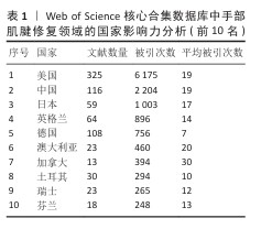
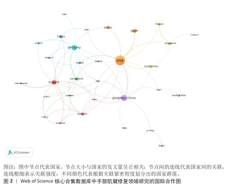
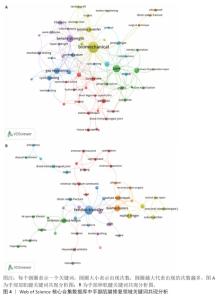
2.2 手部肌腱修复领域研究国家/地区分布 如表1显示,美国在手部肌腱修复领域的影响力最大,总被引次数达6 175,中国在领域内的学术影响力排名第二,总被引次数达2 204,日本则位列第三,总被引次数达1 003。 通过分析手部肌腱修复领域的国际合作现状,发现该领域内具有显著的跨国合作特点(图2)。美国在全球研究网络中占据核心地位,中国、日本、韩国、新加坡等亚洲国家以及与德国、芬兰、瑞典土耳其等欧洲国家建立了紧密的合作关系,尤其是美国与亚洲国家之间的合作最为频繁,形成了强大的科研网络,体现了美国在该领域内的领导作用。 欧洲国家在这一领域的合作也十分活跃,各国之间的科研联系较为均衡,显示出欧洲地区高水平的研究参与度和稳定的跨国合作趋势。此外,中国作为亚洲的主要研究贡献国,与美国和欧洲的合作尤为突出,但在与其他亚洲国家的合作方面仍显不足。 "

"
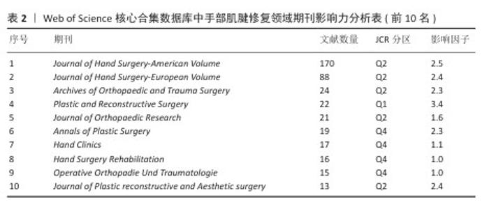
2.3 手部肌腱修复领域研究期刊分布 对手部肌腱修复领域发文量居前10位的期刊进行分析,见表2。《Journal of Hand Surgery-American Volume》的发文量位居首位(170篇),其次是《Journal of Hand Surgery-European Volume》(88篇)和《Archives of Orthopaedic and Trauma Surgery》(24篇)。上述3种期刊的JCR分区均为Q2。值得注意的是,位列Q1分区的期刊《Plastic and Reconstructive Surgery》,亦对该领域研究做出了重要贡献。"

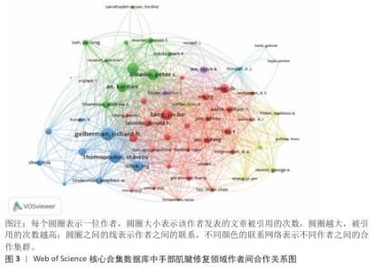
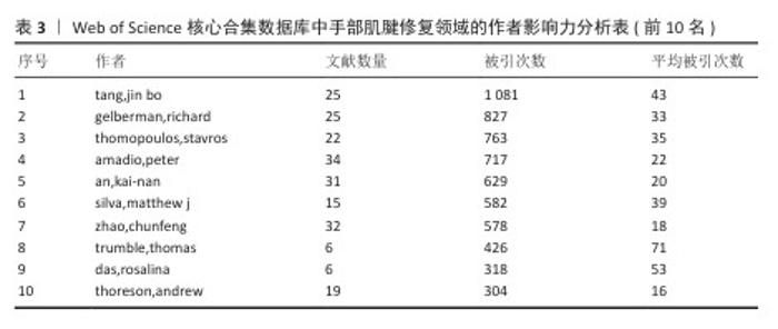
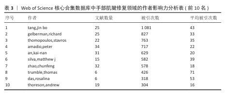
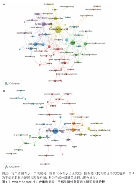
2.4 手部肌腱修复领域主要作者合作 通过VOSviewer对手部肌腱缝合领域的作者合作网络分析表明,该领域内已经形成多个显著的研究合作团体(图3)。Gelberman Richard H.和Zhao Chunfeng分别处于绿色和蓝色模块的核心位置。红色模块以Tang Jinbo为核心,该团队内合作紧密,显示出较高的学术影响力。相比之下,紫色和黄色模块的团队规模较小,但内部合作关系相对稳定。 表3总结了“手部肌腱修复”领域内主要研究者的学术贡献及其影响力。从表中可以看出,tang,jin bo是该研究领域中最具影响力的研究者,其发表的25篇论文总被引次数达到1 081次。紧随其后的是gelberman,rh.和thomopoulos,stavros分别获得了827次和763次的引用。 "
"
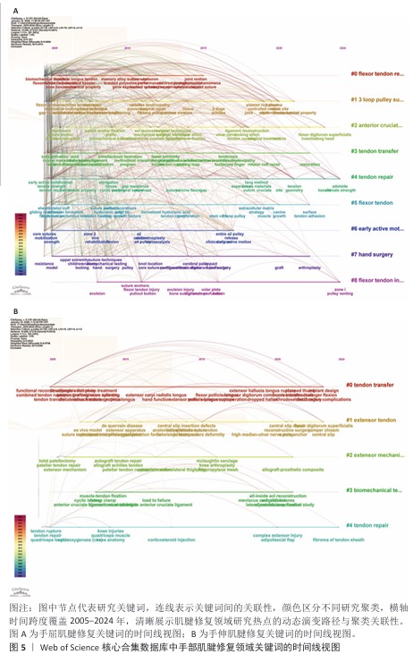
2.5 手部肌腱修复领域研究热点分析 2.5.1 关键词聚类分析 通过对手部屈肌腱和伸肌腱修复领域的关键词共现分析,可以明确识别出这2种肌腱修复的研究热点与核心方向。 (1)手部屈肌腱修复领域关键词共现分析:见图4A。 生物力学性能与修复技术(黄色模块):这个模块的核心关注点是“biomechanics”(生物力学)、“tensile strength”(拉伸强度)、“gap resistance” (抗间隙性)、“cyclic loading”(循环负载)和“barbed suture”(倒刺缝线)。 肌腱修复与功能恢复(绿色模块):这个模块的研究聚焦于“core suture”(核心缝合)、“flexor tendon repair”(屈肌腱修复)、“active mobilization”(主动活动)、“pulley”(滑车)和“zone”(解剖区)。 细胞治疗与生物材料(红色模块):这一部分主要关注“adhesion”(粘连)、“hyaluronic acid”(透明质酸)和“tissue engineering”(组织工程)。 术后并发症与外科技术(蓝色模块):核心话题包括“complications” (并发症)、“surgical technique”(外科技术)、“outcomes”(术后结果)、“thumb”(拇指)和“primary flexor tendon repair”(初级屈肌腱修复)。 动物模型与实验研究(紫色模块):这个模块聚焦于“porcine”(猪)、“ex vivo”(体外实验)等关键词。 (2)手部伸肌腱修复领域关键词共现分析:见图4B。 肌腱移植与修复(蓝色模块):该模块的核心概念有“tendon transfer”(肌腱移 植)、“extensor pollicis longus”(拇长伸肌)和“distal radius fracture”(远端桡骨骨折)。 生物力学与缝合技术(橙色模块):该模块内容包括“biomechanics” (生物力学)、 “suture techniques”(缝合技术)、“grafting”(移植)和“extensor apparatus”(伸肌结构)。 骨关节与肌腱病理(绿色模块):涉及的关键包括“thumb”(拇指)、“de Quervain’s disease”(德奎尔病)和“rheumatoid arthritis”(类风湿关节炎)。 创伤与修复后的并发症(黄色模块):该模块的核心词汇为“mallet finger”(锤状指损伤)、“central slip repair”(中心滑脱修复)。 临床治疗与手术技术(紫色模块):该模块包括“reconstructive surgery”(重建手术)、“wrist”(手腕)等术语。 "

2.5.2 研究热点变迁 利用CiteSpace,通过关键词时间线可视化分析功能分别构建了手部屈肌腱与伸肌腱研究热点的年度演变图。 (1)手部屈肌腱修复领域关键词时间线可视化分析:见图5A。 2005-2010年,研究热点集中于肌腱修复的生物力学性能评价和修复技术的优化,关键词有“biomechanical property”(生物力学性能)、“fiberwire”(纤维丝)、“memory alloy suture wire”(记忆合金缝线)、“core suture”(核心缝合)。2011-2015年,随着研究的深入,研究聚焦于新型修复材料和技术在临床中的应用,研究热点包括“barbed polyester suture”(倒刺缝线)、“platelet-rich plasma”(富血小板血浆)、“stem cell”(干细胞)、“gene expression”(基因表达)。2016-2020年,研究重点逐渐转向术后功能恢复和复杂肌腱损伤的重建,关键词如“early active mobilization”(早期主动活动)、“tendon adhesion”(肌腱粘连)、“tendon transfer”(肌腱移植)和“Tissue engineering”(组织工程)。2021-2024年,屈肌腱修复领域的研究热点逐渐呈现出多元化趋势,基础研究与临床实践的结合更加紧密,关键词如“extracellular matrix”(细胞外基质)、“stem cell”(干细胞)、“tendon adhesion”(肌腱粘连)、“loop pulley system”(滑车系统)和“zone ii”(解剖区域Ⅱ)。 (2)手部伸肌腱修复领域关键词时间线可视化分析:见图5B。 2005-2010年,研究主要集中在伸肌腱修复的功能恢复与重建技术方面,关键词如“splinting”(夹板固定)、“tendon transfer” (肌腱移植)、“functional reconstruction”(功能性重建)、“extensor pollicis longus”(拇长伸肌腱)和“dynamic follow up treatment”(动态随访治疗)。2011-2015年,研究热点逐渐转向修复技术与生物力学性能的分析,关键词如“suture technique”(缝合技术)、“biomechanical evaluation”(生物力学评价)、“central slip”(中间滑移)和“anatomical repair”(解剖学修复)。2016-2020年,研究重点逐渐转向复杂损伤修复的移植物和重建技术,关键词如“graft”(移植)、“autograft tendon repair”(自体肌腱移植修复)、“Adhesion”(粘连)。2021-2024年,研究热点逐渐呈现多样化,基础研究与临床实践的结合更加紧密,关键词如“complex extensor injury”(复杂伸肌腱损伤)、“adipofascial flap”(脂筋膜瓣)、“biomechanical testing”(生物力学测试)、“load to failure”(失效载荷)。 "
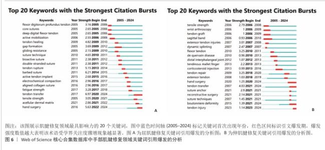
2.5.3 突现词分析 (1)手部屈肌腱修复关键词引用爆发的分析:见图6A。 早期研究主要关注基础生物学和修复技术的改进,例如,“flexor digitorum profundus tendon”(深指屈肌腱)、“gap formation”(间隙形成)、“gliding resistance”(滑动阻力)、“core sutures”(核心缝合)和“suture technique”(缝合技术)是领域内研究热点。中期研究的重点逐渐转向术后功能恢复和生物材料的应用,例如,“bioactive suture”(生物活性缝线)、“barbed suture”(倒刺缝线)、“active tendon implant”(主动肌腱植入物)和“electrochemical compaction”(电化学致密化)是当时研究热点。后期研究方向变得更加多样化,技术创新与临床实践的结合也更加紧密,例如,“tendon transfer”(肌腱移植)、“fatigue strength”(疲劳强度)、“tensile strength”(拉伸强度)、“aligned collagen suture”(对齐的胶原缝线)和“acellular dermal matrix”(无细胞真皮基质)是研究热点。 (2)手部伸肌腱修复关键词引用爆发的分析:见图6B。 早期的研究主要集中在基础生物力学特性和解剖学的探讨,例如,“tensile strength”(拉伸强度)、“wrist arthroscopy”(腕关节镜)、“tendon graft”(肌腱移植物)和“extensor tendon injuries”(伸肌腱损伤)是研究焦点。中期的研究重点逐渐转向术后功能恢复和复杂损伤的修复技术,例如,“flexor tendon”(屈肌腱)、“tendon repair”(肌腱修复)、“tendinous mallet finger”(锤状指肌腱损伤)是当时研究热点。后期的研究方向则更加集中于创新技术和新型材料的临床应用,“hand surgery”(手部手术)、“tendon transfer”(肌腱移植)、“suture anchor”(缝合锚定)、“reconstructive surgery”(重建手术)和“tendon injury”(肌腱损伤)成为关注的热点。 "
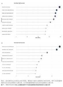
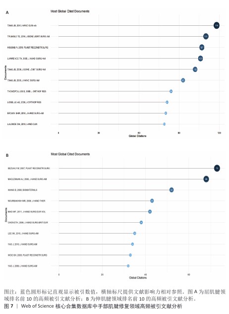
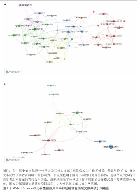
2.5.4 共被引文献分析 手部肌腱修复领域Bibliometrix共被引文献分析显示(图7),屈肌腱领域排名前10的高频被引文献引用频次 均≥79次;相比之下,伸肌腱领域排名前10的高频被引文献最低引用频次则为32次。 为揭示核心文献的共引网络结构,分别选取引用频次≥40次的屈肌腱相关文献以及≥5次的伸肌腱相关文献构建文献共被引网络图(图8)。在屈肌腱修复领域,核心文献节点间存在显著的共引关联,例如:Tang (2013年)与Sandow (2011年)、Cao (2006年)、Miller (2007年) 以及 Gelberman (2017年)等文献形成紧密的共引关系。在伸肌腱修复领域,核心文献节点Gu (2012年)、Bellemere (2015年)、Lu (2013年)与Ulusoy (2006年)、Georgescu (2013年)等同样存在密切的共被引关系。"
"
| [1] SEBASTIN SJ, HO A, KARJALAINEN T, et al. History and evolution of the Kessler repair. J Hand Surg Am. 2013;38(3):552-561. [2] HAUSMANN J, VEKSZLER G, BIJAK M, et al. Biomechanical comparison of modified Kessler and running suture repair in 3 different animal tendons and in human flexor tendons. J Hand Surg Am. 2009;34(1):93-101. [3] HAVULINNA J, LEPPANEN OV, JARVINEN TLN, et al. Comparison of modified Kessler tendon suture at different levels in the human flexor digitorum profundus tendon and porcine flexors and porcine extensors: an experimental biomechanical study. J Hand Surg Eur Vol. 2011;36(8):670-676. [4] REES L, MATTHEWS A, MASOUROS SD, et al. Comparison of 1- and 2-knot, 4-strand, double-modified kessler tendon repairs in a porcine model. J Hand Surg Am. 2009;34(4):705-709. [5] VIINIKAINEN A, GORANSSON H, HUOVINEN K, et al. The strength of the 6-strand modified Kessler repair performed with triple-stranded or triple-stranded bound suture in a porcine extensor tendon model: an ex vivo study. J Hand Surg Am. 2007;32(4):510-517. [6] SHEN H, KORMPAKIS I, HAVLIOGLU N, et al. The effect of mesenchymal stromal cell sheets on the inflammatory stage of flexor tendon healing. Stem Cell Res Ther. 2016;7(1):144. [7] ISHIYAMA N, MORO T, OHE T, et al. Reduction of Peritendinous adhesions by hydrogel containing biocompatible phospholipid polymer MPC for tendon repair. J Bone Joint Surg Am. 2011;93(2): 142-149. [8] WANG B, LIU W, ZHANG Y, et al. Engineering of extensor tendon complex by an ex vivo approach. Biomaterials. 2008;29(20):2954-2961. [9] KIM HM, NELSON G, THOMOPOULOS S, et al. Technical and biological modifications for enhanced flexor tendon repair. J Hand Surg Am. 2010;35(6):1031-1037,1038. [10] MYER C, FOWLER JR. Flexor Tendon Repair: Healing, Biomechanics, and Suture Configurations. Orthop Clin North Am. 2016;47(1):219-226. [11] SU BW, PROTOPSALTIS TS, KOFF MF, et al. The biomechanical analysis of a tendon fixation device for flexor tendon repair. J Hand Surg Am. 2005;30(2):237-245. [12] PARIKH PM, DAVISON SP, HIGGINS JP. Barbed suture tenorrhaphy: an ex vivo biomechanical analysis. Plast Reconstr Surg. 2009;124(5): 1551-1558. [13] HAMADA Y, KATOH S, HIBINO N, et al. Effects of monofilament nylon coated with basic fibroblast growth factor on endogenous intrasynovial flexor tendon healing. J Hand Surg Am. 2006;31(4): 530-540. [14] LEE YJ, RYOO HJ, SHIM H. Prevention of postoperative adhesions after flexor tendon repair with acellular dermal matrix in Zones III, IV, and V of the hand: A randomized controlled (CONSORT-compliant) trial. Medicine (Baltimore). 2022;101(3):e28630. [15] TANG JB, ZHOU X, PAN ZJ, et al. Strong Digital Flexor Tendon Repair, Extension-Flexion Test, and Early Active Flexion: Experience in 300 Tendons. Hand Clin. 2017;33(3):455-463. [16] MORIYA K, YOSHIZU T, MAKI Y, et al. Clinical outcomes of early active mobilization following flexor tendon repair using the six-strand technique: short- and long-term evaluations. J Hand Surg Eur Vol. 2015;40(3):250-258. [17] PAN Z J, QIN J, ZHOU X, et al. Robust thumb flexor tendon repairs with a six-strand M-Tang method, pulley venting, and early active motion. J Hand Surg Eur Vol. 2017;42(9):909-914. [18] OU Y, ZHAN Y, ZHUANG X, et al. A bibliometric analysis of primary immune thrombocytopenia from 2011 to 2021. Br J Haematol. 2023;201(5): 954-970. [19] ALAVANJA G, DAILEY E, MASS DP. Repair of zone II flexor digitorum profundus lacerations using varying suture sizes: a comparative biomechanical study. J Hand Surg Am. 2005; 30(3):448-454. [20] THOMOPOULOS S, KIM HM, DAS R, et al. The effects of exogenous basic fibroblast growth factor on intrasynovial flexor tendon healing in a canine model. J Bone Joint Surg Am. 2010;92(13): 2285-2293. [21] LEE SK, GOLDSTEIN RY, ZINGMAN A, et al. The effects of core suture purchase on the biomechanical characteristics of a multistrand locking flexor tendon repair: a cadaveric study. J Hand Surg Am. 2010;35(7):1165-1171. [22] ZHOU YL, YANG QQ, YAN YY, et al. Gene-Loaded Nanoparticle-Coated Sutures Provide Effective Gene Delivery to Enhance Tendon Healing. Mol Ther. 2019;27(9):1534-1546. [23] RODRIGUEZ REINOSO M, CIVERA M, BURGIO V, et al. 3D printing and testing of rose thorns or limpet teeth inspired anchor device for tendon tissue repair. Acta Bioeng Biomech. 2021;23(4):63-74. [24] THOMOPOULOS S, DAS R, SILVA MJ, et al. Enhanced flexor tendon healing through controlled delivery of PDGF-BB. J Orthop Res. 2009;27(9):1209-1215. [25] TANG JB, CAO Y, ZHU B, et al. Adeno-associated virus-2-mediated bFGF gene transfer to digital flexor tendons significantly increases healing strength. an in vivo study. J Bone Joint Surg Am. 2008;90(5):1078-1089. [26] TRUMBLE TE, VEDDER NB, SEILER JGR, et al. Zone-II flexor tendon repair: a randomized prospective trial of active place-and-hold therapy compared with passive motion therapy. J Bone Joint Surg Am. 2010;92(6):1381-1389. [27] ZHOU YL, YANG QQ, ZHANG L, et al. Nanoparticle-coated sutures providing sustained growth factor delivery to improve the healing strength of injured tendons. Acta Biomater. 2021;124:301-314. [28] MATSUZAKI H, ZAEGEL MA, GELBERMAN RH, et al. Effect of suture material and bone quality on the mechanical properties of zone I flexor tendon-bone reattachment with bone anchors. J Hand Surg Am. 2008;33(5):709-717. [29] WIESKOTTER B, HERBORT M, LANGER M, et al. The impact of different peripheral suture techniques on the biomechanical stability in flexor tendon repair. Arch Orthop Trauma Surg. 2018;138(1):139-145. [30] SANDOW MJ, MCMAHON M. Active mobilisation following single cross grasp four-strand flexor tenorrhaphy (Adelaide repair). J Hand Surg Eur Vol. 2011;36(6):467-475. [31] HIGGINS A, LALONDE DH, BELL M, et al. Avoiding flexor tendon repair rupture with intraoperative total active movement examination. Plast Reconstr Surg. 2010;126(3):941-945. [32] HAHN JM, INCEOGLU S, WONGWORAWAT MD. Biomechanical comparison of Krackow locking stitch versus nonlocking loop stitch with varying number of throws. Am J Sports Med. 2014;42(12): 3003-3008. [33] ZHAO C, SUN Y, KIRK RL, et al. Effects of a lubricin-containing compound on the results of flexor tendon repair in a canine model in vivo. J Bone Joint Surg Am. 2010;92(6):1453-1461. [34] YOUNESI M, KNAPIK DM, CUMSKY J, et al. Effects of PDGF-BB delivery from heparinized collagen sutures on the healing of lacerated chicken flexor tendon in vivo. Acta Biomater. 2017;63:200-209. [35] YOUNESI M, DONMEZ BO, ISLAM A, et al. Heparinized collagen sutures for sustained delivery of PDGF-BB: Delivery profile and effects on tendon-derived cells In-Vitro. Acta Biomater. 2016;41:100-109. [36] BROWN SHM, HENTZEN ER, KWAN A, et al. Mechanical strength of the side-to-side versus Pulvertaft weave tendon repair. J Hand Surg Am. 2010;35(4):540-545. [37] LAWRENCE TM, DAVIS TR. A biomechanical analysis of suture materials and their influence on a four-strand flexor tendon repair. J Hand Surg Am.2005;30(4):836-841. [38] KUSANO N, ZAEGEL MA, PLACZEK JD, et al. Supplementary core sutures increase resistance to gapping for flexor digitorum profundus tendon to bone surface repair - an in vitro biomechanical analysis. J Hand Surg Br. 2005;30(3):288-293. [39] DY CJ, HERNANDEZ-SORIA A, MA Y, et al. Complications after Flexor Tendon Repair: A Systematic Review and Meta-Analysis. J Hand Surg Am. 2012;37(3):543-551.e1. [40] HASHEMI S, PIRMORADI M, RAFATI A, et al. A Human Acellular Dermal Matrix Coated with Zinc Oxide Nanoparticles Accelerates Tendon Repair in Patients with Hand Flexor Tendon Injuries in Zone 5 of the Hand. Bioimpacts. 2024;14(5):27748. [41] YAŞAR B. Encircling Tendon Repair Site with Collagen Sheet in Flexor Zone 2: Retrospective Study. J Orthop Surg Res. 2023;18(1):793. [42] BHAVSAR D, SHETTKO D, TENENHAUS M. Encircling the Tendon Repair Site with Collagen-Gag Reduces the Formation of Postoperative Tendon Adhesions in A Chicken Flexor Tendon Model. J Surg Res. 2010;159(2):765-771. [43] REED ER, HENDRYCKS R, GRAHAM EM, et al. Wrist-Level Tendon Repairs Utilizing a Novel Tendon Stapler Device: an Efficiency and Biomechanical Study. Plast Reconstr Surg. 2024;154(3):582-591. [44] DAGHAN B, CINAR F, YALCIN CE, et al. Morphological, Histological and Biomechanical Comparison of Bone Marrow Aspirate Concentrate, Micro-Fragmented Adipose Tissue and Platelet-Rich Plasma in Prevention of Tendon Adhesion. J Plast Reconstr Aesthet Surg. 2023;87:1-9. [45] TAN S, CHAN C, AHMAD TS, et al. Growth Differentiation Factor 5-Induced Mesenchymal Stromal Cells Enhance Tendon Healing. Tissue Eng Part C Methods. 2024;30(10):431-442. [46] LANE RA, MIGOTSKY N, HAVLIOGLU N, et al. The Effects of NF-κB Suppression on the Early Healing Response Following Intrasynovial Tendon Repair in a Canine Model. J Orthop Res. 2023;41(10): 2295-2304. |
| [1] | Fu Dongsheng, , Aikeremujiang • Muheremu, . Single-cell transcriptome analysis of mesenchymal and epithelial cells from four patients with polydactyly in the GEO public database [J]. Chinese Journal of Tissue Engineering Research, 2025, 29(20): 4379-4388. |
| [2] | Ye Shuming, Xu Chungui, Zhang Jisen, Xu Xinzhong, Xu Youjia, Jing Juehua. Effect of healing of ulnar styloid fracture on joint function after distal radius fracture surgery [J]. Chinese Journal of Tissue Engineering Research, 2020, 24(33): 5321-5325. |
| Viewed | ||||||
|
Full text |
|
|||||
|
Abstract |
|
|||||