[1] ROTH GA, MENSAH GA, JOHNSON CO, et al. Global Burden of Cardiovascular Diseases and Risk Factors, 1990–2019. J Am Coll Cardiol. 2020;76:2982-3021.
[2] LI MX, WEI QQ, MO HL, et al. Challenges and advances in materials and fabrication technologies of small-diameter vascular grafts. Biomate Res. 2023;27(1):58.
[3] ROLL S, MÜLLER-NORDHORN J, KEIL T, et al. Dacron® vs. PTFE as bypass materials in peripheral vascular surgery – systematic review and meta-analysis. BMC Surgery. 2008;8:22.
[4] SPADACCIO C, NAPPI F, AL-ATTAR N, et al. Old Myths, New Concerns: the Long-Term Effects of Ascending Aorta Replacement with Dacron Grafts. Not All That Glitters Is Gold. J Cardiovasc Transl Res. 2016;9: 334-342.
[5] XU H, LIU K, SU Y, et al. Rapid formation of ultrahigh strength vascular graft: Prolonging clotting time micro-dimension hollow vessels with interpenetrating polymer networks. Compos Part B-Eng 2023;250:110456.
[6] XIANG Z, CHEN H, XU B, et al. Gelatin/heparin coated bio-inspired polyurethane composite fibers to construct small-caliber artificial blood vessel grafts. Int J Biol Macromol. 2024;269(Pt 1):131849.
[7] 邹纯才,鄢海燕,王莉丽,等.基于网络药理学分析瓜蒌阿司匹林配伍抗血小板聚集和抗血栓的作用机制研究[J].中国中药杂志, 2019,44(8):1654-1659.
[8] KONG X, HE Y, ZHOU H, et al. Chondroitin Sulfate/Polycaprolactone/Gelatin Electrospun Nanofibers with Antithrombogenicity and Enhanced Endothelial Cell Affinity as a Potential Scaffold for Blood Vessel Tissue Engineering. Nanoscale Res Lett. 2021;16(1):62.
[9] BOSIERS M, DELOOSE K, VERBIST J, et al. Heparin-bonded expanded polytetrafluoroethylene vascular graft for femoropopliteal and femorocrural bypass grafting: 1-year results. J Vasc Surg. 2006;43: 313-318.
[10] WANG W, HU J, HE C, et al. Heparinized PLLA/PLCL nanofibrous scaffold for potential engineering of small‐diameter blood vessel: Tunable elasticity and anticoagulation property. J Biomed Mater Res A. 2015;103:1784-1797.
[11] ASLANI S, KABIRI M, HOSSEINZADEH S, et al. The applications of heparin in vascular tissue engineering. Microvasc Res. 2020;131:104027.
[12] PALUCK SJ, NGUYEN TH, MAYNARD HD. Heparin-Mimicking Polymers: Synthesis and Biological Applications. Biomacromolecules. 2016;17: 3417-3440.
[13] WANG D, XU Y, LI Q, et al. Artificial small-diameter blood vessels: materials, fabrication, surface modification, mechanical properties, and bioactive functionalities. J Mater Chem B. 2020;8:1801-1822.
[14] RICKEL AP, DENG X, ENGEBRETSON D, et al. Electrospun nanofiber scaffold for vascular tissue engineering. Mater Sci Eng C Mater Biol Appl. 2021;129:112373.
[15] XUERONG H, FENG Z, ZIFAN T, et al. Electrospinning of emulsions stabilized by octenylsuccinylated starch and pullulan. Food Hydrocolloids. 2025;158:110482.
[16] İNAN-ÇıNKıR N, AĞÇAM E, ALTAY F, et al. Emulsion electrospinning of zein nanofibers with carotenoid microemulsion: Optimization, characterization and fortification. Food Chem. 2024;430:137005.
[17] LIAN S, LAMPROU D, ZHAO M. Electrospinning technologies for the delivery of Biopharmaceuticals: Current status and future trends. Int J Pharm. 2024;651:123641.
[18] ZHANG C, FENG F, ZHANG H. Emulsion electrospinning: Fundamentals, food applications and prospects. Trends Food Sci Tech. 2018;80: 175-186.
[19] 王培.聚己内酯类生物高分子支架在组织工程领域的应用[J].中国组织工程研究,2021,25(34):5506-5510.
[20] 武小童,何儿,刘来俊,等.基于聚己内酯纤维的组织工程支架研究进展[J].中国生物医学工程学报,2020,39(5):611-620.
[21] 吴鑫,侯磊,许锋.小口径人工血管的研究现状及展望[J].医用生物力学,2024,39(2):355-360.
[22] MENSAH EO, KANWUGU ON, PANDA PK, et al. Marine fucoidans: Structural, extraction, biological activities and their applications in the food industry. Food Hydrocolloids. 2023;142:108784.
[23] WANG F, SCHMIDT H, PAVLESKA D, et al. Crude Fucoidan Extracts Impair Angiogenesis in Models Relevant for Bone Regeneration and Osteosarcoma via Reduction of VEGF and SDF-1. Mar Drugs. 2017; 15:186.
[24] CUI M, LI X, GENG L, et al. Comparative study of the immunomodulatory effects of different fucoidans from Saccharina japonica mediated by scavenger receptors on RAW 264.7 macrophages. Int J Biol Macromol. 2022;215:253-261.
[25] RELIGA P, KAZI M, THYBERG J, et al. Fucoidan inhibits smooth muscle cell proliferation and reduces mitogen-activated protein kinase activity. Eur J Vasc Endovasc Surg. 2000;20(5):419-426.
[26] UMMAT V, SIVAGNANAM SP, RAI DK, et al. Conventional extraction of fucoidan from Irish brown seaweed Fucus vesiculosus followed by ultrasound-assisted depolymerization. Sci Rep. 2024;14(1):6214.
[27] LI Z, WU N, WANG J, et al. Low molecular weight fucoidan alleviates cerebrovascular damage by promoting angiogenesis in type 2 diabetes mice. Int J Biol Macromol. 2022;217:345-355.
[28] AMIN ML, MAWAD D, DOKOS S, et al. Fucoidan- and carrageenan-based biosynthetic poly(vinyl alcohol) hydrogels for controlled permeation. Mater Sci Eng C Mater Biol Appl. 2021;121:111821.
[29] BAI X, ZHANG E, HU B, et al. Study on Absorption Mechanism and Tissue Distribution of Fucoidan. Molecules. 2020;25:1087.
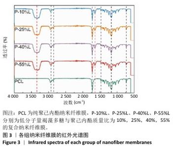
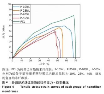
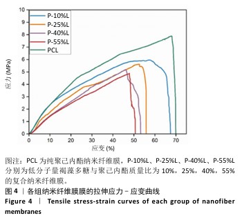
[30] 胡蝶,刘涛,高付蕾,等.聚己内酯-明胶-生物玻璃基不对称润湿性三明治结构复合膜的制备及其性能[J/OL].复合材料学报,1-11[2024-12-24].https://doi.org/10.13801/j.cnki.fhclxb.20240821.008.
[31] PTAK SH, SANCHEZ L, FRETTÉ X, et al. Complementarity of Raman and Infrared spectroscopy for rapid characterization of fucoidan extracts. Plant Methods. 2021;17:130.
[32] 李灿,吕金博,刘会平,等.分级醇沉裙带菜褐藻糖胶及其体外降血糖活性研究[J].食品工业科技,2024,45(8):309-317.
[33] Norouzi SK, Shamloo A. Bilayered heparinized vascular graft fabricated by combining electrospinning and freeze drying methods. Mater Sci Eng C Mater Biol Appl. 2019;94:1067-1076.
[34] LI G, BAO L, HU G, et al. Development and performance evaluation of a novel elastic bacterial nanocellulose/polyurethane small caliber artificial blood vessels. Int J Biol Macromol. 2024;268:131685.
[35] LIU Y, MA Q, WANG Z, et al. Antimicrobial peptide-based pH-responsive Polyvinyl alcohol hydrogel artificial vascular grafts with excellent antimicrobial and hematologic compatibility. Eur Polym J. 2024;221:113554.
[36] STRUCZYŃSKA M, FIRKOWSKA-BODEN I, LEVANDOVSKY N, et al. How Crystallographic Orientation-Induced Fibrinogen Conformation Affects Platelet Adhesion and Activation on TiO2. Adv Healthc Mater. 2023;12(13):e2202508.
[37] NGUYEN AT, SATHE SR, YIM EKF. From nano to micro: topographical scale and its impact on cell adhesion, morphology and contact guidance. J Phys Condens Matter. 2016;28:183001.
[38] AHMED M, RAMOS T, WIERINGA P, et al. Geometric constraints of endothelial cell migration on electrospun fibres. Sci Rep. 2018; 8(1):6386.
[39] ZHOU SY, LI L, XIE E, et al. Small-diameter PCL/PU vascular graft modified with heparin-aspirin compound for preventing the occurrence of acute thrombosis. Int J Biol Macromol. 2023;249:126058.
[40] WANG F, QIN K, WANG K, et al. Nitric oxide improves regeneration and prevents calcification in bio-hybrid vascular grafts via regulation of vascular stem/progenitor cells. Cell Rep. 2022;39:110981.
[41] 徐志伟,谭燕,吴昊,等.小口径人工血管支架材料:问题与前景[J].中国组织工程研究,2014,18(3):452-457.
[42] YIN C, BI Q, CHEN W, et al. Fucoidan Supplementation Improves Antioxidant Capacity via Regulating the Keap1/Nrf2 Signaling Pathway and Mitochondrial Function in Low-Weaning Weight Piglets. Antioxidants (Basel). 2024;13(4):407.
[43] 刘亮,胡高铨,韦昭,等.细菌纳米纤维素/聚多巴胺复合管作为小口径人工血管的潜力[J].中国组织工程研究,2022,26(22): 3535-3542.
[44] MOORE MJ, TAN RP, YANG N, et al. Bioengineering artificial blood vessels from natural materials. Trends Biotechnol. 2022;40:693-707. |