Chinese Journal of Tissue Engineering Research ›› 2026, Vol. 30 ›› Issue (10): 2641-2652.doi: 10.12307/2026.635
Previous Articles Next Articles
Screening of genes related to mitochondrial dysfunction and ferroptosis in atherosclerosis and target prediction of regulatory traditional Chinese medicine
Qi Xiang1, Cao Shan2, Chen Jian1, Zhang Yijia3, Liu Keke2, Xu Zifu1, Liu Wang1, Fu Xiaoxiao1, Yin Xiaolei1
- 1College of Traditional Chinese Medicine (Zhongjing College), 2School of Medicine, 3Academic Affairs Office, Henan University of Chinese medicine, Zhengzhou 450046, Henan Province, China
-
Received:2025-05-06Accepted:2025-06-10Online:2026-04-08Published:2025-09-01 -
Contact:Cao Shan, MD, Professor, Doctoral supervisor, School of Medicine, Henan University of Chinese medicine, Zhengzhou 450046, Henan Province, China -
About author:Qi Xiang, PhD candidate, College of Traditional Chinese Medicine (Zhongjing College), Henan University of Chinese medicine, Zhengzhou 450046, Henan Province, China -
Supported by:Henan Provincial Natural Science Foundation, No. 242300421295 (to CS); National Famous Traditional Chinese Medicine Experts Inheritance Studio Construction Project, No. [2022]75 (to CS); Henan Provincial Science and Technology Research Project, No. 232102310434 (to CS); Major Special Project of Henan Traditional Chinese Medicine Scientific Research, No. 2022ZYZD20 (to CS); Key Project of Henan Traditional Chinese Medicine Scientific Research, No. 2023ZY1031 (to CS)
CLC Number:
Cite this article
Qi Xiang, Cao Shan, Chen Jian, Zhang Yijia, Liu Keke, Xu Zifu, Liu Wang, Fu Xiaoxiao, Yin Xiaolei. Screening of genes related to mitochondrial dysfunction and ferroptosis in atherosclerosis and target prediction of regulatory traditional Chinese medicine[J]. Chinese Journal of Tissue Engineering Research, 2026, 30(10): 2641-2652.
share this article
Add to citation manager EndNote|Reference Manager|ProCite|BibTeX|RefWorks

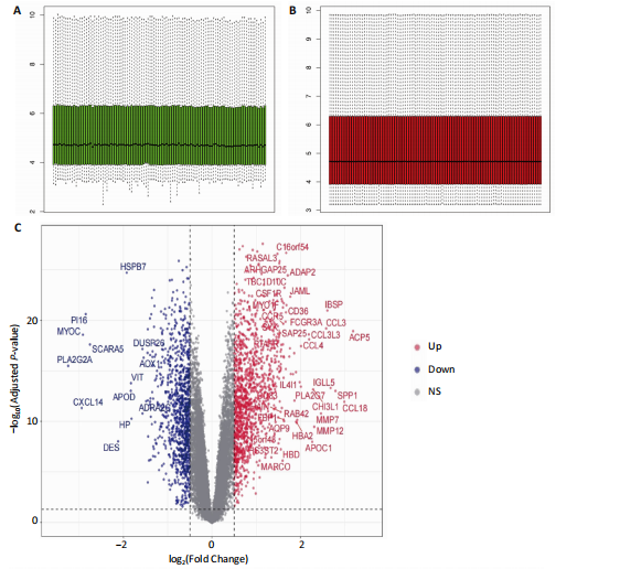
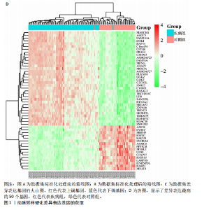
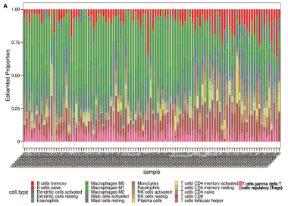
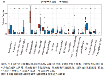
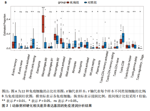
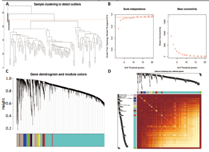
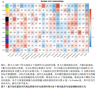
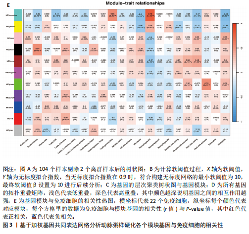
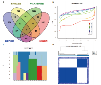
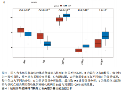
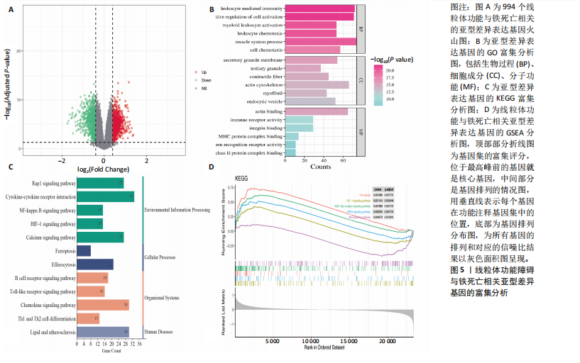
2.1 差异表达基因的识别 通过对动脉粥样硬化数据集进行差异分析后获得2 289个差异表达基因,其中992个基因表达下调,由此推测这些基因在动脉粥样硬化病理进程中受到了抑制;1 296个基因表达上调,反映了这些基因在动脉粥样硬化病理进程中可能被激活,进一步绘制了火山图和热图,见图1。 2.2 免疫浸润细胞分析 免疫浸润分析结果表明,疾病组和正常组之间静息树突状细胞、M0巨噬细胞、活化肥大细胞、静息肥大细胞、单核细胞、中性粒细胞、活化CD4+记忆T细胞、CD8+ T细胞、γδT细胞浸润水平存在显著差异(P < 0.05),见图2,表明以上9种免疫细胞可能在动脉粥样硬化的病理进程中发挥着一定作用。 2.3 加权基因共同表达网络的构建 基于加权基因共表达网络分析GSE100927数据集基因与免疫细胞的相关性,样本聚类后先剔除2个离群样本,见图3A。将软阈值β设置为10,最终鉴定出11个目标模块,见图3B-D。通过热图展示出每个模块和免疫细胞的相关性,结果发现绿色模块与Mast.cells.activated高度正相关(r=0.53,P=8×10⁻⁹),黑色模块与B.cells.memory高度正相关(r=0.54,P=8×10⁻⁹),青绿色模块与Macrophages.M0高度正相关(r=0.79,P=2×10-22),见图3E。将以上3 760个模块基因与线粒体功能障碍相关基因、铁死亡相关基因以及差异表达基因取交集获取5个关键基因,分别为BID、BAX、COX7A1、 LYRM1与MGST1,见图4A。 2.4 亚型基因的差异表达分析 基于5个线粒体功能障碍与铁死亡相关差"
"

"

"
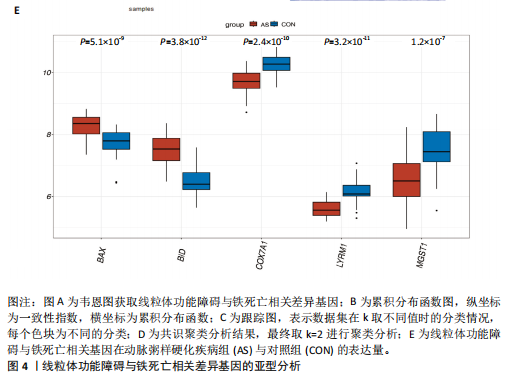
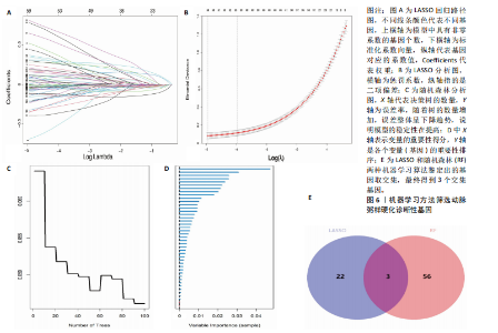
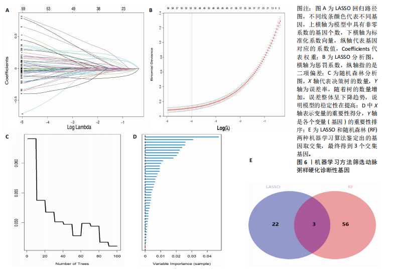
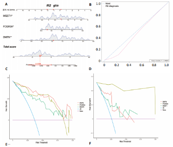
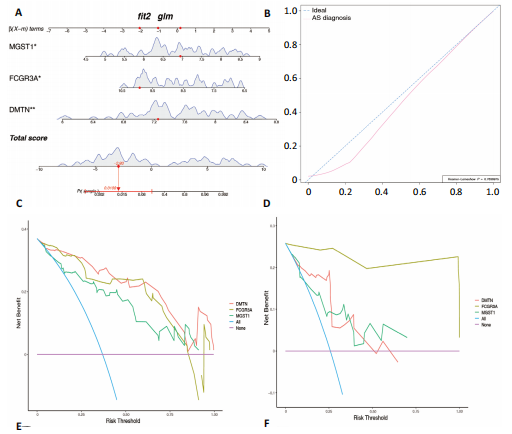
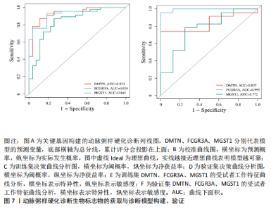
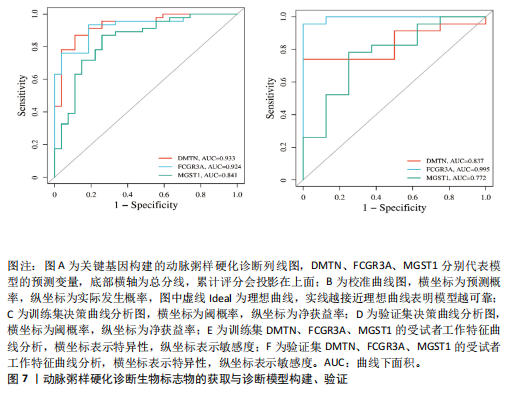
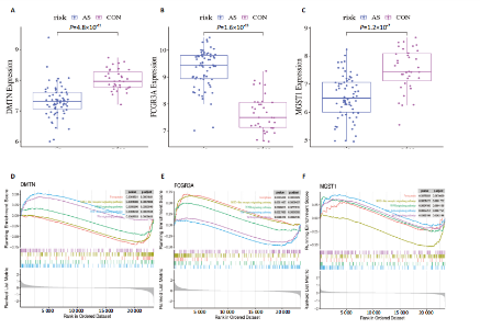
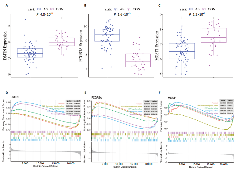
异基因,使用共识聚类算法进行无监督聚类分析,以期识别与动脉粥样硬化疾病组相关的亚型基因,见图4B-E。结合累积分布函数图可以看到聚类簇k值被设置为9,此次研究选取k=2作为聚类结果;结合共识矩阵的热图可以看到k=2时具有清晰明确的分类边界,因此将疾病组的69个动脉粥样硬化样本分为A亚型和B亚型。基于A与B两种亚型基因进行差异分析,最终获取994个线粒体功能与铁死亡相关的亚型差异表达基因,并绘制火山图,见图5A。 2.5 差异表达基因富集分析 994个线粒体功能与铁死亡相关亚型差异表达基因的GO富集分析结果显示,动脉粥样硬化病变涉及白细胞介导的免疫、细胞活化的正调控、白细胞趋化性、肌动蛋白细胞骨架、内吞囊泡、肌动蛋白结合、免疫受体活性、整合素结合、模式识别受体活性等生物学过程,见图5B;KEGG富集分析显示,动脉粥样硬化病变过程中涉及B细胞受体信号通路、脂质与动脉粥样硬化、Rap1信号通路、细胞因子-细胞因子受体相互作用、Toll样受体信号通路、核因子κB信号通路、缺氧诱导因子1信号通路、钙信号通路、铁死亡、趋化因子信号通路、胞葬、Th1和Th2细胞分化,见图5C;GSEA分析显示以上基因与铁死亡信号通路,缺氧诱导因子1信号通路,NOD样受体信号通路,过氧化物酶体增殖物激活受体信号通路的上调有关,与Wnt信号通路的下调有关,由此推测线粒体功能障碍和铁死亡可能受到以上信号通路的调控,并参与到动脉粥样硬化病变过程,见图5D。 2.6 机器学习模型的构建与验证 LASSO回归分析鉴定出59个特征基因,Random Forest鉴定出25个基因,两种算法取交集获得3个诊断生物标志物,分别为DMTN、FCGR3A与MGST1,见图6。基于“rms”包构建动脉粥样硬化诊断列线图,利用校准曲线评估该诊断模型,结果显示患病风险与预测患病风险之间的差异较小,表明该模型预测动脉粥样硬化的发病较为准确,见图7A,B。决策曲线分析结果表明,DMTN、FCGR3A和MGST1曲线在阈概率为0-0.5时净获益率较大,验证集分析结果也证实了以上结论,见图7C,D。受试者工作特征曲线评估结果表明DMTN、FCGR3A和MGST1的曲线下面积值均高于0.7,验证集数据也验证了该结果,见图7E,F。这进一步证明列线图模型可以对早期动脉粥样硬化患者具有较好的诊断能力。 2.7 核心基因的GSEA分析与相关性分析 DMTN、FCGR3A和MGST1基因在疾病组和对照组中的表达量,见图8A-C。GSEA分析发现, DMTN基因低表达时,Wnt信号通路和转化生长因子β信号通路呈现出上调趋势,铁死亡信号通路、NOD样受体信号通路与过氧化物酶体增殖物激活受体信号通路呈现出下调趋势;FCGR3A基因高表达时,Wnt信号通路与转化生长因子β信号通路为下调趋势,铁死亡信号通路、NOD样受体信号通路与过氧化物酶体增殖物激活受体信号通路则为上调趋势;MGST1基因低表达时,Wnt信号通路,转化生长因子β信号"

"
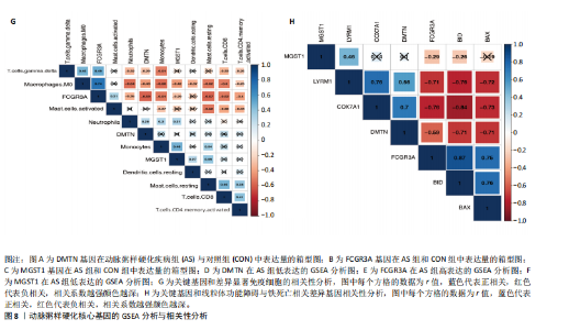
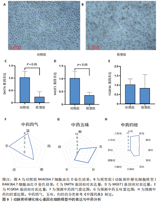
通路、铁死亡信号通路和过氧化物酶体增殖物激活受体信号通路为上调趋势,NOD样受体信号通路则会下调趋势,见图8D-F。 相关性分析显示,FCGR3A基因与M0巨噬细胞(r=0.74)显著正相关,与活化肥大细胞(r=-0.57)显著负相关;DMTN基因与M0巨噬细胞(r=-0.54)显著负相关,与中性粒细胞(r=0.28)显著正相关;MGST1基因与M0巨噬细胞(r=-0.42)显著负相关,与单核细胞(r=0.48)显著正相关,见图8G。Hub基因与线粒体功能障碍铁死亡相关差异基因相关性分析发现,FCGR3A与BID基因(r=0.87)显著正相关,与COX7A1基因(r=-0.76)显著负相关;DMTN与BID基因(r=-0.71)显著负相关,与COX7A1基因(r=-0.70)显著正相关;MGST1与LYRM1基因(r=0.46)显著正相关,与BID基因(r=-0.26)显著负相关,见图8H。 2.8 动脉粥样硬化核心基因在细胞模型中的表达 油红O染色结果显示,与对照组相比,模型组细胞体积有所增大,并出现脂质沉积、细胞泡沫化的病理现象,表明动脉粥样硬化细胞模型构建成功,见图9A,B。qPCR检测结果表明,与对照组相比,模型组RAW264.7细胞中DMTN、MGST1基因表达量显著下降(P < 0.05),FCGR3A基因表达量无明显变化(P > 0.05),见图9C-E。 2.9 调控中药预测结果 此次研究基于DMTN、MGST1预测得到当归、肉桂等16味中药,见表2。经过进一步分析发现以上药物四气以温、平为主,五味以甘、苦味为主,归经主归心经、肝经,见图9F-H。功效分析发现以上药物功效以行气活血、温中补虚为主。"

"
"
"
"
"
"
"


"
| [1] GISTERA A, KETELHUTH D. Lipid-driven immunometabolic responses in atherosclerosis. Curr Opin Lipidol. 2018; 29(5):375-380. [2] DORAN AC. Inflammation Resolution: Implications for Atherosclerosis. Circ Res. 2022;130(1):130-148. [4] QU K, YAN F, QIN X, et al. Mitochondrial dysfunction in vascular endothelial cells and its role in atherosclerosis. Front Physiol. 2022;13:1084604. [4] OREKHOV AN, POZNYAK AV, SOBENIN IA, et al. Mitochondrion as a Selective Target for the Treatment of Atherosclerosis: Role ofMitochondrial DNA Mutations and Defective Mitophagy in the Pathogenesis of Atherosclerosis and Chronic Inflammation. Curr Neuropharmacol. 2020;18(11):1064-1075. [5] ASHAR FN, ZHANG Y, LONGCHAMPS RJ, et al. Association of Mitochondrial DNA Copy Number With Cardiovascular Disease. JAMA Cardiol. 2017;2(11):1247-1255. [6] 刘若楠,王媞,王大新.铁死亡作为动脉粥样硬化治疗靶点最新研究进展[J].临床心血管病杂志,2024,40(4):335-340. [7] 刘磊,贾少晗,于鹏.线粒体在铁死亡中的形态特征和作用[J].中国生物化学与分子生物学报,2023,39(6):769-777. [8] 刘长兴,郭心怡,黄继强,等.基于“脾不散精、痰瘀互结”理论探讨线粒体自噬与动脉粥样硬化的关系[J].中药药理与临床,2025,41(2):95-99. [9] 万宛若,黄丹,徐婉茹,等.中药成分改善动脉粥样硬化及其机制研究进展[J].中草药,2023,54(17):5748-5758. [10] 朱利,吴鸿飞.铁死亡在动脉粥样硬化中的作用及中医药干预研究进展[J].中国实验方剂学杂志,2023,29(2):244-252. [11] 安冬青,吴宗贵.动脉粥样硬化中西医结合诊疗专家共识[J].中国全科医学,2017, 20(5):507-511. [12] CHOI RY, COYNER AS, KALPATHY-CRAMER J, et al. Introduction to Machine Learning, Neural Networks, and Deep Learning. Transl Vis Sci Technol. 2020;9(2):14. [13] RATH S, SHARMA R, GUPTA R, et al. MitoCarta3.0: an updated mitochondrial proteome now with sub-organelle localization and pathway annotations. Nucleic Acids Res. 2021;49(D1): D1541-D1547. [14] ZHOU N, YUAN X, DU Q, et al. FerrDb V2: update of the manually curated database of ferroptosis regulators and ferroptosis-disease associations. Nucleic Acids Res. 2023;51(D1):D571-D582. [15] LANGFELDER P, HORVATH S. WGCNA: an R package for weighted correlation network analysis. BMC Bioinformatics. 2008;9:559. [16] 周云,沃兴德,卢德赵.RAW264.7巨噬细胞源性泡沫细胞模型的建立及鉴定[J].中国动脉硬化杂志,2010,18(9):687-690. [17] 李藤藤,徐东升,李琪,等.淫羊藿苷对ox-LDL诱导的RAW264.7细胞凋亡及炎症的影响[J].中国比较医学杂志,2022, 32(3):9-15. [18] RAITAKARI O, PAHKALA K, MAGNUSSEN CG. Prevention of atherosclerosis from childhood. Nat Rev Cardiol. 2022;19(8): 543-554. [19] SALNIKOVA D, OREKHOVA V, GRECHKO A, et al. Mitochondrial Dysfunction in Vascular Wall Cells and Its Role in Atherosclerosis. Int J Mol Sci. 2021;22(16):8990. [20] FENG X, ZHANG Y, DU M, et al. Identification of diagnostic biomarkers and therapeutic targets in peripheral immune landscape from coronary artery disease. J Transl Med. 2022;20(1):399. [21] 李兴渊,李晓辉,彭广操,等.动脉粥样硬化进展中差异免疫相关基因与免疫细胞的相关性及潜在干预中药的探索[J].中国免疫学杂志,2023,39(12):2553-2559. [22] SUN Y, YANG Z, ZHENG B, et al. A Novel Regulatory Mechanism of Smooth Muscle alpha-Actin Expression by NRG-1/circACTA2/miR-548f-5p Axis. Circ Res. 2017;121(6):628-635. [23] LI HS, ZHOU YN, LI L, et al. HIF-1alpha protects against oxidative stress by directly targeting mitochondria. Redox Biol. 2019; 25:101109. [24] WU X, PAN J, YU JJ, et al. DiDang decoction improves mitochondrial function and lipid metabolism via the HIF-1 signaling pathway to treat atherosclerosis and hyperlipidemia. J Ethnopharmacol. 2023;308:116289. [25] 王雪梅,王怡婷,曹莹,等.线粒体功能调控动脉粥样硬化的研究进展[J].心血管病学进展,2022,43(11):1016-1020. [26] Chen Z, Sun X, Li X, et al. Oleoylethanolamide alleviates hyperlipidaemia-mediated vascular calcification via attenuating mitochondrial DNA stress triggered autophagy-dependent ferroptosis by activating PPARalpha. Biochem Pharmacol. 2023;208:115379. [27] 祁祥,曹珊,段凯旋,等.基于生物信息学预测冠心病心肌梗死的诊断性生物标志物及靶向铜死亡相关基因的中药[J].中药新药与临床药理,2024,35(5):694-705. [28] FENG Y, XU J, SHI M, et al. COX7A1 enhances the sensitivity of human NSCLC cells to cystine deprivation-induced ferroptosis via regulating mitochondrial metabolism. Cell Death Dis. 2022;13(11):988. [29] WEI X, WU Y, PAN H, et al. Proteomics Revealed That Mitochondrial Function Contributed to the Protective Effect of Herba Siegesbeckiae Against Cardiac Ischemia/Reperfusion Injury. Front Cardiovasc Med. 2022;9:895797. [30] NEITEMEIER S, JELINEK A, LAINO V, et al. BID links ferroptosis to mitochondrial cell death pathways. Redox Biol. 2017;12:558-570. Against Cardiac Ischemia/Reperfusion Injury. Front Cardiovasc Med. 2022;9:895797. [31] ITEM F, WUEEST S, LEMOS V, et al. Fas cell surface death receptor controls hepatic lipid metabolism by regulating mitochondrial function. Nat Commun. 2017;8(1):480. [32] JELINEK A, HEYDER L, DAUDE M, et al. Mitochondrial rescue prevents glutathione peroxidase-dependent ferroptosis. Free Radic Biol Med. 2018;117:45-57. [33] 王道鑫,张添光,缪朝玉.血管内皮细胞线粒体氧化应激与动脉粥样硬化[J].药学实践与服务,2023,41(6):329-334. [34] ANIYA Y, IMAIZUMI N. Mitochondrial glutathione transferases involving a new function for membrane permeability transition pore regulation. Drug Metab Rev. 2011;43(2):292-299. [35] KUANG F, LIU J, XIE Y, et al. MGST1 is a redox-sensitive repressor of ferroptosis in pancreatic cancer cells. Cell Chem Biol. 2021;28(6):765-775. [36] GUO Z, WANG L, LIU H, et al. Innate Immune Memory in Monocytes and Macrophages: The Potential Therapeutic Strategies for Atherosclerosis. Cells. 2022;11(24):4072. [37] DUAN M, CHEN H, YIN L, et al. Mitochondrial apolipoprotein A-I binding protein alleviates atherosclerosis by regulating mitophagy and macrophage polarization. Cell Commun Signal. 2022;20(1):60. [38] 马贵萍,陈冉,肖稳康,等.基于DJ-1/GPX4通路探讨加味二至丸对动脉粥样硬化巨噬细胞铁死亡的影响[J].中国病理生理杂志,2024,40(4):627-636. [39] 杜高辉,张青,魏宇淼.中性粒细胞胞外诱捕网在动脉粥样硬化中的研究进展[J].华中科技大学学报(医学版),2023, 52(2):252-256. [40] ZHAO P, LI Y, XU X, et al. Neutrophil extracellular traps mediate cardiomyocyte ferroptosis via the Hippo-Yap pathway to exacerbate doxorubicin-induced cardiotoxicity. Cell Mol Life Sci. 2024; 81(1):122. [41] 张晋锐,吴圣贤,杜雅薇.基于“寒中于暮”探析动脉粥样硬化的病因病机[J].北京中医药大学学报,2024,47(1):92-96. [42] 陈小敏,张健,丁砚兵,等.从络虚毒损探讨症状性颅内动脉粥样硬化性狭窄因机治法[J].北京中医药大学学报,2022, 45(2):135-139. [43] 魏一鸣,王显.论“心肝相关”理论在心系疾病临证治疗中的应用[J].中医杂志, 2020,61(6):493-496. |
| [1] | Liu Anting, Lu Jiangtao, Zhang Wenjie, He Ling, Tang Zongsheng, Chen Xiaoling. Regulation of AMP-activated protein kinase by platelet lysate inhibits cadmium-induced neuronal apoptosis [J]. Chinese Journal of Tissue Engineering Research, 2026, 30(7): 1800-1807. |
| [2] | Wang Zhenze, Liu Fende, Zhang Rui, Li Wujun. Mesenchymal stem cells in treatment of arteriosclerosis obliterans of lower extremities: systematic review and meta-analysis [J]. Chinese Journal of Tissue Engineering Research, 2026, 30(7): 1869-1876. |
| [3] | Chen Yulin, He Yingying, Hu Kai, Chen Zhifan, Nie Sha Meng Yanhui, Li Runzhen, Zhang Xiaoduo , Li Yuxi, Tang Yaoping. Effect and mechanism of exosome-like vesicles derived from Trichosanthes kirilowii Maxim. in preventing and treating atherosclerosis [J]. Chinese Journal of Tissue Engineering Research, 2026, 30(7): 1768-1781. |
| [4] | Lyu Guoqing, Aizimaitijiang·Rouzi, Xiong Daohai. Irisin inhibits ferroptosis in human articular chondrocytes: roles and mechanisms [J]. Chinese Journal of Tissue Engineering Research, 2026, 30(6): 1359-1367. |
| [5] | Lai Jiaming, , Song Yuling, Chen Zixi, Wei Jinghuan, Cai Hao, , Li Guoquan, . Screening of diagnostic markers for endothelial cell Senescence in mice with radiation-induced heart disease and analysis of immune infiltration [J]. Chinese Journal of Tissue Engineering Research, 2026, 30(6): 1450-1463. |
| [6] | Zhang Qian, Huang Dongfeng. Weighted gene co-expression network analysis combined with machine learning to screen and validate biomarkers for osteoarthritis [J]. Chinese Journal of Tissue Engineering Research, 2026, 30(5): 1096-1105. |
| [7] | Zou Rongji, Yu Fangfang, Wang Maolin, Jia Zhuopeng. Triptolide inhibits ferroptosis and improves cerebral ischemia-reperfusion injury in a rat model of cerebral artery occlusion/reperfusion [J]. Chinese Journal of Tissue Engineering Research, 2026, 30(4): 873-881. |
| [8] | Yang Xiao, Bai Yuehui, Zhao Tiantian, Wang Donghao, Zhao Chen, Yuan Shuo. Cartilage degeneration in temporomandibular joint osteoarthritis: mechanisms and regenerative challenges [J]. Chinese Journal of Tissue Engineering Research, 2026, 30(4): 926-935. |
| [9] | Rong Xiangbin, , Zheng Haibo, Mo Xueshen, Hou Kun, Zeng Ping, . Plasma metabolites, immune cells, and hip osteoarthritis: causal inference based on GWAS data from European populations [J]. Chinese Journal of Tissue Engineering Research, 2026, 30(4): 1028-1035. |
| [10] | Gu Fucheng, Yang Meixin, Wu Weixin, Cai Weijun, Qin Yangyi, Sun Mingyi, Sun Jian, Geng Qiudong, Li Nan. Effects of Guilu Erxian Glue on gut microbiota in rats with knee osteoarthritis: machine learning and 16S rDNA analysis [J]. Chinese Journal of Tissue Engineering Research, 2026, 30(4): 1058-1072. |
| [11] | Guan Yujie, Zhao Bin. Application and prospect of artificial intelligence in screening and diagnosis of scoliosis [J]. Chinese Journal of Tissue Engineering Research, 2026, 30(3): 721-730. |
| [12] | Wang Zhipeng, Zhang Xiaogang, Zhang Hongwei, Zhao Xiyun, Li Yuanzhen, Guo Chenglong, Qin Daping, Ren Zhen. A systematic review of application value of machine learning to prognostic prediction models for patients with lumbar disc herniation [J]. Chinese Journal of Tissue Engineering Research, 2026, 30(3): 740-748. |
| [13] | Zhao Feifan, Cao Yujing. An artificial neural network model of ankylosing spondylitis and psoriasis shared genes and machine learning-based mining and validation [J]. Chinese Journal of Tissue Engineering Research, 2026, 30(3): 770-784. |
| [14] | Li Jiagen, Chen Yueping, Huang Keqi, Chen Shangtong, Huang Chuanhong. The construction and validation of a prediction model based on multiple machine learning algorithms and the immunomodulatory analysis of rheumatoid arthritis from the perspective of mitophagy [J]. Chinese Journal of Tissue Engineering Research, 2025, 29(在线): 1-15. |
| [15] | Zhang Yibo, Lu Jianqi, Mao Meiling, Pang Yan, Dong Li, Yang Shangbing, Xiao Xiang. Exploring the causal relationship between rheumatoid arthritis and coronary atherosclerosis: a Mendel randomized study involving serum metabolites and inflammatory factors [J]. Chinese Journal of Tissue Engineering Research, 2025, 29(在线): 1-9. |
| Viewed | ||||||
|
Full text |
|
|||||
|
Abstract |
|
|||||