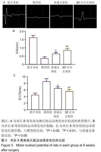
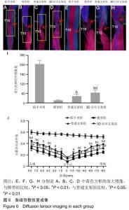
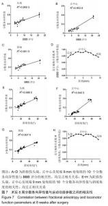
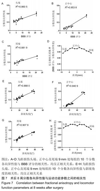
|
[1] FREUND P, CURT A, FRISTON K, et al. Tracking changes following spinal cord injury: insights from neuroimaging. Neuroscientist. 2013;19(2):116-128.
[2] LI G, CHE MT, ZHANG K, et al. Graft of the NT-3 persistent delivery gelatin sponge scaffold promotes axon regeneration, attenuates inflammation, and induces cell migration in rat and canine with spinal cord injury.Biomaterials.2016;83:233-248.
[3] WU GH, SHI HJ, CHE MT, et al. Recovery of paralyzed limb motor function in canine with complete spinal cord injury following implantation of MSC-derived neural network tissue. Biomaterials. 2018;181:15-34.
[4] VAN GOETHEM JW, MAES M, OZSARLAK O, et al. Imaging in spinal trauma. Eur Radiol. 2005;15(3):582-590.
[5] LAI BQ, CHE MT, DU BL, et al. Transplantation of tissue engineering neural network and formation of neuronal relay into the transected rat spinal cord.Biomaterials. 2016;109:40-54.
[6] LI G, CHE MT, ZENG X, et al. Neurotrophin-3 released from implant of tissue-engineered fibroin scaffolds inhibits inflammation, enhances nerve fiber regeneration, and improves motor function in canine spinal cord injury. J Biomed Mater Res A.2018;106(8):2158-2170.
[7] QIU XC, JIN H, ZHANG RY, et al. Donor mesenchymal stem cell-derived neural-like cells transdifferentiate into myelin-forming cells and promote axon regeneration in rat spinal cord transection. Stem Cell Res Ther.2015;6:105.
[8] ZENG X, QIU XC, MA YH, et al. Integration of donor mesenchymal stem cell-derived neuron-like cells into host neural network after rat spinal cord transection. Biomaterials. 2015;53:184-201.
[9] LI J, LEPSKI G. Cell transplantation for spinal cord injury: a systematic review. Biomed Res Int.2013;2013:786475.
[10] OLIVERI RS, BELLO S, BIERING-SØRENSEN F. Mesenchymal stem cells improve locomotor recovery in traumatic spinal cord injury: systematic review with meta-analyses of rat models. Neurobiol Dis.2014;62:338-353.
[11] FAKHOURY M. Spinal cord injury: overview of experimental approaches used to restore locomotor activity. Rev Neurosci. 2015;26(4):397-405.
[12] WANG-LEANDRO A, HOBERT MK, KRAMER S, et al. The role of diffusion tensor imaging as an objective tool for the assessment of motor function recovery after paraplegia in a naturally-occurring large animal model of spinal cord injury. J Transl Med.2018;16(1):258.
[13] KIM JH, SONG SK, BURKE DA, et al. Comprehensive locomotor outcomes correlate to hyperacute diffusion tensor measures after spinal cord injury in the adult rat. Exp Neurol. 2012;235(1): 188-196.
[14] SHANMUGANATHAN K, GULLAPALLI RP, ZHUO J, et al. Diffusion tensor MR imaging in cervical spine trauma. AJNR Am J Neuroradiol. 2008;29(4):655-659.
[15] WANG X, DUFFY P, MCGEE AW, et al. Recovery from chronic spinal cord contusion after Nogo receptor intervention. Ann Neurol. 2011;70(5):805-821.
[16] ZHAO C, RAO JS, PEI XJ, Et al. Longitudinal study on diffusion tensor imaging and diffusion tensor tractography following spinal cord contusion injury in rats. Neuroradiology. 2016;58(6):607-614.
[17] WANG F, HUANG SL, HE XJ, et al. Determination of the ideal rat model for spinal cord injury by diffusion tensor imaging. Neuroreport. 2014;25(17):1386-1392.
[18] YOO WK, KIM TH, HAI DM, et al. Correlation of magnetic resonance diffusion tensor imaging and clinical findings of cervical myelopathy. Spine J.2013;13(8):867-876.
[19] RAO JS, ZHAO C, YANG ZY, et al. Diffusion tensor tractography of residual fibers in traumatic spinal cord injury: a pilot study. J Neuroradiol.2013;40(3):181-186.
[20] HOBERT MK, STEIN VM, DZIALLAS P, et al. Evaluation of normal appearing spinal cord by diffusion tensor imaging, fiber tracking, fractional anisotropy, and apparent diffusion coefficient measurement in 13 dogs. Acta Vet Scand. 2013;55:36.
[21] RASTOGI S, HÖHNE GWH, KELLER A. Efficacy of diffusion tensor anisotropy indices and tractography in assessing the extent of severity of spinal cord injury: an in vitro analytical study in calf spinal cords. Spine J.2012;12(12):1147-1153.
[22] KONOMI T, FUJIYOSHI K, HIKISHIMA K, et al. Conditions for quantitative evaluation of injured spinal cord by in vivo diffusion tensor imaging and tractography: preclinical longitudinal study in common marmosets. Neuroimage.2012;63(4):1841-1853.
[23] LOY DN, KIM JH, XIE M, et al. Diffusion tensor imaging predicts hyperacute spinal cord injury severity. J Neurotrauma. 2007; 24(6):979-990.
[24] ZENG S, LIU L, SHI Y, et al. Characterization of Silk Fibroin/Chitosan 3D Porous Scaffold and In Vitro Cytology.PLoS One. 2015;10(6):e0128658.
[25] 李东,张振辉,郑程程,等.低温3D打印联合冷冻干燥技术制备组织工程骨支架的研究[J].中国修复重建外科杂志,2016,30(3):292-297.
[26] BAZLEY FA, HU C, MAYBHATE A, et al. Electrophysiological evaluation of sensory and motor pathways after incomplete unilateral spinal cord contusion. J Neurosurg Spine. 2012; 16(4):414-423.
[27] CHEN C, ZHAO ML, ZHANG RK, et al. Collagen/heparin sulfate scaffolds fabricated by a 3D bioprinter improved mechanical properties and neurological function after spinal cord injury in rats. J Biomed Mater Res A.2017;105(5):1324-1332.
[28] HAN S, WANG B, JIN W, et al. The linear-ordered collagen scaffold-BDNF complex significantly promotes functional recovery after completely transected spinal cord injury in canine. Biomaterials. 2015;41:89-96.
[29] KUNDU B, RAJKHOWA R, KUNDU SC, et al. Silk fibroin biomaterials for tissue regenerations. Adv Drug Deliv Rev. 2013; 65(4):457-470.
[30] HUANG SL, LIU YX, YUAN GL, et al. Characteristics of lumbar disc herniation with exacerbation of presentation due to spinal manipulative therapy. Medicine (Abingdon). 2015;94(12):e661.
[31] HUANG SL, HE XJ, XIANG L, et al. CT and MRI features of patients with diastematomyelia. Spinal Cord.2014;52(9):689-692.
|