Chinese Journal of Tissue Engineering Research ›› 2016, Vol. 20 ›› Issue (39): 5919-5928.doi: 10.3969/j.issn.2095-4344.2016.39.022
Research progress of biomechanics and finite element analysis of lumbar interspinous devices
Xiong Yang, Yu Xing
- Department of Orthopedics, Dongzhimen Hospital Affiliated to Beijing University of Chinese Medicine, Beijing 100700, China
-
Revised:2016-08-08Online:2016-09-23Published:2016-09-23 -
Contact:Yu Xing, Professor, Doctoral supervisor, Chief physician, Department of Orthopedics, Dongzhimen Hospital Affiliated to Beijing University of Chinese Medicine, Beijing 100700, China -
About author:Xiong Yang, Studying for master’s degree, Department of Orthopedics, Dongzhimen Hospital Affiliated to Beijing University of Chinese Medicine, Beijing 100700, China -
Supported by:the New Century Talent Supporting Project of Education Ministry in 2012, No. NCET-10-0249
CLC Number:
Cite this article
Xiong Yang, Yu Xing. Research progress of biomechanics and finite element analysis of lumbar interspinous devices[J]. Chinese Journal of Tissue Engineering Research, 2016, 20(39): 5919-5928.
share this article
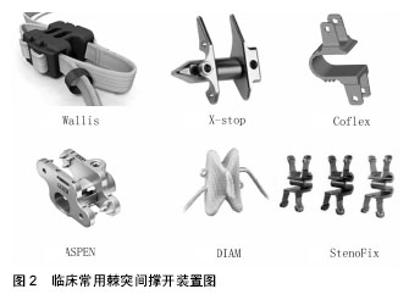
2.1 组成 2.1.1 Wallis系统 Wallis棘突间动态稳定系统是由法国医生Sénégas等[8]1984年开始研发,1986年始用于临床。第1代由钛合金主体和涤纶带两部分组成,仅用于临床实验观察研究,未形成产品推广;第2代由聚醚醚酮材料主体和聚酯纤维带两部分组成,也就是Wallis系统,两条聚酯纤维带分别绕过上下棘突将主体部分固定在棘突间,形成一个“漂浮”装置。 2.1.2 X-Stop系统 X-Stop系统是首个被美国FDA认证运用于临床的棘突间撑开装置[9]。主要由中间椭圆形的撑开主体和两侧翼组成,其材料均为钛合金,坚固的侧翼结合周围韧带可防止其发生移动,在美国临床应用较广,主要用于治疗中度的腰椎管狭窄。 2.1.3 Coflex系统 Coflex棘突间稳定系统从侧面看类似“U”形,上下两侧分别为“夹状”翼,可将整个装置固定于上下棘突间,由于上下固定翼不在同一垂线上,故该装置可同时用于上下多个相邻节段。在“U”形体的上下面分别有1个和3个小齿,以防止装置滑脱移位,整套装置均由钛合金制成。 2.1.4 ASPEN系统 ASPEN系统中间为横行中空圆柱,两侧为坚固翼,各翼均有3个防滑齿和一个固定钉孔,为整套装置在上下棘突间提供了坚固的稳定性,整套装置材料为钛合金。 2.1.5 DIAM系统 DIAM系统与其他棘突间内固定装置相比更具有吸收震动的作用,其主体材料为硅酮,具有一定的弹性,外部以聚乙烯套包绕,两侧的捆绑带将其固定于棘突之间。 2.1.6 StenoFix系统 StenoFix系统与Coflex系统类似,从侧面看似“w”形,该结构可以使其承载更多轴向负荷,上下两侧翼一前一后,因此可同时用于相邻多个节段,其“w”形主体上下与棘突接触面分别有2个和5个小齿,加强固定防止装置滑脱,该装置整体由钛合金制成。各装置如图2所示。"


2.2 生物力学研究 2.2.1 Wallis系统 2007年Lafage等[10]通过体外生物力学检测及有限元分析,评价稳定性受损节段置入Wallis系统后生物力学特性及椎间压力的变化。实验分为3组:完整标本组、稳定性受损标本组(L4/5节段行腰椎间盘切除)、稳定性受损节段置入Wallis系统组,分别测定在轴向负载状况下各组前屈后伸、左右侧屈和轴向旋转活动度。与其他两组对比,Wallis置入节段屈伸稳定性增加、前屈后伸活动度减少,对左右侧屈和轴向旋转活动度无明显影响;有限元分析显示Wallis系统置入节段椎间盘压力有所减少而棘突间撑开装置承重负载有所增加。Ilharreborde等[11]2011年报道尸体标本L3/4置入Wallis系统后生物力学特性,结果显示,置入节段屈伸活动度减少13.8%,而左右侧屈和轴向旋转活动度分别增加6.2%和0.4%。 2.2.2 X-Stop系统 Lindsey等[12]通过尸体生物力学检查评估X-Stop系统置入后对置入节段和邻近节段各向活动度的影响,研究发现,X-Stop置入节段前屈后伸活动度明显降低,侧屈和轴向旋转活动无影响,邻近节段屈伸、轴向旋转及侧向弯曲活动度不受影响,L2-5腰椎整体的前屈活动度减少2°。 Richards等[13]研究了X-Stop置入后屈伸活动时置入节段椎管和椎间孔大小的变化。与完整样本组相比,置入节段伸展位时椎管面积增加18%,椎管矢状径增加10%,椎间孔面积增加约25%(从106mm2到133 mm2),椎间孔宽度增加41%,这说明X-Stop置入后可预防置入节段后伸时椎管和椎间孔狭窄。Lee等[14]对10例接受X-Stop置入的老年腰椎管狭窄患者行MRI检查,发现X-Stop置入后,相应节段椎间孔横截面积增加了36.5%(或22 mm2),椎管面积平均扩大了22%,站立位、中立坐位和后伸坐位之间椎管面积增加程度有显著差异。Siddiqui等[15]对26例老年腰椎管狭窄患者行体位MRI检查发现,X-Stop置入后单节段和双节段椎间孔面积分别增加20%和20%-32%。Wan等[16]也发现X-Stop置入节段后伸位椎间孔面积增加了32.9%(32 mm2),椎间孔宽度增加24.4%,站立和前屈位椎间孔面积仅有微小的变化。 2.2.3 DIAM系统 Phillips等[17]通过体外生物力学评估置入DIAM系统后对部分小关节切除和椎间盘切除节段前屈后伸、侧屈和轴向活动度的影响,研究发现单侧部分小关节切除对相应节段屈伸、侧屈和轴向活动无明显影响,单侧部分小关节切除同时行椎间盘切除则明显增加该节段上述各向活动度,DIAM系统置入后相应节段前屈后伸活动度明显下降(活动度小于完整标本组)、侧屈活动度有所降低(活动度仍大于完整标本组)、轴向活动度无明显变化。Anasetti等[18]在猪腰椎模型中置入不同尺寸大小的DIAM(10 mm和14 mm),观察其对置入节段瞬时旋转中心的影响,DIAM置入后前屈、后伸活动时瞬时旋转中心均发生后移,14 mm尺寸组前屈后伸时瞬时旋转中心的移动明显受限、置入节段处于更大的屈曲中立位(后凸);置入节段未固定捆绑带前屈时瞬时旋转中心位置与正常标本组相似,这提示无捆绑带的棘突撑开装置对前屈的运动力学影响不大。 2.2.4 Coflex系统 Tsai等[19]通过体外生物力学研究评估Coflex系统对部分失稳和完全失稳节段的稳定作用,研究结果显示Coflex系统置入后对后伸活动和轴向旋转活动均有稳定作用,部分失稳和完全失稳节段置入Coflex系统后后伸活动及轴向选择活动范围恢复至完整标本的活动范围,这说明Coflex系统可在屈伸及旋转两个层面提供非刚性固定,可恢复失稳节段正常的后伸及旋转活动。Kettler等[20]对Coflex系统的改进装置-Coflex Rivet(在Coflex系统基础上增加了棘突固定螺钉)进行体外生物力学研究,双侧小关节部分切除和黄韧带切除后置入Coflex Rivet,该装置不但显著增加置入节段前屈后伸方向的稳定性,对轴向旋转及左右侧屈也有明显的稳定效果,作者认为该装置生物力学方面的稳定作用甚至可作为融合术的一种辅助手段。祖丹等[21]通过体外生物力学测定研究Coflex不同置入深度对相邻节段运动范围的影响,Coflex的U形顶端与硬脊膜的距离≤5mm 时,对上下邻近节段前屈、后伸、左右侧弯、左右侧旋 6 个方向运动范围无明显影响。 2.2.5 ASPEN系统 在Karahalios等[22]进行的一项生物力学研究中,将ASPEN装置单独使用与联合前路腰椎体融合使用进行对比。其作者发现,独立ASPEN装置明显减少了屈伸位活动度,对侧弯和轴向旋转活动影响较小。ASPEN装置和前路腰椎体融合的联合使用具有稳定的固定作用,与前路腰椎体融合联合椎弓根钉系统相仿,比前路腰椎体融合联合前路钢板系统更好。其作者指出,当与前路腰椎体融合联合使用时,ASPEN装置可能将成为椎弓根钉系统和前路钢板系统的替代植入物。使用ASPEN装置出现了呈指数水平的改变,其结果是使得椎间孔的高度增加。在邻近节段伸展的补偿可以防止在所有矢状面发生任何严重改变而失衡。 Kaibara等[23]在人尸体脊柱上进行了一项关于ASPEN棘突间固定装置的生物力学研究,将其与经椎间孔融合术及其他后路固定联合使用。单独使用ASPEN装置明显减少了屈伸活动,该结果与联合使用经椎间孔融合术和双边经椎弓根钉系统相似。在侧弯和轴向旋转方面,ASPEN装置伴或不伴经椎间孔融合术相比于双边椎弓根钉系统,显示其稳定性相对较差。经椎间孔融合术联合ASPEN装置和单边螺钉系统提供稳定性与经椎间孔融合术伴双边椎弓根钉相似。其作者指出ASPEN装置可作为椎弓根钉系统的替代。 Techy等[24]进行了一项生物力学研究,将ASPEN装置作为扩大化的椎间融合器或椎弓根钉固定进行评估。作为扩大化椎间融合器,ASPEN装置在置入后,置入节段屈伸位活动度明显减少了74%,但是在侧弯和轴向旋转活动时没有明显改变。当联合单边椎弓根钉后,与对照组相比,显示其活动度有明显减少,在屈伸位减少77%,侧弯时减少55%以及轴向旋转时减少42%。其作者指出,ASPEN装置用于扩大椎间融合器,相比于双边椎弓根钉固定,可以在屈伸位提供稳定作用。然而,在侧弯和旋转位仅提供了微小的稳定作用,除非进一步与椎弓根螺钉联合的扩大。 Gonzalez-Blohm等[25]发表的研究中也得到了相同的结论。在这项研究中,其作者将ASPEN在腰椎减压术后作为独立装置使用,同时联合后路腰椎融合术作为辅助固定,并对其生物力学性能进行了评价。他们认为ASPEN装置可能是适合于在单边椎板切除术后,提供屈伸平衡的装置。后路融合术联合ASPEN装置与椎弓根钉固定在屈伸和轴向旋转等效,但是后路融合联合双边椎弓根钉对于侧弯更加坚固。其作者指出,进一步生物力学和临床证据将更有力支持该棘突间融合装置的单独使用或联合后路椎体融合术作为辅助固定系统而使用。 2.2.6 多置入系统研究 Wilke等[26]对4种不同的棘突间植入物(Coflex、Wallis、DIAM和X-Stop)进行体外生物力学作用评价:24具腰椎样本平均分为4组,依次对各组完整标本、失稳标本及失稳后置入棘突间装置标本进行屈伸、侧弯和轴向旋转活动度测定,失稳标本较完整标本在前屈、后伸、左右侧屈及左右旋转等6个方向的活动度均明显增加。4种装置置入后均能明显降低后伸位置入节段椎间盘内压力,对前屈、左右侧屈及旋转时间盘压力无明显影响;4种装置置入节段后伸活动均可恢复至完整标本水平,Coflex、DIAM和X-Stop对置入节段前屈失稳无明显的稳定作用,Wallis可将置入节段前屈活动度稳定至完整标本水平;4种置入装置对左右侧屈的稳定作用有限,均恢复不到完整标本程度,对旋转运动均无明显稳定作用。Hartmann等[27]通过体外生物力学研究Aperius、In-Space、X-Stop和Coflex 4种不同棘突间装置对置入节段及相邻节段屈伸、侧屈和轴向旋转活动的影响:在无轴向负载情况下,4种装置对置入节段后伸活动均有明显的限制作用,对其他活动无明显影响,相邻节段活动度均有所增加;在轴向负载情况下,这4种装置对置入节段后伸活动也均有明显的稳定作用,对前屈的活动度有一定程度的限制作用,相邻节段的活动度也有所增加。Hirsch等[28]对In-Space、X-Stop、Wallis和DIAM 四种装置分别置入同一腰椎节段,通过尸体生物力学研究发现棘突间撑开增加了置入节段椎间孔面积,但是对邻近下位节段椎间孔可能带来不利影响。Schilling等[29]对 Coflex、Wallis、DIAM以及 InterActiv四种装置一起在人腰椎尸体上进行了生物力学方面的研究,结果显示在腰椎棘突间分别置入以上4种装置后,均可获得相对于完整脊柱状态下大约为50%的活动度,但对脊柱屈曲运动、侧屈运动以及轴向旋转运动方面的限制较小。由此可以看出,虽然各腰椎棘突间撑开装置的外观、材料、设计甚至手术方式都不尽相同,但他们对于腰椎活动度的改善具有相同作用特点:主要限制了腰椎矢状位屈伸活动,对侧弯、旋转运动限制较小,它可以有效减少腰椎不稳的发生。 2.3 有限元分析 2.3.1 Wallis系统 刘瑞等[30]通过三维有限元分析Wallis系统置入后相应节段活动度及应力分布变化,研究发现Wallis系统参与置入节段腰椎不同方向的活动,与置入节段腰椎能很好地匹配,顺应了腰椎整体的运动;Wallis棘突间撑开器置入后可分担椎间盘应力和小关节压力,Wallis系统本身和棘突应力升高,植入物与下位椎体棘突相接触部分的应力最高,提示存在棘突骨折及植入物疲劳性断裂的可能性。 2.3.2 X-Stop系统 姚庆强等[31]根据X-Stop的资料构建了L4/5置入相应棘突间装置的模拟结构,应用三维有限元软件进行分析,结果显示中立位负荷时棘突间装置受力小,后伸活动时可分担椎间盘负荷,同时L5椎弓根负荷增大,作者认为棘突间撑开器可减少后伸活动相应节段椎间盘的负荷,但可能会增加L5椎弓根崩裂的危险。 2.3.3 DIAM系统 王乃国[32]通过有限元分析对比DIAM置入与椎弓根螺钉内固定后相应节段的应力分布变化,与椎弓根螺钉内固定联合小关节融合相比,DIAM通过棘突间隙的撑开,可减小上位椎体小关节、峡部和下位椎体小关节的应力,而DIAM本身及接触部位骨质结构的应力集中并不明显,作者认为在病例选择合适的情况下,DIAM系统可以缓解因腰椎不稳引起的下腰痛。 2.3.4 Coflex系统 陈肇辉等[33]2010年进行了一项Coflex三维有限元分析发现,Coflex棘突间动态固定可保留椎体间部分活动度,可降低置入节段椎间盘应力和小关节压力,对相邻节段活动度及椎间盘应力的影响不大,但可增加相邻上下节段小关节压力;同时发现置入Coflex系统本身及置入节段棘突应力升高,提示有增加棘突骨折及植入物疲劳性断裂的风险。 至今在临床上,腰椎棘突间撑开装置种类繁多,一方面它们限制了腰椎过度屈伸,在棘突间发挥支撑作用,分担了后方椎间盘及关节突关节的压应力,另一方面,一定程度上保留了置入节段活动性,减少了邻近节段活动代偿负荷,降低了邻近节段退变等并发症的风险。新一代棘突间撑开装置也不断出现,其中,Wallis系统经过不断改进已出现第3代-UniWallis系统,其对置入节段生理结构破坏更小,且严格掌握适应证的前提下,可以应用于更多节段,近期俞兴教授完成了国内首例双节段UniWallis系统置入。另一些新装置则采用经皮置入,对组织结构破坏更小,如In-Space、Superion等。棘突间撑开装置作为腰椎动态稳定系统中重要一员,临床做了大量生物力学研究,有限元分析相对较少,但已有的有限元分析中已证明棘突间撑开装置置入前后,置入节段应力分布发生明显变化,有力的支持了生物力学研究结果,同时也发现可能会增加置入节段棘突骨折的风险。目前,棘突间撑开装置短、中期临床疗效与融合相当,同时降低了融合并发症的风险,但其适应证的选择及自身并发症的风险仍使其在临床存在许多争议。在未来,其远期临床疗效仍需要大样本、长期随访结果的支持。"

| [1] Kaner T, Ozer AF. Dynamic stabilization for challenging lumbar degenerative diseases of the spine: a review of the literature. Adv Orthop. 2013,doi: 10.1155/2013/753470. [2] 申洪全,汪洋,柯珍勇.腰椎棘突间分离装置应用进展[J]. 现代医药卫生,2014,30(8):1184-1187. [3] Kabir SM, Gupta SR, Casey AT. Lumbar interspinous spacers: a systematic review of clinical and biomechanical evidence. Spine. 2010;35(25): E1499-E1506. [4] Gazzeri R, Galarza M, Alfieri A. Controversies about interspinous process devices in the treatment of degenerative lumbar spine diseases: past, present, and future. Bio Med Rese Int. 2014, doi: 10.1155/2014/975052. [5] Bonaldi G, Brembilla C, Cianfoni A. Minimally-invasive posterior lumbar stabilization for degenerative low back pain and sciatica. A review. Eur J Radiol. 2015; 84(5): 789-798. [6] Alfieri A, Gazzeri R, Prell J, et al. Role of lumbar interspinous distraction on the neural elements. Neurosurg Rev. 2012;35(4):477-484. [7] Rolfe KW, Zucherman JF, Kondrashov DG, et al. Scoliosis and interspinous decompression with the X-STOP: prospective minimum 1-year outcomes in lumbar spinal stenosis. Spine J. 2010;10(11): 972-978. [8] Sénégas J. Mechanical supplementation by non-rigid fixation in degenerative intervertebral lumbar segments: the Wallis system//Arthroplasty of the Spine. Springer Berlin Heidelberg, 2004: 108-113. [9] 陈宏亮,丁文元.腰椎棘突间非融合技术的研究进展[J]. 中国修复重建外科杂志,2010,24(3): 368-373. [10] Lafage V, Gangnet N, Sénégas J, et al. New interspinous implant evaluation using an in vitro biomechanical study combined with a finite-element analysis. Spine. 2007;32(16): 1706-1713. [11] Ilharreborde B, Shaw MN, Berglund LJ, et al. Biomechanical evaluation of posterior lumbar dynamic stabilization: an in vitro comparison between Universal Clamp and Wallis systems. Eur Spine J. 2011;20(2): 289-296. [12] Lindsey DP, Swanson KE, Fuchs P, et al. The effects of an interspinous implant on the kinematics of the instrumented and adjacent levels in the lumbar spine. Spine. 2003;28(19): 2192-2197. [13] Richards JC, Majumdar S, Lindsey DP, et al. The treatment mechanism of an interspinous process implant for lumbar neurogenic intermittent claudication. Spine. 2005;30(7): 744-749. [14] Lee J, Hida K, Seki T, et al. An interspinous process distractor(X-Stop)for lumbar spinal stenosis in elderly patients: preliminary experiences in 10 consecutive cases. J Spinal Disord Tech. 2004;17(1):72-77. [15] Siddiqui M, Karadimas E, Nicol M, et al. Influence of X-Stop on neural foramina and spinal canal area in spinal stenosis. Spine. 2006;31(25): 2958-2962. [16] Wan Z, Wang S, Kozánek M, et al. Biomechanical evaluation of the X-stop device for surgical treatment of lumbar spinal stenosis. J Spinal Disord Tech. 2012; 25(7): 374. [17] Phillips FM, Voronov LI, Gaitanis IN, et al. Biomechanics of posterior dynamic stabilizing device(DIAM) after facetectomy and discectomy. Spine J. 2006;6(6): 714-722. [18] Anasetti F, Galbusera F, Aziz HN, et al. Spine stability after implantation of an interspinous device: an in vitro and finite element biomechanical study: Laboratory investigation J Neurosurg. 2010;13(5): 568-575. [19] Tsai KJ, Murakami H, Lowery GL, et al. A biomechanical evaluation of an interspinous device (Coflex device) used to stabilize lumbar spine. Paradigm Spine J. 2006;1: 14. [20] Kettler A, Drumm J, Heuer F, et al. Can a modified interspinous spacer prevent instability in axial rotation and lateral bending? A biomechanical in vitro study resulting in a new idea. Clin Biomech. 2008;23(2): 242-247. [21] 祖丹,海涌,云才,等. 腰椎棘突间动态稳定装置Coflex不同置入深度对相邻节段运动范围影响的生物力学研究[J]. 中国脊柱脊髓杂志,2014,24(10): 933-937. [22] Karahalios DG, Kaibara T, Porter RW, et al. Biomechanics of a lumbar interspinous anchor with anterior lumbar interbody fusion: laboratory investigation. J Neurosurg. 2010;12(4): 372-380. [23] Kaibara T, Karahalios DG, Porter RW, et al. Biomechanics of a lumbar interspinous anchor with transforaminal lumbar interbody fixation. World Neurosurg. 2010;73(5): 572-577. [24] Techy F, Mageswaran P, Colbrunn RW, et al. Properties of an interspinous fixation device (ISD) in lumbar fusion constructs: a biomechanical study. Spine J. 2013;13(5): 572-579. [25] Gonzalez-Blohm SA, Doulgeris JJ, Aghayev K, et al. Biomechanical analysis of an interspinous fusion device as a stand-alone and as supplemental fixation to posterior expandable interbody cages in the lumbar spine: Laboratory investigation. J Neurosurg. 2014; 20(2): 209-219. [26] Wilke HJ, Drumm J, Häussler K, et al. Biomechanical effect of different lumbar interspinous implants on flexibility and intradiscal pressure. Eur Spine J. 2008; 17(8): 1049-1056. [27] Hartmann F, Dietz SO, Hely H, et al. Biomechanical effect of different interspinous devices on lumbar spinal range of motion under preload conditions. Arch Orthop Trauma Surg. 2011;131(7): 917-926. [28] Hirsch C, Breque C, Ragot S, et al. Biomechanical study of dynamic changes in L4–L5 foramen surface area in flexion and extension after implantation of four interspinous process devices. Orthop Traumatol Surg Rese. 2015;101(2): 215-219. [29] Schilling C, Pfeiffer M, Grupp TM, et al. The effect of design parameters of interspinous implants on kinematics and load bearing: an in vitro study. Eur Spine J. 2014;23(4):762-771. [30] 刘瑞,徐林,张元智,等. Wallis腰椎非融合系统的有限元分析[J].中国组织工程研究,2012,16(13):2287-2291. [31] 姚庆强,王黎明,蒋纯志,等. 非融合棘突间撑开器治疗腰椎早期退变性腰痛的三维有限元分析[J]. 中国骨与关节损伤杂志,2008,23(4):292-295. [32] 王乃国.腰椎棘突间动力固定的三维有限元分析和前瞻性临床研究[D].中国协和医科大学,2010. [33] 陈肇辉.腰椎棘突间撑开装置的三维有限元分析及临床应用研究[D]. 第二军医大学,2010. [34] 王冰,于秀淳,周银,等.棘突间动态内固定系统在腰椎间盘退变性疾病中的应用及中期随访结果[J].中国骨与关节杂志,2015,14(3):224-229. [35] Zheng S, Yao Q, Cheng L, et al. The effects of a new shape-memory alloy interspinous process device on the distribution of intervertebral disc pressures in vitro. J Biomed Res. 2010;24(2): 115-123. [36] Bowers C, Amini A, Dailey AT, et al. Dynamic interspinous process stabilization: review of complications associated with the X-Stop device. Neurosurg Focus. 2010;28(6): E8. [37] Barbagallo GM, Corbino LA, Olindo G, et al. The “sandwich phenomenon”: a rare complication in adjacent, double-level X-stop surgery: report of three cases and review of the literature. Spine. 2010;35(3): E96-E100. [38] Liu HY, Gu AQ, Zhu ZQ, et al. The efficacy and complication analysis of interspinous dynamic device (Wallis) in patients of degenerative lumbar disease. Zhonghua wai ke za zhi. 2012;50(9): 788-791. [39] Kaulhausen T, Zarghooni K, Stein G, et al. The interspinous spacer: a clinicoanatomical investigation using plastination. Minim Invasive Surg. 2012;2012: 538697. [40] Maida G, Marcati E, Sarubbo S. Heterotopic ossification in vertebral interlaminar/interspinous instrumentation: report of a case. Case Rep Surg. 2012;2012:970642. [41] Barz T, Lange J, Melloh M, et al. Histomorphometric and radiographical changes after lumbar implantation of the PEEK nonfusion interspinous device in the BB. 4S rat model. Spine. 2013;38(5): E263-E269. [42] Epstein N. A review of interspinous fusion devices: High complication, reoperation rates, and costs with poor outcomes. Surg Neurol Int. 2012; 3: 7. [43] Tuschel A, Chavanne A, Eder C, et al. Implant survival analysis and failure modes of the X-Stop interspinous distraction device. Spine. 2013;38(21): 1826-1831. [44] Kim DH, Tantorski M, Shaw J, et al. Occult spinous process fractures associated with interspinous process spacers. Spine. 2011;36(16): E1080-E1085. |
| [1] | Yuan Wei, Zhao Hui, Ding Zhe-ru, Wu Yu-li, Wu Hai-shan, Qian Qi-rong. Association between psychological resilience and acute mental disorders after total knee arthroplasty [J]. Chinese Journal of Tissue Engineering Research, 2017, 21(7): 1015-1019. |
| [2] | Chen Qun-qun, Qiao Rong-qin, Duan Rui-qi, Hu Nian-hong, Li Zhao, Shao Min. Acu-Loc®2 volar distal radius bone plate system for repairing type C fracture of distal radius [J]. Chinese Journal of Tissue Engineering Research, 2017, 21(7): 1025-1030. |
| [3] | Huang Xiang-wang, Liu Hong-zhe. A new low elastic modulus of beta titanium alloy Ti2448 spinal pedicle screw fixation affects thoracic stability: biomechanical analysis [J]. Chinese Journal of Tissue Engineering Research, 2017, 21(7): 1031-1035. |
| [4] | Xie Qiang. Three-dimensional finite element model for biomechanical analysis of stress in knee inversion and external rotation after posterior cruciate ligament rupture [J]. Chinese Journal of Tissue Engineering Research, 2017, 21(7): 1036-1040. |
| [5] | He Ze-dong, Zhao Jing, Chen Liang-yu, Li Ke, Weng Jie. Multilevel finite element analysis on the biological tribology damage of water on bone tissue [J]. Chinese Journal of Tissue Engineering Research, 2017, 21(7): 1041-1045. |
| [6] | Jiang Zi-wei, Huang Feng, Cheng Si-yuan, Zheng Xiao-hui, Sun Shi-dong, Zhao Jing-tao, Cong Hai-chen,Sun Han-qiao, Dong Hang. Design and finite element analysis of digital splint [J]. Chinese Journal of Tissue Engineering Research, 2017, 21(7): 1052-1056. |
| [7] | Wang Fei, Liu Zhi-bin, Tao Hui-ren, Zhang Jian-hua, Li Chang-hong, Cao Qiang, Zheng Jun, Liu Yan-xiong, Qu Xiao-peng. Clinical efficacy of preoperative osteotomy designs using paper-cut technology versus photoshop software for ankylosing spondylitis with kyphosis [J]. Chinese Journal of Tissue Engineering Research, 2017, 21(7): 1057-1063. |
| [8] | Li Peng, Li Hong-wei, Wang Shuang, Wang Hai-zhou. Digital anatomy of lumbar spinous process tilt angle of adults in northeast China: prodinding reference for pedicle screw insertion [J]. Chinese Journal of Tissue Engineering Research, 2017, 21(7): 1064-1068. |
| [9] | Li Hui, Ma Jun-yi, Ma Yuan, Zhu Xu . Establishment of a three-dimensional finite element model of ankylosing spondylitis kyphosis [J]. Chinese Journal of Tissue Engineering Research, 2017, 21(7): 1069-1073. |
| [10] | Ling Guan-han, Ou Zhi-xue, Yao Lan, Wen Li-chun, Wang Guo-xiang, Lin Heng-feng. Establishment of simulating three-dimensional model of China-Japan Friendship Hospital Classification for L type osteonecrosis of the femoral head [J]. Chinese Journal of Tissue Engineering Research, 2017, 21(7): 1074-1079. |
| [11] | Fu Wei-min, Wang Ben-jie. Assessing the degree of necrotic femoral head, and association of blood supply with pathlogical changes: study protocol for a diagnostic animal trial [J]. Chinese Journal of Tissue Engineering Research, 2017, 21(7): 1086-1091. |
| [12] | Zhang Wen-qiang, Ding Qian, Zhang Na. Associations between alpha angle and herniation pit on oblique axial magnetic resonance imaging in asymptomatic hip joints of adults [J]. Chinese Journal of Tissue Engineering Research, 2017, 21(7): 1098-1103. |
| [13] | Sun Xiao-xin1, Zhou Wei2, Zuo Shu-ping3, Liu Hao1, Song Jing-feng1, Liang Chun-yu1. Morphological characteristics for the magnetic resonance imaging assessment of discoid lateral meniscal tears in children [J]. Chinese Journal of Tissue Engineering Research, 2017, 21(7): 1104-1109. |
| [14] | Lin Han-wen, Wen Jun-mao, Huang Chao-yuan, Zhou Chi, Tang Hong-yu. Correlation between the changes in lower limb power line and pain area in the knee osteoarthritis patients: imaging evaluation [J]. Chinese Journal of Tissue Engineering Research, 2017, 21(7): 1110-1114. |
| [15] | Zhang Yun-ge, Song Ke-guan. Periprosthetic osteolysis induced by wear particles: research progress of calcineurin/activated T cell nuclear factor signaling pathway [J]. Chinese Journal of Tissue Engineering Research, 2017, 21(7): 1115-1122. |
| Viewed | ||||||
|
Full text |
|
|||||
|
Abstract |
|
|||||