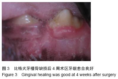
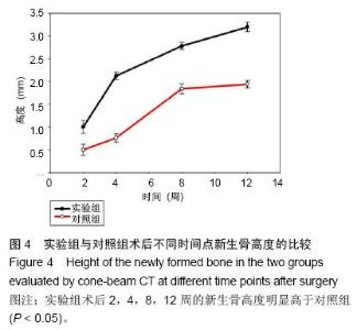
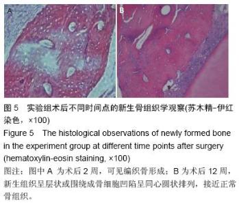
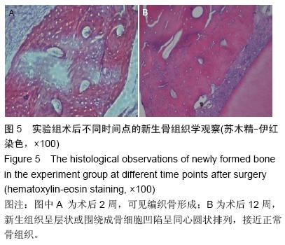
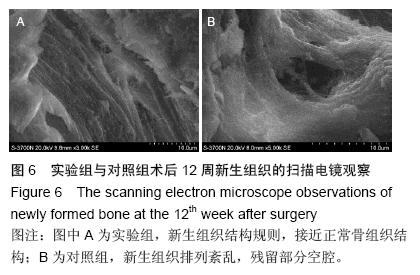
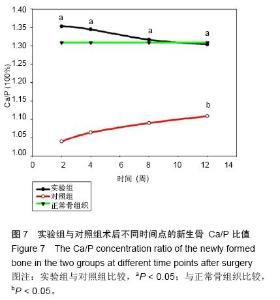
| [1] Tobita M, Mizuno H. Periodontal Disease and Periodontal Tissue Regeneration. Curr Stem Cell Res Ther.2010;5(2):168-174.[2] 鲁琴艳,陈黄琴,黄彬,等. PHBV膜与珊瑚羟基磷灰石联合修复颌骨缺损的研究[J]. 湖北科技学院学报(医学版), 2015,37(3): 204-206, 277. [3] Prabhakaran MP, Vatankhah E, Ramakrishna S, et al. Electrospun aligned PHBV/collagen nanofibers as substrates for nerve tissue engineering. Biotechnol Bioeng. 2013;110(10): 2775-2784.[4] Fan JP, Kalia P, Di Silvio L, et al. In vitro response of human osteoblasts to multi-step sol-gel derived bioactive glass nanoparticles for bone tissue engineering. Mater Sci Eng C Mater Biol Appl. 2014;36: 206-214.[5] Zhang Y, Miron RJ, Li S, et al. Novel MesoPorous BioGlass/silk scaffold containing adPDGF-B and adBMP7 for the repair of periodontal defects in beagle dogs. J Clin Periodontol. 2015; 42(3): 262-271.[6] 陈建洪,黄南楠,唐倩,等.人牙周韧带细胞在SGBG/PHBV三维支架材料上分泌功能的研究[J].中国实用医药, 2010, 5(26):1-2.[7] 唐倩,陈建洪,吴坚,等.多孔复合材料PHBV-SGBG的体外细胞相容性和体内成骨性[J].中华生物医学工程杂志, 2010,16(1):52-57.[8] 唐倩,陈建洪,黄南楠,等.聚羟基丁酸-羟基戊酸共聚酯膜与聚羟基丁酸-羟基戊酸共聚酯-溶胶-凝胶生物活性玻璃复合材料修复骨缺损的研究[J].中华生物医学工程杂志, 2010,16(3): 201-205.[9] 唐倩,陈建洪,黄南楠,等.新型复合骨植入材料的体内成骨性能研究[J].中华口腔医学研究杂志(电子版), 2010,4(3): 13-16.[10] Carlo Reis EC,Borges AP,Araújo MV,et al.Periodontal regeneration using abilayered PLGA/calcium phosphate construct. Bioaterials. 2011;32(35): 9244-9253.[11] Kosen Y,Miyaji H,Kato A,at al.Application of collagen hydrogel/sponge scaffold facilitates periodontal wound healing in classⅡ furcation defects in beagle dogs. J Periodontal Res.2012;47(5):626-634.[12] Kato A,Miyaji H,Kosen Y,et al.Periodontal Healing by Implantation of Collagen Hydrogel-sponge Composite in One-wall Infrabony Defects in Beagle Dogs.J Oral Tissue Engin.2010;8(1):39-46.[13] Lee JS,Park WY,Cha JK,et al.Periodontal tissue reaction to customized nano-hydroxyapatite block scaffold in one-wall intrabony defect: a histologic study in dogs.J Periodontal Implant Sci.2012;42(2):50-58.[14] Matsuura T,Akizuki T,Hoshi S,et al.Effect of a tunnel-structured β-tricalcium phosphate graft material on periodontal regeneration: A pilot study in a canine one-wall intrabony defect model.J Periodontal Res. 2014; 50(3):347-355.[15] 郑贵成,梁焕友,吴志玲,等.仿生型改性胶原膜的动物学实验研究[J].中华口腔医学研究杂志(电子版), 2012,6(3): 245-250. [16] Mopur JM,Devi TR,Ali SM,et al.Clinical and radiographic evaluation of regenerative potential of GTR membrane (Biomesh) along with alloplastic bone graft (Biograft) in the treatment of periodontal intrabony defects.J Contemp Dent Pract.2013;14(3):434-439.[17] Chen FM,Jin Y.Periodontal tissue engineering and regeneration: current approaches and expanding opportunities.Tissue Eng Part B Rev.2010;16:219-255[18] Chen FM,Sun HH,Lu H,et al.Stem cell-delivery therapeutics for periodontal tissue regeneration. Biomaterials.2012;33(27):6320-6344.[19] Ivanovski S,Vaquette C,Gronthos S,et al.Multiphasic scaffolds for periodontal tissue engineering.J Dent Res. 2014;93(12):1212-1221.[20] Galler KM,D'Souza RN.Tissue engineering approaches for regenerative dentistry. Regen Med. 2011;6(1):111-124.[21] Biazar E,Heidari Keshel S,Pouya M.Behavioral evaluation of regenerated rat sciatic nerve by a nanofibrous PHBV conduit filled with Schwann cells as artificial nerve graft.Cell Commun Adhes. 2013; 20(5): 93-103.[22] Sundaramurthi D,Krishnan UM,Sethuraman S.Biocompatibility of poly(3-hydroxybutyrate-co-3- hydroxyvalerate) (PHBV) nanofibers for skin tissue engineering.J Biomed Nanotechnol. 2013;9(8): 1383-1392.[23] Kumarasuriyar A,Jackson RA,Grondahl L,et al.Poly (beta-hydroxybutyrate-co-beta-hydroxyvalerate) supports in vitro osteogenesis. Tissue Eng. 2005; 11(7-8):1281-1295.[24] Biazar E,Keshel SH. Chitosan-cross-linked nanofibrous PHBV nerve guide for rat sciatic nerve regeneration across a defect bridge.ASAIO J. 2013; 59(6):651-659.[25] Zhou M,Yu U.Cartilage tissue engineering using PHBV and PHBV/Bioglass scaffolds. Mol Med Rep. 2014; 10(1):508-514.[26] Wu J,Xue K,Li H,et al.Improvement of PHBV scaffolds with bioglass for cartilage tissue engineering.PLoS One.2013;8(8):e71563.[27] Jones JR,Lin S,Yue S,et al.Bioactive glass scaffolds for bone regeneration and their hierarchical characterization.Proc Inst Mech Eng H. 2010;224(12): 1373-1387.[28] Jones JR.Review of bioactive glass: from Hench to hybrids.Acta Biomater. 2013;9(1):4457-4486. [29] Lee JH,Kang MS,Mahapatra C,et al.Effect of Aminated Mesoporous Bioactive Glass Nanoparticles on the Differentiation of Dental Pulp Stem Cells.PLoS One. 2016;11(3):e0150727.[30] Hulsen DJ,Geurts J,van Gestel NA,et al.Mechanical behaviour of Bioactive Glass granules and morselized cancellous bone allograft in load bearing defects.J Biomech.2016.pii: S0021-9290(16)30234-2. [31] Li J,Zhai D,Lv F,et al. Preparation of copper-containing bioactive glass/eggshell membrane nanocomposites for improving angiogenesis, antibacterial activity and wound healing.Acta Biomater.2016.pii: S1742-7061 (16)30095-2. [32] Das I,De G,Hupa L,et al.Porous SiO2 nanofiber grafted novel bioactive glass-ceramic coating: A structural scaffold for uniform apatite precipitation and oriented cell proliferation on inert implant.Mater Sci Eng C Mater Biol Appl.2016;62:206-214. [33] Kankare J,Lindfors NC.Reconstruction of Vertebral Bone Defects using an Expandable Replacement Device and Bioactive Glass S53P4 in the Treatment of Vertebral Osteomyelitis: Three Patients and Three Pathogens.Scand J Surg.2016.pii: 1457496915626834. Epub ahead of print][34] Lyu X,Li Z,Wang H,et al.Bioactive glass 45S5-silk fibroin membrane supports proliferation and differentiation of human dental pulp stem cells. Zhonghua Kou Qiang Yi Xue Za Zhi.2015; 50(12): 725-730. [35] Ummadi JG,Downs CJ,Joshi VS,et al.Carbon-Based Solid-State Calcium Ion-Selective Microelectrode and Scanning Electrochemical Microscopy: A Quantitative Study of pH-Dependent Release of Calcium Ions from Bioactive Glass.Anal Chem.2016;88(6):3218-3226. [36] Hiltunen AK,Skogman ME,Rosenqvist K,et al.Bioactive glass combined with bisphosphonates provides protection against biofilms formed by the periodontal pathogen Aggregatibacter actinomycetemcomitans.Int J Pharm. 2016;501(1-2):211-220. [37] Leung CC,Palomo L,Griffith R,et al.Accuracy and reliability of cone-beam computed tomography for measuring alveolar bone height and detecting bony dehiscences and fenestrations.Am J Orthod Dentofacial Orthop.2010;137(4 Suppl):S109-S119.[38] Hohlweg-Majert B,Metzger MC,Kummer T,et al. Morphometric analysis - Cone beam computed tomography to predict bone quality and quantity.J Craniomaxillofac Surg.2011; 39(5):330-334. |