Chinese Journal of Tissue Engineering Research ›› 2015, Vol. 19 ›› Issue (14): 2283-2290.doi: 10.3969/j.issn.2095-4344.2015.14.026
Previous Articles Next Articles
Bone marrow microenvironment controls the “fate” of hematopoietic stem cells
Du Hua, Shi Ying-xu
- Department of Laboratory Medicine, Affiliated Hospital of Inner Mongolia Autonomous Region, Hohhot 010050, Inner Mongolia Autonomous Region, China
-
Revised:2015-03-07Online:2015-04-02Published:2015-04-02 -
Contact:Shi Ying-xu, M.D., Assistant researcher, Department of Laboratory Medicine, Affiliated Hospital of Inner Mongolia Autonomous Region, Hohhot 010050, Inner Mongolia Autonomous Region, China -
About author:Du Hua, Master, Lecturer, Department of Laboratory Medicine, Affiliated Hospital of Inner Mongolia Autonomous Region, Hohhot 010050, Inner Mongolia Autonomous Region, China -
Supported by:the “One Million” Project of Inner Mongolia Medical University, No. YKD2012KJBW006; the Higher Research Project in Inner Mongolia Autonomous Region, No. NJZY12145
CLC Number:
Cite this article
Du Hua, Shi Ying-xu . Bone marrow microenvironment controls the “fate” of hematopoietic stem cells [J]. Chinese Journal of Tissue Engineering Research, 2015, 19(14): 2283-2290.
share this article

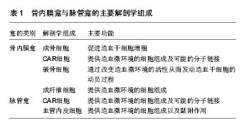
2.1 造血微环境 造血干细胞定位的区域被称为造血微环境-龛(Niche),这一概念最早由Schofield提出[4]。关于造血干细胞微环境的研究有很多,但造血微环境的解剖学定位仍不明确。目前人们普遍接受的是造血干细胞在骨髓腔内存在两类造血微环境:骨内膜造血微环境[5-7],脉管造血微环境[7],有人甚至还称他们为造血干细胞的硬件与软件[8]。解剖学上骨内膜造血微环境主要由成骨细胞、CAR(CXCL12-abundant reticular)细胞、破骨细胞、成纤维细胞组成。脉管造血微环境主要由CAR细胞和血管内皮细胞组成(表1)。 骨内膜造血微环境与脉管造血微环境对造血干细胞的存在、功能维持十分重要。形态学研究可以发现造血干细胞驻留在骨髓腔内,主要分布于骨髓腔骨小梁区的骨内膜附近,而分化较明显的造血祖细胞多分布于骨髓的中央区[9-12],其中约有57%的CD150+CD48- CD41-Lin-细胞分布于骨小梁附近,约14%的细胞分布于骨内膜区,约29%的细胞分布于血窦区[10]。归巢实验发现,尾静脉移植造血干细胞15 h后,可以观察到约60%的细胞定位到骨小梁区骨内膜上,由此可以看出骨内膜具有重要的作用。但骨内膜是否是构成造血微环境的主要部分,以及骨内膜造血微环境是如何建立并对造血干细胞进行调节?目前研究主要认为有3种方式:骨内膜通过细胞-细胞直接相互作用,将造血干细胞黏附在成骨细胞表面然后对造血干细胞进行调节;骨内膜细胞通过释放相关可溶性因子将造血干细胞吸引到骨内膜附近,从而对其进行调节;单独的骨内膜造血微环境不具有使造血干细胞靠近自己的能力,需要第三方细胞的参与、协调造血干细胞靠近骨内膜表面。但骨内膜造血微环境是以何种方式来建立以及如何进行调控仍需要进一步研究。但可以肯定的是,骨内膜细胞对于造血干细胞的维护与调节起重要作用。这些位置是否就是造血微环境,还是一过性的或临时的造血干细胞停留位置,仍需进一步的证明。 通过实验发现,人与鼠的成骨细胞能分泌大量的细胞因子促进造血干细胞的增殖,首次证明了参与骨形成的细胞具有支持干细胞活性的能力[13-15]。骨内膜表面血管化程度较高,表明血管内皮细胞可能参与调控造血干细胞。体外实验发现成体内非造血系统的血管内皮细胞不能够支持造血干细胞[5],而骨髓血窦血管内皮细胞和其他器官内的血窦血管内皮细胞相比功能及表型完全不同[16]。骨髓血窦血管内皮细胞通过表达细胞因子、黏附分子参与造血干细胞的动员、归巢及移植过程[17-18]。在胚胎发育过程中脉管细胞对于造血干细胞的发育以及增殖起到重要作用。脉管系统细胞与造血干细胞均从共同的胚胎期血管周样祖细胞分化而来。因此,胚胎期的造血与血管的发生发展密不可分,在胚胎期造血期,没有骨组织来参与造血微环境的构成,此时的造血干细胞只能定位于脉管或脉管周围。这些胚胎期脉管极可能是一些特化的脉管结构参与早期造血微环境的构成,由此推测脉管也可能参与并调节成体造血干细胞。脉管细胞是否参与到造血干细胞的造血微环境组成仍不确定。研究发现成体内的脉管细胞也可能参与造血干细胞的造血微环境的构成。在成体内的髓外部分发现脉管细胞极可能参与髓外造血微环境的构成,因为发生髓外造血时可在肝、脾组织内观察到造血干细胞在血窦附近大量存在。而在骨髓内也可以发现大量的造血干细胞驻留在脉管附近,约有60%的造血干细胞驻留在脉管骨髓血窦附近,只有约20%的造血干细胞靠近骨内膜,其他造血干细胞散落于骨髓腔内部。培养实验发现,血管内皮细胞类似于成骨细胞能够维持造血干细胞,而且对于体内造血功能也是必须的。 Morrison研究报道,仅43%的造血干细胞/造血祖细胞定位于骨内膜造血微环境与脉管造血微环境中[9],因此人们推测在骨髓腔内可能还存在其他类型的造血微环境。例如Suda等研究发现骨髓腔低氧区的分布与造血干细胞的分布密切相关,低氧环境能够有效上调细胞周期抑制因子的表达,从而对造血干细胞处于静止期起到重要作用[19]。其他实验也发现,活性氧家族水平决定造血干细胞的活性及类型,静止期的造血干细胞具有长期自我更新的能力,而增殖期的造血干细胞只具有短期的自我更新能力[20-22]。通过有限的现有知识推测骨髓腔内可能存在以下几类造血微环境,参与调节造血干细胞的维持、增殖以及定位过程[9,23]。第一类造血微环境:脉管或血管周边细胞以及骨内膜细胞共同形成一个靠近骨内膜的造血微环境,两类细胞一同参与调控造血干细胞。第二类造血微环境:两类细胞分别在空间上形成相互独立的两个造血微环境,两个造血微环境对造血干细胞具有相同的调控作用,只是位置不同而已。第三类造血微环境:这两类细胞分别在空间及功能上形成相互独立的两个造血微环境,骨内膜造血微环境参与造血干细胞的自我更新及增殖,脉管造血微环境参与造血干细胞的动员、归巢及移植过程。第四类造血微环境:骨髓腔内某些或某类未知的细胞,参与构成特化的造血微环境。 2.2 造血微环境中的调控途径 由于组成造血微环境的细胞能够通过释放调控信号(细胞因子、趋化因子、黏附因子等物质)使造血干细胞定位于造血微环境之内,并使造血干细胞处于休眠状态,平衡自我更新与分化状态使造血干细胞维持相对稳态。造血微环境与造血干细胞间的突触联合作用对于造血干细胞的稳态起到至关重要的作用。细胞-细胞以及细胞-基质的黏附在生物体、器官和组织的形态和发育中起着非常重要的作用,由此可以设想,大量的黏附分子协同细胞外基质将造血干细胞尽可能的向造血微环境的组成细胞拉近,使造血干细胞与造血微环境细胞在相互靠近或接触过程中发生相互作用,最终通过黏附分子将造血干细胞与造血微环境紧密连接在一起,形成有效的配体-受体作用胞间隙。因为大多数分泌的信号分子多是与细胞表面或细胞外基质结合,所以扩散的不是很远,由此认为造血微环境对于造血干细胞调控的前提是二者必须靠近或接触,在此过程中黏附分子起重要作用。造血微环境通过细胞-细胞、细胞-可溶性因子与细胞-细胞外基质3种模式对造血干细胞进行调控。 2.2.1 细胞与细胞(Cell-Cell)间黏附过程中起主要作用的分子通路 ①VCAM-1/VLA-4途径:VLA-4(very late antigen 4)是细胞黏附分子(cell adhesion molecules,CAM)整合素家族成员,由α4β1异源二聚体组成,也称为整联蛋白α4β1或CD49d/CD29,所有的单个核造血细胞都有表达。它与纤连蛋白以及免疫球蛋白超家族分子VCAM-1相结合,与VCAM-1的亲合力大约是与纤连蛋白的4倍,并介导靶细胞在机体内的迁移过程。VCAM-1也被称为CD106,含有6个或7个免疫球蛋白结构域,主要由被激活的血管内皮细胞分泌。通过阻断VLA-4/VACAM-1途径可以降低造血祖细胞向骨髓腔约50%的归巢效率[24-25]。相对的,阻断Integrin(CD49d)与VLA4的作用,有利于造血微环境中的造血干细胞进入外周血,而且有趣的是,迁移的造血干细胞会低表达VLA-4[26-27]。通过条件性敲除VLA-4或VACAM-1,可以有效的抑制造血干细胞的迁移以及在骨髓腔内的定位[28]。此外,VLA-4也可通过识别纤连蛋白的CS1区EILDVPST序列(关键序列为LDV)将纤连蛋白作为配体并与之结合,作用同VLA-5,通过细胞-细胞外基质模式来对造血干细胞的迁移进行调控。目前的研究报道推测VlA-4可能参与的介导过程:首先通过VLA-4和VLA-5与细胞外基质作用,使干细胞穿过细胞外基质,并顺着SDF-1浓度梯度迁移至骨髓腔内的干细胞龛附近。然后,通过VLA-4/VACAM-1相互作用协助造血细胞黏附并定位于有利于发育的微环境中,促使造血干细胞和基质细胞形成聚合体,完成造血干细胞的归巢和迁移过程。②N-cadherin—N-cadherin途径:神经性钙黏附蛋白(neural calcium-dependent adhesion protein,N-cadherin)最早于1982年由Grunwald等在鸡的神经视网膜中发现[29],是一类细胞黏附分子,主要通过与其他N-cadherin分子以同型嗜同种的方式相互结合来发挥生物学功能。N-cadherin也可以与其他cadherin分子以异型嗜同种或异型嗜异种的方式结合[30]。N-cadherin与FGF受体直接相互作用,参与调节轴突的生长、突触的形成以及可塑性。FGF受体还参与调控N-cadherin相关细胞的运动以及癌细胞的迁移过程。在基因敲除实验中发现,缺乏N-cadherin表达的小鼠在妊娠初期便死亡,这些胚胎表现为心脏发育不全、神经管与体节畸形。通过重新表达N-cadherin对这些缺陷有一定程度的修复作用[31]。全长的N-cadherin或其可溶性胞外功能区都能够促进细胞黏附或细胞的迁移,抑制其表达,就能够有效的抑制细胞的黏附与转移,甚至是肿瘤细胞[32]。N-cadherin在早期的CD34+CD19+造血干细胞中表达,参与调节早期造血干细胞在骨髓腔内的静息状态以及自我更新的调控过程[33]。N-cadherin对于造血干细胞的功能及作用一直存在着争论,主要是N-cadherin是否在造血干细胞定位于骨髓腔骨内膜造血微环境过程中起作用。2004年,Toshio Suda等指出相对于其他cadherin家族成员,N-cadherin既在造血干细胞表达也在成骨细胞中表达,N-cadherin以及β1-integrin在造血干细胞与成骨细胞发生相互作用中起重要作用。抑制N-cadherin 与 Integrins会导致造血干细胞丧失自我更新能力[34]。2007年, Morrison通过PCR法、抗体以及β-galactosidase染色,检测出高度纯化后的造血干细胞中未发现N-cadherin的表达。照射后的小鼠体内只能检测到N-cadherin阴性细胞中有造血干细胞的活性[35]。2008年,Linheng Li提出非造血干细胞的骨髓细胞高度表达N-cadherin, 称为N-cadherinhi,这些细胞不能够重建造血。而表达量居中N-cadherinint和低表达N-cadherinlo的细胞是long term HSC(LT-HSC),其中N-cadherinlo的造血干细胞具有较强的造血重建能力[36]。2009年,Morrison通过对N-cadherin基因敲除鼠的研究发现,N-cadherin的缺失并不影响造血干细胞的维持与造血功能,也不会影响骨髓细胞的组成、各系分化、祖细胞的克隆形成能力、造血干细胞的比例以及造血重建的能力[37]。2010年,Toshio Suda通过RNAi干扰N-cadherin基因的表达,发现N-cadherin能够调节细胞造血干细胞黏附于骨髓腔造血微环境,对于骨髓移植后的造血功能重建起重要作用[38]。③Jagged-1-Notch途径,Notch信号通路是一重要的进化保守通路,参与多种细胞功能性活动,如细胞增殖、分化、凋亡、干细胞的维持与自我更新。Notch对于T细胞的早期发育至关重要[39],过表达Notch能够引起T淋巴细胞白血病(T-ALL),但在造血干细胞中Notch信号通路高度活化。在体外造血干细胞分化时Notch被下调;抑制造血干细胞的Notch信号通路,表现为促分化。体内抑制造血干细胞的Notch信号通路表现为耗竭作用[40]。强化Notch1以及Notch4能够增强造血干细胞的自我更新能力,并抑制分化[41],增强Notch的下游分子Hes1的表达,能够增强造血干细胞在体内的长期自我更新能力[42],甚至使原始的造血祖细胞永生化[43]。这些都表明,对于造血干细胞,Notch途径是维持LT-HSCs细胞自我更新的重要分子。有趣的是,有研究发现敲除Notch或存在Notch的拮抗物时,造血干细胞仍能够通过非活化的Jagged-1与骨髓基质细胞相结合,所以Mancini等[44]认为Notch信号通路对于造血的发生不是必需的。这样就产生了相反的研究结果,这一现象可以解释为哺乳动物Notch通路较多,存在4种受体5种配体,在不同的研究前提条件下,得出的结果可能有所不同[45]。④SCF—Kit途径:小鼠10q以及人的12q22-12q24上的Steel(Sl)基因座基因编码干细胞因子(stem cell factor,SCF),在RNA水平通过选择性剪接表达两种蛋白,即膜结合型干细胞因子和分泌型干细胞因子。这两类干细胞因子都是以c-Kit作为受体来行使生物学功能,c-Kit像其配体一样也有剪接变体。干细胞因子与c-Kit结合,促使c-Kit二聚化,激活多条信号通路,如PI3K、JAK/STAT pathway、Src家族、Ras–Raf–MAP 级联激酶[46]。干细胞因子相对于造血干细胞是一类趋化因子,通过两种方式促进造血干细胞与造血微环境黏附,首先膜结合型干细胞因子与c-Kit直接结合,然后通过c-Kit途径上调造血干细胞与β1integrins、VLA-4和VLA-5、胞外纤连蛋白的亲和力。分泌型干细胞因子对于造血干细胞主要有两方面的作用,体外实验证实干细胞因子能够促进造血干细胞的自我更新[47];体内试验发现干细胞因子对于造血干细胞的存活起重要作用。在体外培养体系中添加干细胞因子能够有效地使造血干细胞的长期造血能力维持数周[48]。c-kit是一类酪氨酸激酶受体,c-kit在约70%的CD34+造血干细胞/造血祖细胞以及在体外具有长期造血能力的细胞中表达,巨核细胞也有表达。通过抗体阻断实验发现,封闭c-Kit可引起造血干细胞数量减少[49],而敲除实验中,剔除干细胞因子后发现支持造血干细胞再生活性的微环境严重受损[50]。正常状态下,c-Kit在造血干细胞发育过程中的表达水平是一定的,与造血干细胞是否进入细胞周期以及在周期内的状态无关[51]。由此认为正常情况下,机体内的造血干细胞由于趋化因子分泌型干细胞因子的作用,使细胞向富含干细胞因子的造血微环境靠近,最终与造血微环境细胞膜上的配体结合,进一步促进造血干细胞与其他黏附分子结合。在Angela Colmone小鼠模型白血病环境中,干细胞因子是高表达的,高表达的干细胞因子能够吸引正常造血干细胞进入白血病造血微环境,进一步抑制造血干细胞的功能[52]。 2.2.2 细胞与可溶性调控因子(Cell-Soluble regulator)起主要作用的分子通路 ①TPO-MPL:血小板生成素受体(myeloproliferative leukemia virus oncogene,MPL)也称为CD110是Ⅰ类细胞因子受体,最初被认为是骨髓组织增殖白血病的癌基因。MPL的配体血小板生成素(thrombopoietin,TPO)于1994年克隆得到,定位于3q26-27,全长8 kb,是调节巨核细胞的生成以及血小板生成最主要的细胞因子,它的生物学效应由其受体c-Mpl介导。血小板生成素主要由骨髓基质细胞、肝脏和肾脏合成,通过与Mpl结合,促使Mpl形成同源二聚体,激活一系列信号通路,包括JAK/STAT、Ras/MAPK、PI3K/Akt等。JAK2与STAT5磷酸化促进细胞增殖,MAPK途径与细胞分化凋亡密切相关。TPO-MPL途径在成体造血的早期阶段起重要作用。MPL的表达有利于造血干细胞保持长期自我更新的能力[53],而照射后的小鼠移植Mpl-/-造血干细胞是不能长期造血的[54],但高表达Mpl可以富集造血干细胞[55]。血小板生成素是一种重要的早期活化细胞因子,在体外与Flt3和干细胞因子一同使用,能够维持造血干细胞存活以及增殖[56]。血小板生成素在体内对于促进造血干细胞的增殖非常重要[57]。在基因敲除鼠(TPO −/−或 mpl−/−)中,这两个基因的缺失都会使90%的血小板下降,而且会导致造血祖细胞减少,体外CFC试验可见CFU-Mixture减少。通过竞争移植可以观察到mpl−/−能够降低LinloSca-1+的细胞数[58]。②ANG-1-Tie2:血管生成素1(angiopoietin-1,Ang-1)也是一类血管内皮细胞所特有的酪氨酸激酶配体,类似于血管内皮生长因子。血管生成素家族包括Ang-1、Ang-2、Ang-3、Ang-4四种分子,它们共同的特异性受体为Tie-2。Ang-1首次由Davis等[59]在1996年从COS细胞利用分子克隆技术中发现,由498个氨基酸所组成,位于染色体8q22-q23,编码Ang-1的cDNA有3个结构域。Ang-1/Tie2对于造血干细胞的功能可以归为以下几类:Tie2有助于使静息状态的造血干细胞黏附于骨表面的成骨细胞上;骨髓内的Ang-1主要由成骨细胞表达;通过Ang-1/Tie2信号通路可以使造血干细胞紧紧黏附于骨内膜细胞,同时体外实验显示这种依赖Tie2的模式还可以维持造血干细胞处于未分化状态;在体内Ang-1通过阻止细胞分裂来维持造血干细胞的功能;在体外LTC-IC试验中,Ang-1激活Tie2后,能够促进鹅卵石样克隆的形成,而在体内有助于造血干细胞长期连续移植并维持其干性[60]。③CXCL12—CXCR4:趋化因子CXCL12 又称基质细胞衍生因子1(SDF-1)是一种小分子的细胞因子,属于趋化因子蛋白家族。它有两种形式,SDF-1α/CXCL12a和SDF-1β/CXCL12b[61]。趋化因子CXCL12对淋巴细胞有强烈的趋化作用并在发育中起重要作用[62]。在胚胎发育中,CXCL12引导造血干细胞从胎儿肝脏到骨髓的迁徙,敲除CXCL12常常可致小鼠胚胎死亡[63],受体是CXCR4[64]。最近发现CXCL12还可以与CXCR7受体结合[65]。大多数的造血干细胞都与高表达CXCL12的细胞相结合,这类高表达CXCL12的细胞称为CXCL12-abundant reticular细胞(CAR)。CAR 细胞主要分布于血窦周围以及骨内膜附近,造血干细胞通过CXCR4与CXCL12相结合,这一信号通路在成体骨髓内以自分泌的模式维持干细胞功能过程中起重要作用。其原因可能是CXCL12能够维持造血干细胞的存活以及自我更新[66]。体外,CXCL12与白细胞介素6、干细胞因子协调作用于造血干细胞可提高其移植效率[67]。此外,CXCL12可能通过充当周期抑制因子来抑制造血干细胞进入周期,从而实现维持干细胞池[68]。④BMPs/TGF-β-TGF-βR:转化生长因子β (transforming growth factor-β,TGF-β)是一组新近发现的调节细胞生长和分化的转化生长因子β超家族。一般在细胞分化活跃的组织中转化生长因子β表达水平较高,如成骨细胞、肾脏、骨髓和胎肝的造血细胞。对于造血系统来说,骨形态发生蛋白在不同的物种的造血发育过程中都起关键性的调控作用,同时骨形态发生蛋白对于早期造血是必需的。在体外培养过程中,骨形态发生蛋白4具有维持成年人造血干细胞的作用,低浓度的骨形态发生蛋白4就可以诱导造血祖细胞增殖、分化[69]。而体外培养时,骨形态发生蛋白4对造血干细胞无促增殖作用。在体内骨形态发生蛋白信号通路有助于造血干细胞的增殖[70]。⑤IFNα-IFNαR:1957年英国科学家Isaacs和Lindenmann首先发现了病毒干扰现象,即病毒感染的细胞能产生一种因子,作用于其他细胞,从而起到干扰病毒的复制,因而命名为干扰素。体内IFNα作用于造血干细胞能够促进STAT1与PKB/Akt的磷酸化,最终引起Sca-1上调,通过活化STAT1与Sca-1信号通路可以介导造血干细胞进入细胞周期,促进造血干细胞增殖[71],这些增殖的造血干细胞能够被5-FU杀死。造血干细胞缺失IFNα/β的受体、STAT1或Sca-1,也对IFNα的刺激不敏感。 2.2.3 细胞与细胞外基质(Cell-ECM)间黏附过程中起主要作用的分子通路 ①细胞外基质—整合素:整合素(Integrin)属于细胞表面受体,它与细胞外基质(extracellular matrix,ECM)相互作用。在细胞与细胞外基质的结合应答的过程中,整合素蛋白家族是起主要介导作用的细胞表面受体。整合素受体由α和β亚基构成, 造血干细胞/造血祖细胞表达β1-和β2-整合素。由整合素/细胞外基质相互作用介导的信号传导在细胞对生长因子信号的应答反应中是必不可少的,可以调控细胞增殖、细胞骨架重排和细胞存活所需的其他应答。在与细胞外基质的黏附性相互作用后,可以激活多个细胞内信号通路。β1-整合素对于造血干细胞的归巢起重要作用,敲除β1-整合素的造血干细胞在移植后不能够归巢到骨髓腔,而会停留在细胞周期内[72]。研究发现,人源造血干细胞/造血祖细胞只表达整合素α9β1,细胞-细胞黏附实验发现,造血干细胞/造血祖细胞通过整合素α9β1黏附于成骨细胞,通过抗体阻断整合素α9β1的功能,可以有效抑制造血干细胞/造血祖细胞的增殖以及体外分化[73]。而整合素α6与α4也参与调控胎肝造血干细胞/造血祖细胞的归巢过程[74]。这些研究都表明,整合素对于造血干细胞/造血祖细胞的归巢起重要作用。②细胞外基 质—VLA-5:VLA-5像VLA-4一样既可以与VCAM-1结合,也可以与细胞外基质中的fibronectin 或osteopontin结合[75]。VLA-5与fibronectin能够促进造血干细胞表达特异的转录因子来维持干细胞[76]。此外,VLA-5也会影响造血祖细胞的黏附作用。 2.2.4 其他重要分子 Pten参与调控多条信号通路,调节造血干细胞的自我更新。敲除Pten虽然能够在短期内促进造血干细胞的增殖,但最终却导致造血干细胞的耗竭。移植缺失Pten的造血干细胞不能够长期维持多系的造血发生,只能进行短期的造血[77]。 Wnt信号通路与Notch信号通路一样,也能够促进造血干细胞的自我更新。Duncan等[78]认为它们通过不同的机制参与调节Wnt信号通路,诱导细胞增殖,促进LSK细胞的发育,而Notch则维持造血干细胞不处于分化状态。 低氧是骨髓腔的一个特质,造血干细胞停留于骨髓腔内氧浓度的最低处。这说明氧分压也会对造血干细胞的功能有影响,低氧有利于造血干细胞处于静止期[79]。骨桥蛋白负调节骨髓腔内造血干细胞的数量,敲除骨桥蛋白能提高造血干细胞的数量。照射后敲除骨桥蛋白大鼠移植正常造血干细胞也能够增加造血干细胞的数量。此外,骨桥蛋白也是造血干细胞处于静息状态的负调控因子[80]。"
| [1] Till JE, McCulloch EA. A direct measurement of the radiation sensitivity of normal mouse bone marrow cells. Radiat Res. 1961;14:213-222. [2] Kiel MJ, Yilmaz OH, Iwashita T, et al. SLAM family receptors distinguish hematopoietic stem and progenitor cells and reveal endothelial niches for stem cells. Cell. 2005;121(7): 1109-1121. [3] Hoover-Plow J, Gong Y. Challenges for heart disease stem cell therapy.Vasc Health Risk Manag. 2012;8:99-113. [4] Schofield R. The relationship between the spleen colony-forming cell and the haemopoietic stem cell. Blood Cells. 1978;4(1-2):7-25. [5] Winkler IG, Sims NA, Pettit AR, et al. Bone marrow macrophages maintain hematopoietic stem cell (HSC) niches and their depletion mobilizes HSCs. Blood. 2010;116(23): 4815-4828. [6] Boisset JC, van Cappellen W, Andrieu-Soler C, et al. In vivo imaging of haematopoietic cells emerging from the mouse aortic endothelium. Nature. 2010 4;464(7285):116-120. [7] Bertrand JY, Chi NC, Santoso B, et al. Haematopoietic stem cells derive directly from aortic endothelium during development. Nature. 2010;464(7285):108-111. [8] Garrett RW, Emerson SG. Bone and blood vessels: the hard and the soft of hematopoietic stem cell niches. Cell Stem Cell. 2009;4(6):503-506. [9] Kiel MJ, Morrison SJ. Uncertainty in the niches that maintain haematopoietic stem cells. Nat Rev Immunol. 2008;8(4): 290-301. [10] Butler JM, Nolan DJ, Vertes EL, et al. Endothelial cells are essential for the self-renewal and repopulation of Notch-dependent hematopoietic stem cells. Cell Stem Cell. 2010;6(3):251-264. [11] Chow A, Lucas D, Hidalgo A, et al. Bone marrow CD169+ macrophages promote the retention of hematopoietic stem and progenitor cells in the mesenchymal stem cell niche. J Exp Med. 2011;208(2):261-271. [12] Sipkins DA, Wei X, Wu JW, et al. In vivo imaging of specialized bone marrow endothelial microdomains for tumour engraftment. Nature. 2005;435(7044):969-973. [13] Christopher MJ, Rao M, Liu F, et al. Expression of the G-CSF receptor in monocytic cells is sufficient to mediate hematopoietic progenitor mobilization by G-CSF in mice. J Exp Med. 2011;208(2):251-260. [14] Kobayashi H, Butler JM, O'Donnell R,et al. Angiocrine factors from Akt-activated endothelial cells balance self-renewal and differentiation of haematopoietic stem cells. Nat Cell Biol. 2010;12(11):1046-1056. [15] Méndez-Ferrer S, Michurina TV, Ferraro F, et al. Mesenchymal and haematopoietic stem cells form a unique bone marrow niche. Nature. 2010;466(7308):829-834. [16] Omatsu Y, Sugiyama T, Kohara H,et al. The essential functions of adipo-osteogenic progenitors as the hematopoietic stem and progenitor cell niche. Immunity. 2010;33(3):387-399. [17] Ehninger A1, Trumpp A.The bone marrow stem cell niche grows up: mesenchymal stem cells and macrophages move in. J Exp Med. 2011;208(3):421-428. [18] Gertz MA. Current status of stem cell mobilization. Br J Haematol. 2010;150(6):647-662. [19] Ito K, Hirao A, Arai F, et al. Reactive oxygen species act through p38 MAPK to limit the lifespan of hematopoietic stem cells. Nat Med. 2006;12(4):446-451. [20] Tothova Z, Kollipara R, Huntly BJ, et al. FoxOs are critical mediators of hematopoietic stem cell resistance to physiologic oxidative stress. Cell. 2007;128(2):325-339. [21] Trumpp A, Essers M, Wilson A. Awakening dormant haematopoietic stem cells. Nat Rev Immunol. 2010;10(3): 201-209. [22] Takizawa H, Regoes RR, Boddupalli CS, et al. Dynamic variation in cycling of hematopoietic stem cells in steady state and inflammation. J Exp Med. 2011;208(2):273-284. [23] Grassinger J, Haylock DN, Williams B, et al. Phenotypically identical hemopoietic stem cells isolated from different regions of bone marrow have different biologic potential. Blood. 2010;116(17):3185-3196. [24] Bari S, Chu PP, Lim A, et al. Protective role of functionalized single walled carbon nanotubes enhance ex vivo expansion of hematopoietic stem and progenitor cells in human umbilical cord blood. Nanomedicine. 2013;9(8):1304-1316. [25] Jung Y, Wang J, Havens A, et al. Cell-to-cell contact is critical for the survival of hematopoietic progenitor cells on osteoblasts. Cytokine. 2005;32(3-4):155-162. [26] Papayannopoulou T. Current mechanistic scenarios in hematopoietic stem/progenitor cell mobilization. Blood. 2004;103(5):1580-1585. [27] Zohren F, Toutzaris D, Klärner V, et al. The monoclonal anti-VLA-4 antibody natalizumab mobilizes CD34+ hematopoietic progenitor cells in humans. Blood. 2008;111(7): 3893-3895. [28] Scott LM, Priestley GV, Papayannopoulou T. Deletion of alpha4 integrins from adult hematopoietic cells reveals roles in homeostasis, regeneration, and homing. Mol Cell Biol. 2003;23(24):9349-9360. [29] Grunwald GB, Pratt RS, Lilien J. Enzymic dissection of embryonic cell adhesive mechanisms. III. Immunological identification of a component of the calcium-dependent adhesive system of embryonic chick neural retina cells. J Cell Sci. 1982;55:69-83. [30] Chiba H, Ataka K, Iba K, et al. Diabetes impairs the interactions between long-term hematopoietic stem cells and osteopontin-positive cells in the endosteal niche of mouse bone marrow. Am J Physiol Cell Physiol. 2013;305(7): C693-703. [31] Arai F, Hosokawa K, Toyama H, et al. Role of N-cadherin in the regulation of hematopoietic stem cells in the bone marrow niche. Ann N Y Acad Sci. 2012;1266:72-77. [32] Bromberg O, Frisch BJ, Weber JM, et al. Osteoblastic N-cadherin is not required for microenvironmental support and regulation of hematopoietic stem and progenitor cells. Blood. 2012;120(2):303-313. [33] Greenbaum AM, Revollo LD, Woloszynek JR, et al. N-cadherin in osteolineage cells is not required for maintenance of hematopoietic stem cells. Blood. 2012; 120(2):295-302. [34] Arai F, Hirao A, Ohmura M, et al. Tie2/angiopoietin-1 signaling regulates hematopoietic stem cell quiescence in the bone marrow niche. Cell. 2004;118(2):149-161. [35] Kiel MJ, Radice GL, Morrison SJ. Lack of evidence that hematopoietic stem cells depend on N-cadherin-mediated adhesion to osteoblasts for their maintenance. Cell Stem Cell. 2007;1(2):204-217. [36] Haug JS, He XC, Grindley JC, et al. N-cadherin expression level distinguishes reserved versus primed states of hematopoietic stem cells. Cell Stem Cell. 2008;2(4):367-379. [37] Kiel MJ, Acar M, Radice GL, et al. Hematopoietic stem cells do not depend on N-cadherin to regulate their maintenance. Cell Stem Cell. 2009;4(2):170-179. [38] Hosokawa K, Arai F, Yoshihara H, et al. Knockdown of N-cadherin suppresses the long-term engraftment of hematopoietic stem cells. Blood. 2010;116(4):554-563. [39] Zhang K, Zhang YQ, Ai WB, et al. Hes1, an important gene for activation of hepatic stellate cells, is regulated by Notch1 and TGF-β/BMP signaling.World J Gastroenterol. 2015; 21(3):878-887. [40] Kim AD, Melick CH, Clements WK, et al. Discrete Notch signaling requirements in the specification of hematopoietic stem cells. EMBO J. 2014;33(20):2363-2373. [41] Gerhardt DM, Pajcini KV, D'altri T, et al. The Notch1 transcriptional activation domain is required for development and reveals a novel role for Notch1 signaling in fetal hematopoietic stem cells. Genes Dev. 2014;28(6):576-593. [42] Tie G, Messina KE, Yan J, et al. Hypercholesterolemia induces oxidant stress that accelerates the ageing of hematopoietic stem cells. J Am Heart Assoc. 2014;3(1): e000241. [43] Tang Y, Bai H, Urs S, et al. Notch1 activation in embryonic VE-cadherin populations selectively blocks hematopoietic stem cell generation and fetal liver hematopoiesis. Transgenic Res. 2013;22(2):403-410. [44] Mancini SJ, Mantei N, Dumortier A, et al. Jagged1-dependent Notch signaling is dispensable for hematopoietic stem cell self-renewal and differentiation. Blood. 2005;105(6): 2340-2342. [45] Chen S, Xu L, Lin N, et al. Activation of Notch1 signaling by marrow-derived mesenchymal stem cells through cell-cell contact inhibits proliferation of hepatic stellate cells. Life Sci. 2011;89(25-26):975-981. [46] Zeng Y, Broxmeyer HE, Staser K, et al. Pak2 regulates hematopoietic progenitor cell proliferation, survival and differentiation. Stem Cells. 2015 Jan 13. doi: 10.1002/stem.1951. [Epub ahead of print]. [47] Zandstra PW, Conneally E, Petzer AL, et al. Cytokine manipulation of primitive human hematopoietic cell self-renewal. Proc Natl Acad Sci U S A. 1997;94(9):4698-4703. [48] Vegh P, Winckler J, Melchers F. Long-term "in vitro" proliferating mouse hematopoietic progenitor cell lines. Immunol Lett. 2010;130(1-2):32-35. [49] Ogawa M, Matsuzaki Y, Nishikawa S, et al. Expression and function of c-kit in hemopoietic progenitor cells. J Exp Med. 1991;174(1):63-71. [50] McCulloch EA, Siminovitch L, Till JE, et al. The cellular basis of the genetically determined hemopoietic defect in anemic mice of genotype Sl-Sld.Blood. 1965;26(4):399-410. [51] Kent D, Copley M, Benz C, et al. Regulation of hematopoietic stem cells by the steel factor/KIT signaling pathway. Clin Cancer Res. 2008;14(7):1926-1930. [52] Colmone A, Amorim M, Pontier AL, et al. Leukemic cells create bone marrow niches that disrupt the behavior of normal hematopoietic progenitor cells. Science. 2008;322 (5909):1861-1865. [53] Chou FS, Mulloy JC. The thrombopoietin/MPL pathway in hematopoiesis and leukemogenesis. J Cell Biochem. 2011; 112(6):1491-1498. [54] Du J, Wang J, Kong G, et al. Signaling profiling at the single-cell level identifies a distinct signaling signature in murine hematopoietic stem cells. Stem Cells. 2012;30(7):1447-1454. [55] Terskikh AV, Miyamoto T, Chang C, et al. Gene expression analysis of purified hematopoietic stem cells and committed progenitors. Blood. 2003;102(1):94-101. [56] Link KA, Chou FS, Mulloy JC. Core binding factor at the crossroads: determining the fate of the HSC. J Cell Physiol. 2010;222(1):50-56. [57] Olson TS, Caselli A, Otsuru S, et al. Megakaryocytes promote murine osteoblastic HSC niche expansion and stem cell engraftment after radioablative conditioning. Blood. 2013; 121(26):5238-5249. [58] Kidd S, Bueso-Ramos C, Jagan S, et al. In vivo expansion of the megakaryocyte progenitor cell population in adult CD26-deficient mice. Exp Hematol. 2011;39(5):580-590. [59] Davis S, Aldrich TH, Jones PF, et al. Isolation of angiopoietin-1, a ligand for the TIE2 receptor, by secretion-trap expression cloning. Cell. 1996;87(7):1161-1169. [60] Yuan J, Fang W, Lin A, et al. Angiopoietin-2/Tie2 signaling involved in TNF-α induced peritoneal angiogenesis. Int J Artif Organs. 2012;35(9):655-662. [61] Zhou ZH, Karnaukhova E, Rajabi M, et al. Oversulfated chondroitin sulfate binds to chemokines and inhibits stromal cell-derived factor-1 mediated signaling in activated T cells. PLoS One. 2014;9(4):e94402. [62] Bleul CC, Fuhlbrigge RC, Casasnovas JM, et al. A highly efficacious lymphocyte chemoattractant, stromal cell-derived factor 1 (SDF-1). J Exp Med. 1996;184(3):1101-1109. [63] Tzeng YS, Li H, Kang YL, et al. Loss of Cxcl12/Sdf-1 in adult mice decreases the quiescent state of hematopoietic stem/progenitor cells and alters the pattern of hematopoietic regeneration after myelosuppression. Blood. 2011;117(2): 429-439. [64] Bleul CC, Farzan M, Choe H, et al. The lymphocyte chemoattractant SDF-1 is a ligand for LESTR/fusin and blocks HIV-1 entry. Nature. 1996;382(6594):829-833. [65] Wang H, Beaty N, Chen S, et al. The CXCR7 chemokine receptor promotes B-cell retention in the splenic marginal zone and serves as a sink for CXCL12. Blood. 2012;119(2): 465-468. [66] Itkin T, Lapidot T. SDF-1 keeps HSC quiescent at home. Blood. 2011;117(2):373-374. [67] Konuma T, Takahashi S. Cord Blood Transplantation in Adults with Acute Leukemia[M]. INTECH Open Access Publisher, 2011. [68] Nishino T, Osawa M, Iwama A. New approaches to expand hematopoietic stem and progenitor cells. Expert Opin Biol Ther. 2012;12(6):743-756. [69] Zhang K, Zhang YQ, Ai WB, et al. Hes1, an important gene for activation of hepatic stellate cells, is regulated by Notch1 and TGF-β/BMP signaling. World J Gastroenterol. 2015; 21(3): 878-887. [70] Shiozawa Y, Jung Y, Ziegler AM, et al. Erythropoietin couples hematopoiesis with bone formation. PLoS One. 2010;5(5): e10853. [71] Essers MA, Offner S, Blanco-Bose WE, et al. IFNalpha activates dormant haematopoietic stem cells in vivo. Nature. 2009;458(7240):904-908. [72] Brakebusch C, Fillatreau S, Potocnik AJ, et al. Beta1 integrin is not essential for hematopoiesis but is necessary for the T cell-dependent IgM antibody response. Immunity. 2002;16(3): 465-477. [73] Schreiber TD, Steinl C, Essl M, et al. The integrin alpha9beta1 on hematopoietic stem and progenitor cells: involvement in cell adhesion, proliferation and differentiation. Haematologica. 2009;94(11):1493-1501. [74] Qian H, Georges-Labouesse E, Nyström A, et al. Distinct roles of integrins alpha6 and alpha4 in homing of fetal liver hematopoietic stem and progenitor cells. Blood. 2007;110(7): 2399-2407. [75] Wilson A, Trumpp A. Bone-marrow haematopoietic-stem-cell niches. Nat Rev Immunol. 2006;6(2):93-106. [76] Dao MA, Nolta JA. Cytokine and integrin stimulation synergize to promote higher levels of GATA-2, c-myb, and CD34 protein in primary human hematopoietic progenitors from bone marrow. Blood. 2007;109(6):2373-2379. [77] Zhang J, Grindley JC, Yin T, et al. PTEN maintains haematopoietic stem cells and acts in lineage choice and leukaemia prevention. Nature. 2006;441(7092):518-522. [78] Duncan AW, Rattis FM, DiMascio LN, et al. Integration of Notch and Wnt signaling in hematopoietic stem cell maintenance. Nat Immunol. 2005;6(3):314-322. [79] Parmar K, Mauch P, Vergilio JA, et al. Distribution of hematopoietic stem cells in the bone marrow according to regional hypoxia. Proc Natl Acad Sci U S A. 2007;104(13): 5431-5436. [80] Nilsson SK, Johnston HM, Whitty GA, et al. Osteopontin, a key component of the hematopoietic stem cell niche and regulator of primitive hema topoietic progenitor cells. Blood. 20055;106(4):1232-1239. |
| [1] | Lin Fang, Jin Jing-jun, Zhang Tao, Ji Bin-feng, Chen Yong. Proliferation of hematopoietic stem cells differentiated from embryonic stem cells via sustained Wnt pathway activation [J]. Chinese Journal of Tissue Engineering Research, 2014, 18(6): 880-887. |
| [2] | Wang A-xian, Tang Li, Liang Yuan, Ji Hai-ning, Wu Jun-jie, Ding Yin. Effects of human bone marrow cells-derived extracellular matrix on the proliferation of human periodontal ligament stem cells [J]. Chinese Journal of Tissue Engineering Research, 2014, 18(6): 938-943. |
| [3] | Liang Yuan, Sui Ke, Shang Feng-qing, Tang Li, Wang A-xian, Ji Hai-ning, Ding Yin. Construction of endothelial progenitor cells/bone marrow mesenchymal stem cells composite sheets [J]. Chinese Journal of Tissue Engineering Research, 2014, 18(41): 6561-6566. |
| [4] | Liu Chen, Wang Sheng-hao, Wang Yi-bin, Tang Xue-bin, Yang Hui-lin, Li Bin. Biocompatibility of annulus fibrosus-derived stem cells with porcine decellularized annulus fibrosus matrix [J]. Chinese Journal of Tissue Engineering Research, 2014, 18(41): 6655-6660. |
| Viewed | ||||||
|
Full text |
|
|||||
|
Abstract |
|
|||||