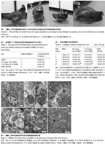
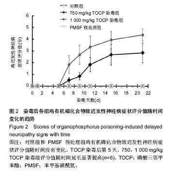
| [1] 王芳,岳凤霞,杨守芳.乐果诱导的迟发性神经病大鼠模型的实验研究[J].中国实用神经疾病杂志,2007,10(2):7-9.
[2] Jokanovic M, Kosanovic M, Brkic D, et al. Organophosphate induced delayed polyneuropathy in man: an overview. Clin Neurol Neurosurg. 2011;113(1):7-10.
[3] Mou DL, Wang YP, Song JF, et al. Triorthocresyl phosphate-induced neuronal losses in lumbar spinal cord of hens-an immunohistochemistry and ultrastructure study. Int J Neurosci. 2006;116(11):1303-1316.
[4] Chang PA. Effect of tri-o-cresyl phosphate on cytoskeleton in human neuroblastoma SK-N-SH cell. Mol Cell Biochem. 2006; 290(7):143-150.
[5] Morgan JP, Penovich P. Jamaica ginger paralysis. Forty-seven-year follow-up. Arch Neurol. 1978;35(8):530- 532.
[6] Song F, Yan Y, Zhao X, et al. Phenylmethylsulfonyl fluoride protects against the degradation of neurofilaments in tri-ortho-cresyl phosphate (TOCP) induced delayed neuropathy. Toxicology. 2009;262(3):258-264.
[7] Piao FY, KitabatakeI M, Xie XK, et al. Effects of various post-treatment by phenylmethylsulfonyl fluoride on delayed neurotoxicity induced by leptophos. J Toxicol Sci. 1995;20(5): 609-617.
[8] Piao FY, Xie XK, Yamamoto H, et al. Effect of phenylmethylsulfonyl fluoride administration prior to and following leptophos administration on electrolyte concentration and enzyme activity in hen serum. Environ Health Prev Med. 1996;1(1):44-50.
[9] Veronesi B, Padilla S. Phenylmethylsulfonyl fluoride protects rats from Mipafox-induced delayed neuropathy. Toxicol Appl Pharmacol. 1985;81(2):258-264.
[10] Carrington CD, Brown HR, Abou-donia MB. Histopathological assessment of triphenyl phosphite neurotoxicity in the hen. Neurotoxicology. 1988;9(2):223-233.
[11] 傅真真,张振玲,刘毅.鸡有机磷中毒迟发性神经病的模型建立及病理研究[J].中国工业医学杂志,2009,22(2):95-97.
[12] Piao FY, Yamauchi T, Ma N. The effect of Calcicol as calcium tonic on delayed neurotoxicity induced by organophosphorus compounds. Toxicol Lett. 2003;143(1):65-71.
[13] Abou-donia MB. Organophosphorus ester-induced delayed neurotoxicity. Annu Rev Pharmacol Toxicol. 1981;21(1): 511-548.
[14] Moretto A, Capodicasa E, Lotti M. Clinical expression of organophosphate-induced delayed polyneuropathy in rats. Toxicol Lett. 1992;63(1):97-102.
[15] 中华人民共和国国家质量监督检验检疫总局,中国国家标准化管理委员会.化学品(有机磷化合物) 急性染毒的迟发性神经毒性试验方法[M].北京:中国标准出版社,2008.
[16] Dearfield KL, Angela AE, Cimino MC, et al. Considerations in the U.S. environmental protection agency's testing approach for mutagenicity. Mutat Res. 1991;258(3):259-283.
[17] Abou-Donia MB, Lapadula DM. Mechanisms of organophosphorus ester-induced delayed neurotoxicity: type I and type II. Annu Rev Pharmacol Toxicol. 1990;30:405-440.
[18] Honarato DE, Oliveira G, Moreira V, et al. Organophosphate induced delayed neuropathy in genetically dissimilar chickens: studies with tri-ortho-cresyl phosphate (TOCP) and trichlorfon. Toxicol Lett. 2002;136(2):143-150.
[19] Johnson MK. Organophosphorus and other inhibitors of brain 'neurotoxic esterase' and the development of delayed neurotoxicity in hens. Biochem J. 1970;120(3):523-531.
[20] Carrington CD, Abou-Donia MB. The correlation between the recovery rate of neurotoxic esterase activity and sensitivity to organophosphorus-induced delayed neurotoxicity. Toxicol Appl Pharmacol. 1984;75(2):350-357.
[21] Abou-Donia MB, Suwita E, Nomeir AA. Absorption, distribution, and elimination of a single oral dose of [14C]tri-o-cresyl phosphate in hens. Toxicology. 1990;61(1): 13-25.
[22] Somkuti SG, Abou-Donia MB. Disposition, elimination, and metabolism of tri-o-cresyl phosphate following daily oral administration in Fischer 344 male rats. Arch Toxicol. 1990; 64(7):572-579.
[23] Tanaka D Jr, Bursian SJ, Lehning E. Selective axonal and terminal degeneration in the chicken brainstem and cerebellumfollowing exposure to bis(1-methylethyl) phosphorofluoridate (DFP). Brain Res. 1990;519(1-2): 200-208.
[24] Tanaka D Jr, Bursian SJ. Degeneration patterns in the chicken central nervous system induced by ingestion of the organophosphorus delayed neurotoxin tri-ortho-tolyl phosphate. A silver impregnation study. Brain Res. 1989; 484(1-2):240-256.
[25] Lehning EJ, Tanaka DJr, Bursian SJ. Triphenyl phosphite and diisopropylphosphorofluoridate produce separate and distinct axonal degeneration patterns in the central nervous system of the rat. Fundam Appl Toxicol. 1996;29(1):110-118.
[26] Jensen KF, Lapadula DM, Anderson JK, et al. Anomalous phosphorylated neurofilament aggregations in central and peripheral axons of hens treated with tri-ortho-cresyl phosphate (TOCP). J Neurosci Res. 1992;33(3):455-460.
[27] Massicotte C, Jortner BS, Ehrich M. Morphological effects of neuropathy-inducing organophosphorus compounds in primary dorsal root ganglia cell cultures. Neurotoxicology. 2003;24(6):787-796.
[28] Xin X, Zeng T, Dou DD, et al. Changes of mitochondrial ultrastructures and function in central nervous tissue of hens treated with tri-ortho-cresyl phosphate (TOCP). Hum Exp Toxicol. 2011;30(8):1062-1072.
[29] Glynn P. Neuropathy target esterase and phospholipid deacylation. Biochim Biophys Acta. 2005;1736(2):87-93.
[30] Chang PA, Wu YJ. Neuropathy target esterase: an essential enzyme for neural development and axonal maintenance. Int J Biochem Cell Biol. 2010;42(5):573-575.
[31] Choudhary S, Verma SK, Raheja G, et al. The L-type calcium channel blocker nimodipine mitigates cytoskeletal proteins phosphorylation in dichlorvos-induced delayed neurotoxicity in rats. Basic Clin Pharmacol Toxicol. 2006;98(5):447-455.
[32] El-Fawal HA, McCain WC. Antibodies to neural proteins in organophosphorus-induced delayed neuropathy (OPIDN) and its amelioration. Neurotoxicol Teratol. 2008;30(3):161-166.
[33] Li XH, Wu YJ. Characteristics of lysophosphatidylcholine- induced Ca2+ response in human neuroblastoma SH-SY5Y cells. Life Sci. 2007;80(9):886-892.
[34] Wu YJ, Li M, Li YX, et al. Verapamil abolished the enhancement of protein phosphorylation of brainstem mitochondria and synaptosomes from the hens dosed with tri-ortho-cresyl phosphate. Environ Toxicol Pharmacol. 2007; 24(1):67-71.
[35] Emerick GL, Peccinini RG, de Oliveira GH. Organophosphorus-induced delayed neuropathy: a simple and efficient therapeutic strategy. Toxicol Lett. 2010;192(2): 238-244.
[36] Mou DL, Wang YP, Song JF, et al. Triorthocresyl phosphate-induced neuronal losses in lumbar spinal cord of hens--an immunohistochemistry and ultrastructure study. Int J Neurosci. 2006;116(11):1303-1316.
[37] Song F, Yan Y, Zhao X, et al. Neurofilaments degradation as an early molecular eventin tri-ortho-cresyl phosphate(TOCP) induced delayed neuropathy. Toxicology. 2009;258(2-3): 94-100.
[38] 窦丹丹,宋永福,辛星,等.苯甲基磺酰氟预处理磷酸三邻甲苯酯染毒母鸡对其脊髓中神经丝的影响[J].中华劳动卫生职业病杂志, 2010,28(4):250-254.
[39] Liu, Z, Lenardo MJ. Reactive oxygen species regulate autophagy through redox-sensitive proteases. Dev Cell. 2007; 12(4):484-485. |