Chinese Journal of Tissue Engineering Research ›› 2026, Vol. 30 ›› Issue (14): 3597-3608.doi: 10.12307/2026.657
Previous Articles Next Articles
Hard template construction of nano-beta-tricalcium phosphate and nano-hydroxyapatite root canal sealing materials
Li Qian1, 2, Qumanguli · Abudukelimu1, 2, Shao Ziyu1, 2, Hu Yang1, 2
- 1Xinjiang Uygur Autonomous Region, China; 2Xinjiang Uygur Autonomous Region Institute of Stomatology, Urumqi 830054, Xinjiang Uygur Autonomous Region, China
-
Received:2025-07-04Accepted:2025-07-22Online:2026-05-18Published:2025-09-10 -
Contact:Hu Yang, Associate chief physician, Associate professor, Department of Prosthodontics and Implantology, First Affiliated Hospital (Affiliated Stomatological Hospital) of Xinjiang Medical University, Urumqi 830054, Xinjiang Uygur Autonomous Region, China; Xinjiang Uygur Autonomous Region Institute of Stomatology, Urumqi 830054, Xinjiang Uygur Autonomous Region, China -
About author:Li Qian, MS, Physician, Department of Prosthodontics and Implantology, First Affiliated Hospital (Affiliated Stomatological Hospital) of Xinjiang Medical University, Urumqi 830054, Xinjiang Uygur Autonomous Region, China; Xinjiang Uygur Autonomous Region Institute of Stomatology, Urumqi 830054, Xinjiang Uygur Autonomous Region, China -
Supported by:Xinjiang Uygur Autonomous Region Key Research & Development Task Special Project, No. 2016B03049-2 (to HY); Shanghai Baoshan District Health Commission Scientific Research Project Plan Task Book, No. BSZK-2023-BP02(01) (to HY)
CLC Number:
Cite this article
Li Qian, Qumanguli · Abudukelimu, Shao Ziyu, Hu Yang. Hard template construction of nano-beta-tricalcium phosphate and nano-hydroxyapatite root canal sealing materials[J]. Chinese Journal of Tissue Engineering Research, 2026, 30(14): 3597-3608.
share this article
Add to citation manager EndNote|Reference Manager|ProCite|BibTeX|RefWorks
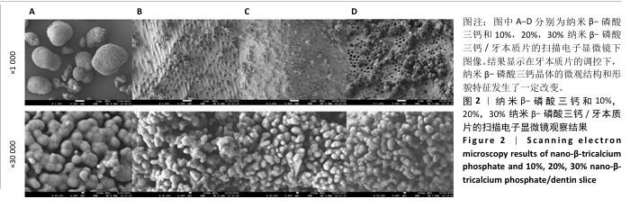
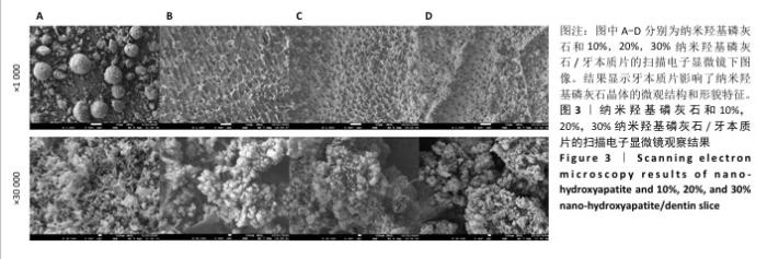
2.1 各组样品扫描电子显微镜观察结果 见图2,3。 扫描电子显微镜观察结果显示,在×1 000镜下纳米β-磷酸三钙组颗粒呈形状和大小各异的团聚体,在×30 000镜下纳米β-磷酸三钙组颗粒为大小均匀的不规则多边形,呈排列较致密的结晶样结构。对于10%,20%,30%纳米β-磷酸三钙/牙本质片组,在×1 000镜下纳米β-磷酸三钙颗粒均匀附着于牙本质小管内及周围胶原纤维蛋白网络框架中,随着纳米β-磷酸三钙浓度的增加,颗粒附着变多,簇状团聚体逐渐增大但不规则;在×30 000镜下纳米β-磷酸三钙颗粒形态呈现为短棒状、球状,排列呈三维多级团簇状,形似“石榴籽”,见图2。说明在牙本质片的调控下,纳米β-磷酸三钙晶体的微观结构和形貌特征发生了一定改变。 扫描电子显微镜观察结果显示,在×1 000镜下纳米羟基磷灰石组颗粒团聚为大小不一的球状,表面粗糙;在×30 000镜下纳米羟基磷灰石颗粒为棒状、针状,排列呈“珊瑚状”,分布较均匀。对于10%,20%,30%纳米羟基磷灰石/牙本质片组,在×1 000镜下纳米羟基磷灰石颗粒呈团簇状均匀附着于牙本质小管内及周围胶原纤维蛋白网络框架中,随着纳米羟基磷灰石浓度的增加,颗粒附着增加;在×30 000镜下纳米羟基磷灰石颗粒为短棒状、球状,排列变紧密,间隙变小,呈“雪花状”,见图3。说明牙本质片影响了纳米羟基磷灰石晶体的微观结构和形貌特征。 "
"
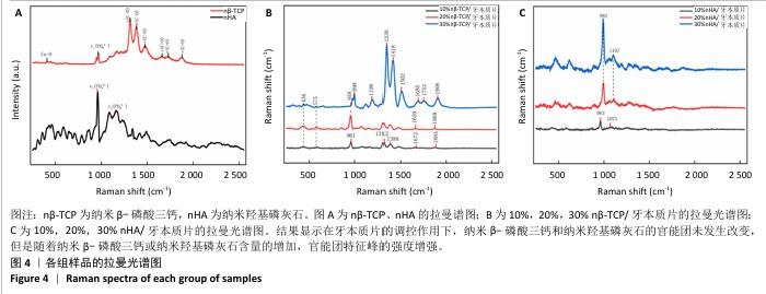
2.2 各组样品拉曼光谱检测结果 见图4。 拉曼光谱检测结果显示,与纳米β-磷酸三钙组相比, 10%,20%,30%纳米β-磷酸三钙/牙本质片显示出相同位置的特征峰:961,958,998 cm-1处PO43-的对称伸缩振动峰,1 312,1 388 cm-1和1 198-1 502 cm-1范围内PO43-中P-O伸缩振动峰及PO43-中的P=O伸缩振动峰。与纳米羟基磷灰石组相比,10%,20%,30%纳米羟基磷灰石/牙本质片组中963,992 cm-1处PO43-的对称伸缩振动峰和1 107 cm-1处的反对称伸缩振动峰位置相同。以上结果说明它们的振动模式在能量上是相同的,可能对应着相同的官能团,即在牙本质片的调控作用下,纳米β-磷酸三钙和纳米羟基磷灰石颗粒的官能团未发生改变,而且随着纳米β-磷酸三钙和纳米羟基磷灰石含量的增加,特征峰的强度依次增强,表明材料官能团的含量增加,原因可能是牙本质片改变了纳米β-磷酸三钙和纳米羟基磷灰石的微观分子结构,影响振动强度,进而增强拉曼光谱强度。 "
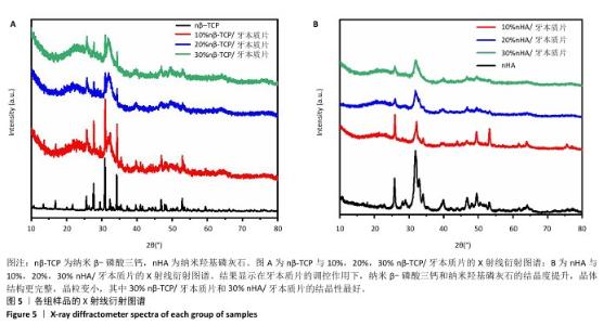
.3 各组样品X射线衍射检测结果 见图5。 X射线衍射检测结果显示,纳米β-磷酸三钙组和纳米羟基磷灰石组显示出清晰且尖锐的衍射峰,表明它们具有高度的结晶性,纳米β-磷酸三钙的特征衍射峰主要出现在2θ的约26°,29°,31°,34°等处,对应于纳米β-磷酸三钙的典型晶面反射;纳米羟基磷灰石的特征衍射峰主要集中在2θ的25.9°,31.7°,32.9°和 34.1°等处,对应于纳米羟基磷灰石的典型晶面反射[如(002)(211)(112)等]。10%,20%,30% 纳米羟基磷灰石/牙本质片组和10%,20%,30% 纳米羟基磷灰石/牙本质片组特征衍射峰位置没有发生变化,说明材料的晶体的主要元素及化学键未发生改变;但随着纳米β-磷酸三钙和纳米羟基磷灰石浓度的增高,特征衍射峰强度逐渐增强、宽度逐渐增宽,表明纳米β-磷酸三钙和纳米羟基磷灰石晶体的结晶度可能逐渐增高,晶体结构越来越完整,晶粒逐渐变小。 "
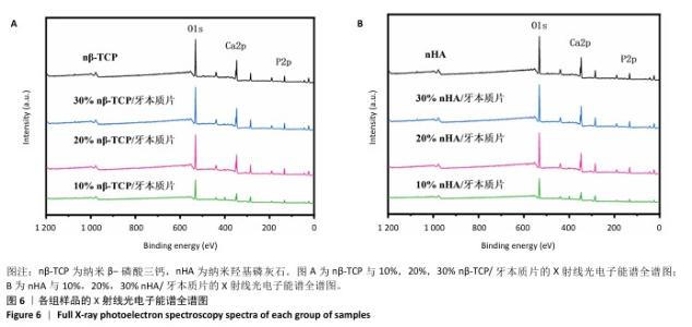
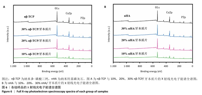
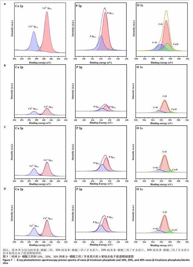
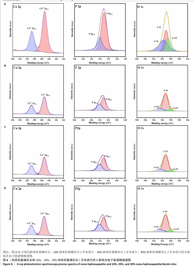
2.4 各组样品X射线光电子能谱检测结果 见图6-8。 X射线光电子能谱图显示,8组呈现出O 1s(532 eV)、Ca 2p(348 eV)及P 2p(132 eV)特征峰,这是纳米β-磷酸三钙和纳米羟基磷灰石成分的直接反映,可见纳米β-磷酸三钙和纳米羟基磷灰石材料表面元素组成无改变。在Ca 2p谱中,观察到347 eV (2p3/2)和350 eV(2p1/2)处的典型双峰分裂结构以及P 2p谱在133 eV(2p3/2)和134 eV(2p1/2)处的分峰现象,这些特征源于钙离子的自旋轨道耦合效应和磷酸根基团中P-O键的存在。在O 1s谱发现了3个不同化学环境的氧物种构成峰形,在各纳米β-磷酸三钙/牙本质片组中,Ca-O键特征峰源自磷酸三钙晶体结构中的钙氧配位,C-O键和C=O键特征峰则与牙本质胶原蛋白中的羟基基团、羧基或材料表面吸附的含氧有机物有关;在各纳米羟基磷灰石/牙本质片组中,Ca-O键特征峰源自纳米羟基磷灰石晶体结构中的钙氧八面体配位;C-O键特征峰与胶原纤维中的羟基基团相关;而532 eV处的C=O键特征峰可能来源于胶原蛋白羧基或材料表面吸附的含氧有机物,这种氧化学态的多样性揭示了纳米β-磷酸三钙和纳米羟基磷灰石与牙本质胶原纤维网络表面复杂的界面相互作用,纳米β-磷酸三钙和纳米羟基磷灰石的部分化学态发生改变,表明牙本质会对纳米β-磷酸三钙和纳米羟基磷灰石晶体的表面微观形态和结构产生一定的调控作用。 "
"
"
| [1] 曾建树.根管治疗根尖周炎感染的应用与有效性研究[J].当代医学,2019,25(1):89-91. [2] LI GH, NIU LN, ZHANG W, et al. Ability of new obturation materials to improve the seal of the root canal system: a review. Acta Biomater. 2014;10(3):1050-1063. [3] GIACOMINO CM, WEALLEANS JA, KUHN N, et al. Comparative Biocompatibility and Osteogenic Potential of Two Bioceramic Sealers. J Endod. 2019;45(1):51-56. [4] ÖZDEMIR O, KOÇAK S, HAZAR E, et al. Dentinal tubule penetration of gutta-percha with syringe-mix resin sealer using different obturation techniques: A confocal laser scanning microscopy study. Aust Endod J. 2022;48(2):258-265. [5] 杨勤,童方丽,杨明,等.根管封闭剂iRoot SP对椭圆形根管根尖封闭性能的评价[J]. 实用医学杂志,2016,32(14):2309-2312. [6] WUERSCHING SN, DIEGRITZ C, HICKEL R, et al. A comprehensive in vitro comparison of the biological and physicochemical properties of bioactive root canal sealers. Clin Oral Investig. 2022;26(10):6209-6222. [7] 杨习良,郑天霞,杨诺亚,等.3种根管封闭剂体外细胞相容性、促成骨性及抗菌性的实验研究[J].口腔医学研究,2022,38(11): 1071-1075. [8] LI Y, LI B, LAI S, et al. The biocompatibility, penetrability, sealing ability, and antibacterial properties of iRoot SP compared to AH plus: An In Vitro evaluation. Arch Oral Biol. 2025;172:106188. [9] SAGHIRI MA, KARAMIFAR K, NATH D, et al. A novel polyurethane expandable root canal sealer. J Endod. 2021;47(4):612-620. [10] OSTAPIUK M, TARCZYDŁO J, KAMIŃSKA K, et al. Compressive Strength Testing of Glass-Fibre-Reinforced Tooth Crown Tissues After Endodontic Treatment. Ann Biomed Eng. 2024;52(2):318-326. [11] MARVANIYA J, AGARWAL K, MEHTA DN, et al. Minimal Invasive Endodontics: A Comprehensive Narrative Review. Cureus. 2022;14(6): e25984. [12] KOMABAYASHI T, COLMENAR D, CVACH N, et al. Comprehensive review of current endodontic sealers. Dent Mater J. 2020;39(5):703-720. [13] ZHOU HM, SHEN Y, ZHENG W, et al. Physical properties of 5 root canal sealers. J Endod. 2013;39(10):1281-1286. [14] HAJI TH, SELIVANY BJ, SULIMAN AA. Sealing ability in vitro study and biocompatibility in vivo animal study of different bioceramic based sealers. Clin Exp Dent Res. 2022;8(6):1582-1590. [15] KWAK SW, KOO J, SONG M, et al. Physicochemical Properties and Biocompatibility of Various Bioceramic Root Canal Sealers: In Vitro Study. J Endod. 2023;49(7):871-879. [16] QUARESMA SAL, ALVES DOS SANTOS GN, SILVA-SOUSA AC, et al. Physicochemical properties of calcium silicate cement based endodontic sealers. J Mech Behav Biomed Mater. 2024;151:106400. [17] ŠIMUNDIĆ MUNITIĆ M, POKLEPOVIĆ PERIČIĆ T, UTROBIČIĆ A, et al. Antimicrobial efficacy of commercially available endodontic bioceramic root canal sealers: A systematic review. PLoS One. 2019; 14(10):e0223575. [18] SATO A, YANAGI T, YAMAGUCHI Y, et al. Effect of DNA/protamine complex paste on bone augmentation of the mandible: A pilot study on dogs. Arch Oral Biol. 2020;115:104729. [19] HARB SV, BASSOUS NJ, DE SOUZA TAC, et al. Hydroxyapatite and β-TCP modified PMMA-TiO2 and PMMA-ZrO2 coatings for bioactive corrosion protection of Ti6Al4V implants. Mater Sci Eng C Mater Biol Appl. 2020;116:111149. [20] KAMAL M, ANDERSSON L, TOLBA R, et al. Bone regeneration using composite non- demineralized xenogenic dentin with beta-tricalcium phosphate in experimental alveolar cleft repair in a rabbit model. J Transl Med. 2017;15(1):263. [21] 李倩,曲曼姑丽•阿布都克力木,胡杨. β-TCP在口腔医学领域的研究进展[J].今日健康,2024(11):142-144. [22] 裘吉雨,英司奇,刘湘,等.BMP9转染的hPDLSCs与HA-TCP支架复合体促进牙槽骨再生的实验研究[J].重庆医学,2020,49(17): 2849-2856. [23] REN-JIE XU, JIN-JIN MA, YU X, et al. A biphasic calcium phosphate/acylated methacrylate gelatin composite hydrogel promotes osteogenesis and bone repair. Connect Tissue Res. 2023;64(5):445-456. [24] 王建平,丛琳. 纳米羟基磷灰石在牙髓炎根管治疗中的临床应用[J].黑龙江医药科学,2015,38(4):67-69. [25] 王继春,杜春艳,陈云凤.纳米羟基磷灰石在牙髓炎根管治疗中的临床应用与疗效分析[J].世界最新医学信息文摘(连续型电子期刊),2017,17(49):46,48. [26] TAVARES FJTM, SOARES PIP, SILVA JC, et al. Preparation and In Vitro Characterization of Magnetic CS/PVA/HA/pSPIONs Scaffolds for Magnetic Hyperthermia and Bone Regeneration. Int J Mol Sci. 2023; 24(2):1128. [27] TRZASKOWSKA M, VIVCHARENKO V, BENKO A, et al. Biocompatible nanocomposite hydroxyapatite-based granules with increased specific surface area and bioresorbability for bone regenerative medicine applications. Sci Rep. 2024;14(1):28137. [28] 曲曼姑丽•阿布都克力木,李倩,胡杨. 纳米羟基磷灰石在牙体牙髓病学中的研究进展[J].今日健康,2024(11):187-189. [29] 邵子瑜,李倩,曲曼姑丽•阿布都克力木,等.三种比例双相磷酸钙的制备及性能表征[J].中国组织工程研究,2026,30(8):1952-1961. [30] CAO Y, MEI ML, LI QL, et al. Agarose hydrogel biomimetic mineralization model for the regeneration of enamel prismlike tissue. ACS Appl Mater Interfaces. 2014;6(1):410-420. [31] LIN K, XIA L, GAN J, et al. Tailoring the nanostructured surfaces of hydroxyapatite bioceramics to promote protein adsorption, osteoblast growth, and osteogenic differentiation. ACS Appl Mater Interfaces. 2013;5(16):8008-8017. [32] LI L, MAO C, WANG J, et al. Bio-Inspired Enamel Repair via Glu-Directed Assembly of Apatite Nanoparticles: an Approach to Biomaterials with Optimal Characteristics. Adv Mater. 2011;23(40):4695-4701. [33] CHANDRA P, SINGH V, SINGH S, et al. Assessment of Fracture Resistances of Endodontically Treated Teeth Filled with Different Root Canal Filling Systems. J Pharm Bioallied Sci. 2021;13(Suppl 1): S109-S111. [34] WANG YH, LIU SY, DONG YM. In vitro evaluation of the impact of a bioceramic root canal sealer on the mechanical properties of tooth roots. J Dent Sci. 2024;19(3):1734-1740. [35] WANG Z, FLORCZYK SJ. Freeze-FRESH: A 3D Printing Technique to Produce Biomaterial Scaffolds with Hierarchical Porosity. Materials (Basel). 2020;13(2):354. [36] QIN K, PARISI C, FERNANDES FM. Recent advances in ice templating: from biomimetic composites to cell culture scaffolds and tissue engineering. J Mater Chem B. 2021;9(4):889-907. [37] 周延民,杨亿鑫.冰模板技术及其在口腔医学领域的应用进展[J].口腔医学研究,2023,39(9):767-770. [38] Ozaki M, Sakashita S, Hamada Y, et al. Peptides for Silica Precipitation: Amino Acid Sequences for Directing Mineralization. Protein Pept Lett. 2018;25(1):15-24. [39] 俞磊,张巍,秦毅,等. 贻贝启发接枝骨形态发生蛋白2成骨活性肽的介孔生物玻璃修复股骨髁缺损[J]. 中国组织工程研究, 2025,29(22):4629-4638. [40] JANAIRO JIB, SAKAGUCHI T, MINE K, et al. Synergic Strategies for the Enhanced Self-Assembly of Biomineralization Peptides for the Synthesis of Functional Nanomaterials. Protein Pept Lett. 2018;25(1):4-14. [41] LUO X, ZHANG S, LUO B, et al. Engineering collagen fiber templates with oriented nanoarchitecture and concerns on osteoblast behaviors. Int J Biol Macromol. 2021;185:77-86. [42] ZHANG W, ZHENG Y, LIU H, et al. A non-invasive monitoring of USPIO labeled silk fibroin/hydroxyapatite scaffold loaded DPSCs for dental pulp regeneration. Mater Sci Eng C Mater Biol Appl. 2019;103:109736. [43] XU X, CHEN X, LI J. Natural protein bioinspired materials for regeneration of hard tissues. J Mater Chem B. 2020;8(11):2199-2215. [44] CHEN X, LIU Y, YANG J, et al. The synthesis of hydroxyapatite with different crystallinities by controlling the concentration of recombinant CEMP1 for biological application. Mater Sci Eng C Mater Biol Appl. 2016;59:384-389. [45] KAMMOUN R, BEHETS C, MANSOUR L, et al. Mineral features of connective dental hard tissues in hypoplastic amelogenesis imperfecta. Oral Dis. 2018;24(3):384-392. [46] HUANG F, SHEN Q, ZHAO J. Growth and differentiation of neural stem cells in a three-dimensional collagen gel scaffold. Neural Regen Res. 2013;8(4):313-319. [47] BANGLMAIER RF, SANDER EA, VANDEVORD PJ. Induction and quantification of collagen fiber alignment in a three-dimensional hydroxyapatite-collagen composite scaffold. Acta Biomater. 2015;17: 26-35. [48] ABDEL-RAHMAN RM, VISHAKHA V, KELNAR I, et al. Synergistic performance of collagen-g-chitosan-glucan fiber biohybrid scaffold with tunable properties. Int J Biol Macromol. 2022;202:671-680. [49] HOSSAIN M, JEONG JH, SULTANA T, et al. A composite of polymethylmethacrylate, hydroxyapatite, and β-tricalcium phosphate for bone regeneration in an osteoporotic rat model. J Biomed Mater Res B Appl Biomater. 2023;111(10):1813-1823. [50] FU M, LI J, LIU M, et al. Sericin/Nano-Hydroxyapatite Hydrogels Based on Graphene Oxide for Effective Bone Regeneration via Immunomodulation and Osteoinduction. Int J Nanomedicine. 2023; 18:1875-1895. [51] SOARES Í, SOTELO L, ERCEG I, et al. Improvement of Metal-Doped β-TCP Scaffolds for Active Bone Substitutes via Ultra-Short Laser Structuring. Bioengineering (Basel). 2023;10(12):1392. [52] PINHEIRO ALB, Soares LGP, Marques AMC, et al. Biochemical changes on the repair of surgical bone defects grafted with biphasic synthetic micro-granular HA + β-tricalcium phosphate induced by laser and LED phototherapies and assessed by Raman spectroscopy. Lasers Med Sci. 2017;32(3):663-672. [53] TAN N, SABALIC-SCHOENER M, NGUYEN L, et al. β-Tricalcium Phosphate-Loaded Chitosan-Based Thermosensitive Hydrogel for Periodontal Regeneration. Polymers (Basel). 2023;15(20):4146. [54] UMA MAHESHWARI S, GOVINDAN K, RAJA M, et al. Preliminary studies of PVA/PVP blends incorporated with HAp and β-TCP bone ceramic as template for hard tissue engineering. Biomed Mater Eng. 2017;28(4):401-415. [55] REYES-GASGA J, MARTÍNEZ-PIÑEIRO EL, RODRÍGUEZ-ÁLVAREZ G, et al. XRD and FTIR crystallinity indices in sound human tooth enamel and synthetic hydroxyapatite. Mater Sci Eng C Mater Biol Appl. 2013; 33(8):4568-74. [56] XU M, QIN M, ZHANG X, et al. Porous PVA/SA/HA hydrogels fabricated by dual-crosslinking method for bone tissue engineering. J Biomater Sci Polym Ed. 2020;31(6):816-831. [57] ZHU S, SUN H, MU T, et al. Cellulose nano-dispersions enhanced by ultrasound assisted chemical modification drive osteoblast proliferation and differentiation in PVA/HA bone tissue engineering scaffolds. Int J Biol Macromol. 2024;279(Pt 4):135571. [58] ZHANG K, LIU Y, ZHAO Z, et al. Magnesium-Doped Nano-Hydroxyapatite/Polyvinyl Alcohol/Chitosan Composite Hydrogel: Preparation and Characterization. Int J Nanomedicine. 2024;19: 651-671. [59] CHOY CS, LEE WF, LIN PY, et al. Surface Modified β-Tricalcium phosphate enhanced stem cell osteogenic differentiation in vitro and bone regeneration in vivo. Sci Rep. 2021;11(1):9234. [60] Minetti E, Palermo A, Malcangi G, et al. Dentin, Dentin Graft, and Bone Graft: Microscopic and Spectroscopic Analysis. J Funct Biomater. 2023;14(5):272. [61] lSHEMARY AZ, CHEIKH L, ARDAKL SS. Extraction and degradation rate analysis of calcium phosphate from diverse fish Bones: A comparative study. J Saudi Chem Soc. 2024;28(3).doi:10.1016/j.jscs.2024.101859. |
| [1] | Shi Xiaonan, Wu Xuan, Zhang Daxu, Hu Jingjing, Zheng Yazhe, Liu Yutong, Zhao Shuo, Li Weilong, Ye Shujun, Wang Jingyi, Yan Li. Preparation and characterization of 3D printed microstructured silk fibroin scaffold for liver injury repair [J]. Chinese Journal of Tissue Engineering Research, 2026, 30(14): 3618-3625. |
| Viewed | ||||||
|
Full text |
|
|||||
|
Abstract |
|
|||||