Chinese Journal of Tissue Engineering Research ›› 2013, Vol. 17 ›› Issue (49): 8468-8473.doi: 10.3969/j.issn.2095-4344.2013.49.002
Previous Articles Next Articles
Red blood cell lysis isolation and culture of rabbit bone marrow mesenchymal stem cells in vitro
Zhang Wei-dong, Zhang Fang-biao, Shi Hong-can, Tan Rong-bang, Ye Gang, Li Guang-yu, Pan Shu, Sun Fei
- Medical College of Yangzhou University, Yangzhou 225001, Jiangsu Province, China
-
Revised:2013-09-14Online:2013-12-03Published:2013-12-03 -
Contact:Shi Hong-can, M.D., Chief physician, Doctoral supervisor, Medical College of Yangzhou University, Yangzhou 225001, Jiangsu Province, China shihongcan@hotmail.com -
About author:Zhang Wei-dong★, Studying for master’s degree, Medical College of Yangzhou University, Yangzhou 225001, Jiangsu Province, China Zhangw20062008@126.com -
Supported by:the National Natural Science Foundation of China, No. 30672080*, 30972968*, 81170014*; the High-level Personnel Fund for the “Six Talents Peak” of Jiangsu Province, No. 2009128*; Jiangsu Province “Blue Project” Academic Leaders Fund, No. 20081215*
CLC Number:
Cite this article
Zhang Wei-dong, Zhang Fang-biao, Shi Hong-can, Tan Rong-bang, Ye Gang, Li Guang-yu, Pan Shu, Sun Fei. Red blood cell lysis isolation and culture of rabbit bone marrow mesenchymal stem cells in vitro[J]. Chinese Journal of Tissue Engineering Research, 2013, 17(49): 8468-8473.
share this article
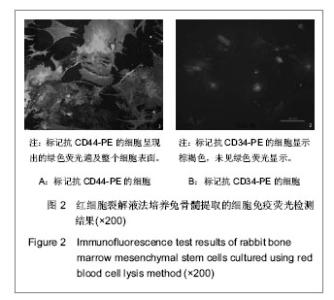
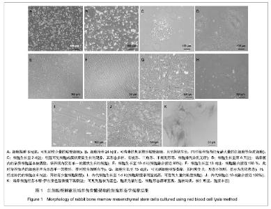
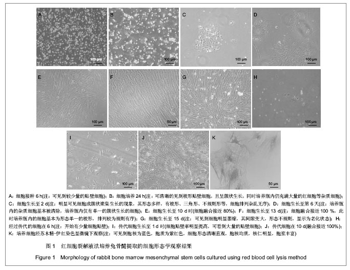
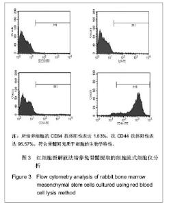
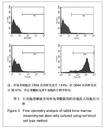
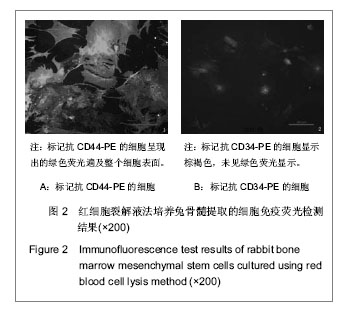
形态学观察与苏木精-伊红染色:刚接种入细胞培养瓶内,细胞悬液中主要以小圆点形红细胞为主,有少量的单个核细胞。恒温培养箱内培养6 h即可见到较少量的贴壁细胞,见图1A。24 h左右时可清晰的见到梭形贴壁细胞,且呈簇状生长,同时培养瓶内仍充满大量的红细胞等杂质细胞,见图1B。2 d后经过换液,培养瓶内的杂质细胞大部分被清除,此时可更加明显的看到细胞成簇状聚集生长的现象,其形态多样,有梭形、三角形、不规则形等,细胞排列杂乱无序,见图1C。经过2次换液,第6天时细胞培养瓶内的杂质细胞基本被清除,培养瓶内仅有单一的簇状生长的细胞,见图1D。当细胞生长至10 d时,细胞融合接近80%,见图1E。细胞生长至13 d时,细胞融合接近100%,此时培养瓶内的细胞基本为形态单一的梭形,排列较为规则有序,见图1F。当细胞生长至15 d左右时,可见到细胞明显萎缩,其间隙变大,形态不规则,显示为老化状态,见图1G。经过传代的细胞在6 h左右即开始有少量细胞贴壁,见图1H。传代细胞生长至1 d时,其细胞贴壁率明显提高,可看到大量的贴壁细胞,见图1I。在10 d左右融合接近100%,见图1J。 苏木精-伊红染色后显微镜下可见到胞核为蓝色,胞质为紫红色的细胞,此时细胞形态更清晰直观,胞核均质,核仁明显,胞浆丰富,见图1K。 细胞免疫荧光:细胞免疫荧光显示标记抗CD44-PE的细胞呈现出的绿色荧光遍及整个细胞表面,标记抗CD34-PE的细胞显示棕褐色,未见绿色荧光显示,见图2,提示该细胞CD44抗原表达阳性,CD34抗原表达阴性。"
| [1] Jones E,McGonagle D.Human bone marrow mesenchymal stem cells in vivo. Rheumatology (Oxford). 2008; 47(02): 126-131.[2] Zhang W,Li Z,Huang Q,et al.Effects of a hybrid micro/nanorod topography-modified titanium implant on adhesion and osteogenic differentiation in rat bone marrow mesenchymal stem cells. Int J Nanomedicine. 2013;8:257-265.[3] Jungebluth P, Alici E, Baiguera S, et al.Tracheobronchial transplantation with a stem-cell-seeded bioartificial nanocomposite: a proof-of-concept study. Lancet. 2011; 378(9808):1997-2004.[4] Karagianni M,Brinkmann I,Kinzebach S,et al. A comparative analysis of the adipogenic potential in human mesenchymal stromal cells from cord blood and other sources. Cytotherapy. 2013; 15(1):76-88.[5] Sun Z, Wang Y, Gong X, et al. Secretion of rat tracheal epithelial cells induces mesenchymal stem cells to differentiate into epithelial cells. Cell Biol Int. 2012; 36(2): 169-175.[6] Zhao JW,Zhang MR,Ji QY,et al.The Role of Slingshot-1L (SSH1L) in the Differentiation of Human Bone Marrow Mesenchymal Stem Cells into Cardiomyocyte-Like Cells. Molecules. 2012; 17(12): 14975-14994.[7] Mohana Kumar B, Lee WJ, Lee YM, et al. 296 in vitro differentiation of porcine bone marrow-derived mesenchymal stem cells into hepatocyte-like cells. Reprod Fertil Dev. 2012; 25(1): 295-296.[8] Tan SL, Ahmad TS, Selvaratnam L, et al. Isolation, characterization and the multi-lineage differentiation potential of rabbit bone marrow-derived mesenchymal stem cells. J Anat. 2013; 222(4):437-450.[9] Peterbauer-Scherb A, van Griensven M, Meinl A, et al. Isolation of pig bone marrow mesenchymal stem cells suitable for one-step procedures in chondrogenic regeneration. J Tissue Eng Regen Med. 2010; 4(6): 485-490.[10] Huang Q, Wang YD, Wu T, et al. Preliminary separation of the growth factors in platelet-rich plasma: effects on the proliferation of human marrow-derived mesenchymal stem cells. Chin Med J. 2009; 122(1): 83-87. [11] 徐海艇,徐晓斐,王健,等.组织工程方法修复羊腭裂骨缺损的实验研究[J]. 中华实验外科杂志, 2012, 29(2): 315-317.[12] 李志营,步星耀,张圣旭,等.自体骨髓干细胞动员移植与手术移植治疗脊髓损伤的比较[J].中国组织工程研究与临床康复, 2009, 13(45): 8911-8916.[13] Li L, Lü G, Wang YF, et al. Glial cell-derived neurotrophic factor mRNA expression in a rat model of spinal cord injury following bone marrow stromal cell transplantation. Neural Regen Res 2008;3(10):1056-1059.[14] Peng Y, Zhang QM, You H, et al. Growth-associated protein 43 and neural cell adhesion molecule expression follow-ing bone marrow-derived mesenchymal stem cell transplantation in a rat model of ischemic brain injury. Neural Regen Res. 2010;5(13):975-980.[15] Williams AR, Hatzistergos KE, Addicott B, et al. Enhanced effect of combining human cardiac stem cells and bone marrow mesenchymal stem cells to reduce infarct size and to restore cardiac function after myocardial infarction. Circulation. 2013; 127(2): 213-223.[16] Jones EA, English A, Kinsey SE, et al. Optimization of a flow cytometry-based protocol for detection and phenotypic characterization of multipotent mesenchymal stromal cells from human bone marrow. Cytometry B Clin Cytom. 2006; 70(6): 391-399.[17] Horn P, Bork S, Diehlmann A, et al. Isolation of human mesenchymal stromal cells is more efficient by red blood cell lysis. Cytotherapy. 2008; 10(7): 676-685.[18] Horn P, Bork S, Wagner W. Standardized isolation of human mesenchymal stromal cells with red blood cell lysis. Methods Mol Biol. 2011; 698: 23-35.[19] Lucarelli E, Fini M, Beccheroni A, et al. Stromal stem cells and platelet-rich plasma improve bone allograft integration. Clin Orthop Relat Res. 2005; 435: 62-68.[20] 翁玄,朱勇军,张健,等.不同分离方法及培养条件对兔骨髓间充质干细胞生物活性的影响[J].中国组织工程研究与临床康复, 2010, 14(10): 1775-1779.[21] Lee JW, Gupta N, Serikov V, et al. Potential application of mesenchymal stem cells in acute lung injury. Expert Opin Biol Ther. 2009; 9(10): 1259-1270.[22] Jarocha D, Lukasiewicz E, Majka M. Adventage of mesenchymal stem cells (MSC) expansion directly from purified bone marrow CD105+ and CD271+ cells. Folia Histochem Cytobiol. 2008; 46(3): 307-314. |
| [1] | Kong Desheng, He Jingjing, Feng Baofeng, Guo Ruiyun, Asiamah Ernest Amponsah, Lü Fei, Zhang Shuhan, Zhang Xiaolin, Ma Jun, Cui Huixian. Efficacy of mesenchymal stem cells in the spinal cord injury of large animal models: a meta-analysis [J]. Chinese Journal of Tissue Engineering Research, 2020, 24(在线): 3-. |
| [2] | Chen Qiang, Zhuo Hongwu, Xia Tian, Ye Zhewei . Toxic effects of different-concentration isoniazid on newborn rat osteoblasts in vitro [J]. Chinese Journal of Tissue Engineering Research, 2020, 24(8): 1162-1167. |
| [3] | Chen Jinsong, Wang Zhonghan, Chang Fei, Liu He. Tissue engineering methods for repair of articular cartilage defect under special conditions [J]. Chinese Journal of Tissue Engineering Research, 2020, 24(8): 1272-1279. |
| [4] | Liu Chundong, Shen Xiaoqing, Zhang Yanli, Zhang Xiaogen, Wu Buling. Effects of strontium-modified titanium surfaces on adhesion, migration and proliferation of bone marrow mesenchymal stem cells and expression of bone formation-related genes [J]. Chinese Journal of Tissue Engineering Research, 2020, 24(7): 1009-1015. |
| [5] | Lin Ming, Pan Jinyong, Zhang Huirong. Knockout of NIPBL gene down-regulates the abilities of proliferation and osteogenic differentiation in mouse bone marrow mesenchymal stem cells [J]. Chinese Journal of Tissue Engineering Research, 2020, 24(7): 1002-1008. |
| [6] | Zhang Wen, Lei Kun, Gao Lei, Li Kuanxin. Neuronal differentiation of rat bone marrow mesenchymal stem cells via lentivirus-mediated bone morphogenetic protein 7 transfection [J]. Chinese Journal of Tissue Engineering Research, 2020, 24(7): 985-990. |
| [7] | Wu Zhifeng, Luo Min. Biomechanical analysis of chemical acellular nerve allograft combined with bone marrow mesenchymal stem cell transplantation for repairing sciatic nerve injury [J]. Chinese Journal of Tissue Engineering Research, 2020, 24(7): 991-995. |
| [8] | Huang Yongming, Huang Qiming, Liu Yanjie, Wang Jun, Cao Zhenwu, Tian Zhenjiang, Chen Bojian, Mai Xiujun, Feng Enhui. Proliferation and apoptosis of chondrocytes co-cultured with TDP43 lentivirus transfected-human umbilical cord mesenchymal stem cells [J]. Chinese Journal of Tissue Engineering Research, 2020, 24(7): 1016-1022. |
| [9] | Qin Xinyu, Zhang Yan, Zhang Ningkun, Gao Lianru, Cheng Tao, Wang Ze, Tong Shanshan, Chen Yu. Elabela promotes differentiation of Wharton’s jelly-derived mesenchymal stem cells into cardiomyocyte-like cells [J]. Chinese Journal of Tissue Engineering Research, 2020, 24(7): 1046-1051. |
| [10] | Liu Mengting, Rao Wei, Han Bing, Xiao Cuihong, Wu Dongcheng. Immunomodulatory characteristics of human umbilical cord mesenchymal stem cells in vitro [J]. Chinese Journal of Tissue Engineering Research, 2020, 24(7): 1063-1068. |
| [11] |
Cen Yanhui, Xia Meng, Jia Wei, Luo Weisheng, Lin Jiang, Chen Songlin, Chen Wei, Liu Peng, Li Mingxing, Li Jingyun, Li Manli, Ai Dingding, Jiang Yunxia.
Baicalein inhibits the biological behavior of hepatocellular
carcinoma stem cells by downregulation of Decoy receptor 3 expression |
| [12] | Zhang Peigen, Heng Xiaolai, Xie Di, Wang Jin, Ma Jinglin, Kang Xuewen. Electrical stimulation combined with neurotrophin 3 promotes proliferation and differentiation of endogenous neural stem cells after spinal cord injury in rats [J]. Chinese Journal of Tissue Engineering Research, 2020, 24(7): 1076-1082. |
| [13] | Huang Cheng, Liu Yuanbing, Dai Yongping, Wang Liangliang, Cui Yihua, Yang Jiandong. Transplantation of bone marrow mesenchymal stem cells overexpressing glial cell line derived neurotrophic factor gene for spinal cord injury [J]. Chinese Journal of Tissue Engineering Research, 2020, 24(7): 1037-1045. |
| [14] | Han Bo, Yang Zhe, Li Jing, Zhang Mingchang . Regulation of limbal stem cells via Wnt signaling in the treatment of limbal stem cell deficiency [J]. Chinese Journal of Tissue Engineering Research, 2020, 24(7): 1057-1062. |
| [15] | Li Jia, Tang Ying, Zhu Qi, Zhang Yanping, Zhou Peigang, Gu Yongchun. Transplantation of human stem cells from the apical papilla for treating dextran sulfate sodium-induced experimental colitis [J]. Chinese Journal of Tissue Engineering Research, 2020, 24(7): 1069-1075. |
| Viewed | ||||||
|
Full text |
|
|||||
|
Abstract |
|
|||||
