Chinese Journal of Tissue Engineering Research
Previous Articles Next Articles
Culture of rabbit’s articular chondrocytes using type Ⅱ collagenase enzyme digestion method
Yan Hu, Su You-xin, Lin Xue-yi, Chen Bao-jun, Zhou Bi-hong, Zhang Qing
- Fujian University of Traditional Chinese Medicine, Fuzhou 350122, Fujian Province, China
-
Revised:2013-09-15Online:2013-12-10Published:2013-12-10 -
Contact:Su You-Xin, M.D., Professor, Doctoral supervisor, Fujian University of Traditional Chinese Medicine, Fuzhou 350122, Fujian Province, China suyouxin777@hotmail.com -
About author:Yan Hu☆, Studying for doctorate, Fujian University of Traditional Chinese Medicine, Fuzhou 350122, Fujian Province, China abcdyh@hotmail.com -
Supported by:the National Natural Science Foundation of China, No. 81072828*
CLC Number:
Cite this article
Yan Hu, Su You-xin, Lin Xue-yi, Chen Bao-jun, Zhou Bi-hong, Zhang Qing. Culture of rabbit’s articular chondrocytes using type Ⅱ collagenase enzyme digestion method[J]. Chinese Journal of Tissue Engineering Research.
share this article
Add to citation manager EndNote|Reference Manager|ProCite|BibTeX|RefWorks
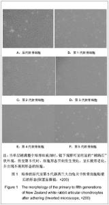
2.1 分离培养的新西兰大白兔软骨细胞的形态 见图1。"

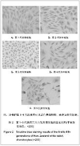
倒置显微镜下观察见分离培养的原代软骨细胞在贴壁前为圆球形,呈悬浮状态,12 h部分贴壁,绝大部分在24 h时贴壁,部分细胞开始伸展,细胞体积较小,均匀散在生长,72 h可以传代,第1至3代细胞48 h可传代,见图1。 当单层铺满整个培养皿底面时,镜下观察可呈明显的“铺路石”状外观。传至第5代时,细胞形态开始发生变化,呈长梭形老化,并出现不规则形态的细胞,生长速度减慢,传代周期延长,见图1。 2.2 传代培养的新西兰大白兔软骨细胞的鉴定结果 第1代至第5代关节软骨细胞经甲苯胺蓝染色后,胞内均可见蓝紫色异染颗粒,细胞核染成深蓝色,细胞周围有少量浅蓝色异染颗粒,细胞见图2。"

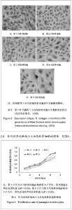
2.3 传代培养的新西兰大白兔软骨细胞中Ⅱ型胶原的表达 免疫组化染色显示,Ⅱ型胶原主要分布在细胞质和细胞膜上呈阳性表达(黄褐色),第1-5代软骨细胞中均表达Ⅱ型胶原,见图3。"

| [1] 邱贵兴,荣国威.骨科学[M].北京:中国协和医科大学出版社,2012: 506.[2] 余振华,朱顺叶,张珑娟. 大鼠胫骨生长板软骨细胞的分离与培养鉴定[J]. 中华实验外科杂志,2008,25(10):1309-1310.[3] 胡志俊,胡波,唐德志,等.兔膝关节软骨细胞的分离培养及形态学特征[J].中国组织工程研究与临床康复,2010,14(46): 8555-8558.[4] 中华人民共和国科学技术部. 关于善待实验动物的指导性意见. 2006-09-30.[5] Isogai N, Kusuhara H, Ikada Y, et al. Comparison of different chondrocytes for use in tissue engineering of cartilage model structures. Tissue Eng. 2006;12(4):691-703.[6] Alsalameh S, Amin R, Gemba T, et al. Identification of mesenchymal progenitor cells in normal and osteoarthritic human articular cartilage. Arthritis Rheum. 2004;50(5): 1522-1532.[7] Musumeci G, Loreto C, Carnazza ML, et al. OA cartilage derived chondrocytes encapsulated in poly(ethylene glycol) diacrylate (PEGDA) for the evaluation of cartilage restoration and apoptosis in an in vitro model. Histol Histopathol. 2011; 26(10):1265-1278.[8] Heckmann L, Fiedler J, Mattes T, et al. Mesenchymal progenitor cells communicate via alpha and beta integrins with a three-dimensional collagen type I matrix. Cells Tissues Organs. 2006;182(3-4):143-154.[9] 张洪,江捍平,王大平,等.关节制动制作骨性关节炎动物模型的探讨[J].中国现代医学杂志,2006,16(12):1843-1844.[10] 韦宋谱,张晓钢,丁道芳,等.一种获取具有旺盛增殖力的大鼠软骨细胞培养方法[J].上海中医药杂志,2012,46(7):8-12.[11] 苏晓川,王义生,郭艳幸,等.成人关节软骨细胞增殖与人参皂甙Rg1的促进作用[J].中国组织工程研究,2012,16(33):6116-6120.[12] 刘伯龄,邹吉林,梁珪清,等.透骨消痛颗粒对关节软骨细胞wnt/β-连环蛋白信号通路的调控[J].中国组织工程研究与临床康复, 2011,15(46):8574-8578.[13] 张世华,张学祥.补肾壮骨胶囊对兔膝骨关节炎软骨ADAMTS-4表达的影响[J].辽宁中医药大学学报,2012,14(2);7-8.[14] 王良兵,何成山,周吕云,等.关节注射结合中药辩证治疗膝骨性关节炎[J].中华全科医学,2011,9(3);418-419.[15] 辛晓东,戴七一,覃学流,等.逍遥散对兔膝关节软骨细胞凋亡影响的实验研究[J].中国中医骨伤科杂志,2011,19(1);4-5.[16] Claus S, Aubert-Foucher E, Demoor M, et al. Chronic exposure of bone morphogenetic protein-2 favors chondrogenic expression in human articular chondrocytes amplified in monolayer cultures. J Cell Biochem. 2010;111 (6):1642-1651.[17] 李文辉,侯筱魁.软骨组织工程种子细胞及其培养方法[J].中华创伤骨科杂志,2003,5(3):244-246.[18] 颉玉欣,张进贵,郭晓华,等.大鼠剑突软骨细胞的体外分离与培养[J].河北医科大学学报,2007,28(1):24-27.[19] Ye K, Felimban R, Moulton SE, et al. Bioengineering of articular cartilage: past, present and future. Regen Med. 2013; 8(3):333-349. [20] Surrao DC, Khan AA, McGregor AJ, et al. Can microcarrier-expanded chondrocytes synthesize cartilaginous tissue in vitro? Tissue Eng Part A. 2011;17(15-16):1959-1967. [21] Hoenig E, Leicht U, Winkler T, et al. Mechanical properties of native and tissue-engineered cartilage depend on carrier permeability: a bioreactor study. Tissue Eng Part A. 2013; 19(13-14):1534-1542. [22] 范学敏,李鹏翠,丁娟,等.玻璃化冻存对兔关节软骨细胞存活率及生物学活性的影响[J].中国骨科临床与基础研究杂志,2012, 4(4): 275-280.[23] 陈北北.体外培养软骨细胞与生长因子的加入[J].中国组织工程研究与临床康复,2010,46(16):8568-8571. [24] Allen JL, Cooke ME, Alliston T. ECM stiffness primes the TGFβ pathway to promote chondrocyte differentiation. Mol Biol Cell. 2012;23(18):3731-3742. [25] Responte DJ, Natoli RM, Athanasiou KA. Collagens of articular cartilage: structure, function, and importance in tissue engineering. Crit Rev Biomed Eng. 2007;35(5): 363-411.[26] Albrecht C, Schlegel W, Eckl P, et al. Alterations in CD44 isoforms and HAS expression in human articular chondrocytes during the de- and re-differentiation processes. Int J Mol Med. 2009;23(2):253-259.[27] 刘军,戴刚,杨柳.骨关节炎软骨细胞的体外分离培养[J].中国组织工程研究与临床康复,2008,12(46):9045-9048.[28] Chockalingam PS, Varadarajan U, Sheldon R, et al. Involvement of protein kinase Czeta in interleukin-1beta induction of ADAMTS-4 and type 2 nitric oxide synthase via NF-kappaB signaling in primary human osteoarthritic chondrocytes. Arthritis Rheum. 2007;56(12):4074-4083.[29] Ho LJ, Lin LC, Hung LF, et al. Retinoic acid blocks pro-inflammatory cytokine-induced matrix metalloproteinase production by down-regulating JNK-AP-1 signaling in human chondrocytes. Biochem Pharmacol. 2005;70(2):200-208.[30] Prasadam I, van Gennip S, Friis T, et al. ERK-1/2 and p38 in the regulation of hypertrophic changes of normal articular cartilage chondrocytes induced by osteoarthritic subchondral osteoblasts. Arthritis Rheum. 2010;62(5):1349-1360. [31] 赵强,王一洲.兔膝关节骨性关节炎模型的建立及软骨细胞的分离、培养[J].天津中医药,2013,30(3):168-170.[32] 周强,李起鸿,戴刚,等.原代骺板软骨细胞的高效分离和体外培养观察[J].中华创伤杂志,2003,19(5):293-296. [33] 周强,李起鸿,戴刚,等.三步酶消化法高效分离兔原代关节软骨细胞及体外培养观察[J].中华外科杂志,2005,43(8):522-526.[34] 丁万军,刘世清,贺斌,等.大鼠膝关节软骨细胞体外培养去分化时间的实验研究[J]. 武汉大学学报:医学版,2012,33(1):47-49.[35] 田峰,崔学生,张帅,等.改良分步消化法培养原代软骨细胞[J].中国组织工程研究与临床康复,2011,15(20):3633-3635.[36] 刘振峰,方锐,孟庆才.Ⅱ型胶原酶消化法可短时间获得大量纯化大鼠关节软骨细胞[J].中国组织工程研究与临床康复,2011, 15(50):9323-9326. [37] 刘小荣,张笠,高武,等.新西兰兔关节软骨分离培养与鉴定的实验研究[J].国际检验医学杂志,2012,33(19):2307-2312.[38] 张艳,柴岗,刘伟,等.体外培养过程中去分化的人软骨细胞基因表达谱的变化[J].中华整形外科杂志,2007,23(4):331-334.[39] Ryu JH, Chun JS. Opposing roles of WNT-5A and WNT-11 in interleukin-1beta regulation of type II collagen expression in articular chondrocytes. J Biol Chem. 2006;281(31): 22039- 22047. [40] 张莉,郭全义,眭翔,等.羊软骨细胞在生物反应器中的培养和扩增[J].中国矫形外科杂志,2006,14(6):446-448. [41] Fosang AJ, Beier F. Emerging Frontiers in cartilage and chondrocyte biology. Best Pract Res Clin Rheumatol. 2011; 25(6):751-766. [42] Dressler MR, Butler DL, Boivin GP. Effects of age on the repair ability of mesenchymal stem cells in rabbit tendon. J Orthop Res. 2005;23(2):287-293.[43] Dorotka R, Bindreiter U, Vavken P, et al. Behavior of human articular chondrocytes derived from nonarthritic and osteoarthritic cartilage in a collagen matrix. Tissue Eng. 2005;11(5-6):877-886.[44] Hsieh YS, Yang SF, Lue KH, et al. Upregulation of urokinase-type plasminogen activator and inhibitor and gelatinase expression via 3 mitogen-activated protein kinases and PI3K pathways during the early development of osteoarthritis. J Rheumatol. 2007;34(4):785-793.[45] Kuhne M, John T, El-Sayed K, et al. Characterization of auricular chondrocytes and auricular/articular chondrocyte co-cultures in terms of an application in articular cartilage repair. Int J Mol Med. 2010;25(5):701-708.[46] Schuh E, Hofmann S, Stok K, et al. Chondrocyte redifferentiation in 3D: the effect of adhesion site density and substrate elasticity. J Biomed Mater Res A. 2012;100(1): 38-47. [47] Wilson R, Diseberg AF, Gordon L, et al. Comprehensive profiling of cartilage extracellular matrix formation and maturation using sequential extraction and label-free quantitative proteomics. Mol Cell Proteomics. 2010;9(6): 1296-1313. [48] Wilson R, Norris EL, Brachvogel B, et al. Changes in the chondrocyte and extracellular matrix proteome during post-natal mouse cartilage development. Mol Cell Proteomics. 2012;11(1):M111.014159. [49] 郭雄明,薛霞,谢忠忱.兔血清制备及兔来源细胞培养实验[J].实验动物科学与管理,2005,22(4):10-12.[50] 曾令员,王世东,贺冬冬,等.同种异体血清体外培养兔膝关节软骨细胞的增殖[J].中国组织工程研究,2012,16(15):2661-2664.[51] 何莉芳,刘静.一种简易的小牛血清制备方法[J].生物学杂志, 2002, 19(4):39-40.[52] Chua KH, Aminuddin BS, Fuzina NH, et al. Insulin-transferrin-selenium prevent human chondrocyte dedifferentiation and promote the formation of high quality tissue engineered human hyaline cartilage. Eur Cell Mater. 2005;9:58-67.[53] 黄爱兵,邱勇,孙光权,等.人髂骨生长板软骨细胞的体外培养及鉴定[J].中国矫形外科杂志,2008,16(11):856-859.[54] Hoshiba T, Lu H, Yamada T, et al. Effects of extracellular matrices derived from different cell sources on chondrocyte functions. Biotechnol Prog. 2011;27(3):788-795.[55] Lin Z, Fitzgerald JB, Xu J, et al. Gene expression profiles of human chondrocytes during passaged monolayer cultivation. J Orthop Res. 2008;26(9):1230-1237.[56] Park K, Min BH, Han DK, et al. Quantitative analysis of temporal and spatial variations of chondrocyte behavior in engineered cartilage during long-term culture. Ann Biomed Eng. 2007;35(3):419-428. [57] Saris DB, Vanlauwe J, Victor J, et al. Characterized chondrocyte implantation results in better structural repair when treating symptomatic cartilage defects of the knee in a randomized controlled trial versus microfracture. Am J Sports Med. 2008;36(2):235-246. [58] Weinstein T, Evron Z, Trebicz-Geffen M, et al. β-D-xylosides stimulate GAG synthesis in chondrocyte cultures due to elevation of the extracellular GAG domains, accompanied by the depletion of the intra-pericellular GAG pools, with alterations in the GAG profiles. Connect Tissue Res. 2012; 53(2):169-179. [59] Kenzaki K, Tsuchikawa K, Kuwahara T. An immunohistochemical study on the localization of type II collagen in the developing mouse mandibular condyle. Okajimas Folia Anat Jpn. 2011;88(2):49-55.[60] Fukui N, Ikeda Y, Tanaka N, et al. αvβ5 integrin promotes dedifferentiation of monolayer-cultured articular chondrocytes. Arthritis Rheum. 2011;63(7):1938-1949. |
| [1] | Huang Yongming, Huang Qiming, Liu Yanjie, Wang Jun, Cao Zhenwu, Tian Zhenjiang, Chen Bojian, Mai Xiujun, Feng Enhui. Proliferation and apoptosis of chondrocytes co-cultured with TDP43 lentivirus transfected-human umbilical cord mesenchymal stem cells [J]. Chinese Journal of Tissue Engineering Research, 2020, 24(7): 1016-1022. |
| [2] | Wang Guoliang, Li Yanlin, Xiang Yaoyu, Jia Di, Li Canzhang, He Lu. MicroRNA expression profiles of chondrocytes in osteoarthritis induced by stromal cell derived factor 1 [J]. Chinese Journal of Tissue Engineering Research, 2020, 24(31): 4948-4953. |
| [3] | Li Yuanqi, Lin Hai, Luo Hongrong, Zhang Xingdong. Relationship between mitochondrial autophagy and chondrogenesis of bone marrow mesenchymal stem cells [J]. Chinese Journal of Tissue Engineering Research, 2020, 24(31): 4954-4960. |
| [4] |
Li Jia, Wang Zhihui, Wu Di, Zhao Yang.
Mesenchymal stem cell-derived exosomes in the treatment of orthopedic diseases: roles and prospects [J]. Chinese Journal of Tissue Engineering Research, 2020, 24(25): 4068-4072. |
| [5] | Yang Fan, Liu Baoyi, Cao Meng, Zhu Xiaoshu, Zhang Yu, Qin Kairong, Zhao Dewei. Basic fibroblast growth factors protect chondrocytes by antagonizing extracellular inflammatory factors [J]. Chinese Journal of Tissue Engineering Research, 2020, 24(23): 3621-3626. |
| [6] | Ye Nan, Huang Jian, Wang Dan. Important roles of growth hormone-releasing peptide in bone growth and development [J]. Chinese Journal of Tissue Engineering Research, 2020, 24(20): 3226-3233. |
| [7] | Chen Hongyu, Han Hao, Chen Wei, Zhang Qiang . Effect of intermittent hydrostatic pressure on early cartilage differentiation of ATDC5 cells [J]. Chinese Journal of Tissue Engineering Research, 2020, 24(14): 2153-2157. |
| [8] | Qiu Hao, Chen Shimou, Weng Zheng, Zhu Yun, Liu Dun, Chen Yu. Platelet-rich plasma interferes with changes in articular cartilage and synovium of rabbits with knee osteoarthritis [J]. Chinese Journal of Tissue Engineering Research, 2020, 24(14): 2205-2210. |
| [9] | Liu Tingting, Han Changxu, Wang Guoqiang. New insight into cell transplantation for repairing intervertebral discs [J]. Chinese Journal of Tissue Engineering Research, 2020, 24(1): 154-158. |
| [10] | Wang Yang, Song Zhuoyue, Li Guangheng. Synergistic promotion of inflammatory chondrocyte reverse differentiation by the combination of curcumin and magnesium sulfate [J]. Chinese Journal of Tissue Engineering Research, 2019, 23(9): 1410-1415. |
| [11] | Wang Canli1, 2, Jiao Zhili2, Sun Yong1, 2, Sun Song3 . Comparison of the effects of two kinds of platelet-rich fibrins on the proliferation activity of human gingival fibroblasts [J]. Chinese Journal of Tissue Engineering Research, 2019, 23(7): 1007-1012. |
| [12] | Liu Meilin, Fu Songtao, Wang Peisen, Hao Ran, Chang Bingmei, Hou Xiuying. Liposome prepared from human umbilical cord mesenchymal stem cell conditioned medium for repair of skin wound in rats [J]. Chinese Journal of Tissue Engineering Research, 2019, 23(5): 734-740. |
| [13] | Liu Jiajia, Li Ping, Deng Qi, Liu Huijuan. Overexpression of heparin-binding epidermal growth factor-like growth factor in Prrx1+ bone marrow mesenchymal stem cells inhibits mouse skeletal development [J]. Chinese Journal of Tissue Engineering Research, 2019, 23(29): 4623-4628. |
| [14] | Ma Dujun1, Peng Liping1, Yu Tian1, Cao Yafei1, Gao Kun1, Xiao Wei1, Hu Liekui2, Xu Wenming1. Influence of Achyranthes bidentata saponins on the repair of cartilage and hypoxia-inducible factor 1 signaling pathway in a rabbit model of osteoarthritis [J]. Chinese Journal of Tissue Engineering Research, 2019, 23(27): 4332-4337. |
| [15] | Wei Yu, Wei Min. In vitro isolation and culture of chondrocytes from human osteoarthritis patients [J]. Chinese Journal of Tissue Engineering Research, 2019, 23(25): 4056-4061. |
| Viewed | ||||||
|
Full text |
|
|||||
|
Abstract |
|
|||||