Chinese Journal of Tissue Engineering Research ›› 2025, Vol. 29 ›› Issue (26): 5543-5548.doi: 10.12307/2025.732
Previous Articles Next Articles
An experimental method for simultaneously extracting the dura mater and deep cervical lymph nodes
Shen Zilong, Wu Mingjie, Chen Xiaojing, Zhou Xibin, Zhou Chunxiang
- College of Traditional Chinese Medicine, Nanjing University of Chinese Medicine, Nanjing 210000, Jiangsu Province, China
-
Received:2024-08-05Accepted:2024-09-11Online:2025-09-18Published:2025-02-21 -
Contact:Zhou Chunxiang, PhD, Professor, Doctoral supervisor, School of Chinese Medicine, Nanjing University of Chinese Medicine, Nanjing 210000, Jiangsu Province, China -
About author:Shen Zilong, Master’s candidate, School of Chinese Medicine, Nanjing University of Chinese Medicine, Nanjing 210000, Jiangsu Province, China -
Supported by:the High-Level Traditional Chinese Medicine Key Discipline Construction Project of the National Administration of Traditional Chinese Medicine, No. [2023]85 (to ZCX); National Natural Science Foundation of China (General Program), No. 82074504 (to ZCX); Jiangsu Graduate Research and Practice Innovation Program, No. KYCX24_2194 (to SZL)
CLC Number:
Cite this article
Shen Zilong, Wu Mingjie, Chen Xiaojing, Zhou Xibin, Zhou Chunxiang. An experimental method for simultaneously extracting the dura mater and deep cervical lymph nodes[J]. Chinese Journal of Tissue Engineering Research, 2025, 29(26): 5543-5548.
share this article
Add to citation manager EndNote|Reference Manager|ProCite|BibTeX|RefWorks

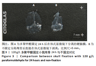
2.1 实验动物数量分析 实验选用ICR小鼠30只,全部进入结果分析。 2.2 颈深淋巴结定位 把5 μL 10%埃文斯蓝溶液以1 μL/min注射入小鼠小脑延髓池,15 min后小鼠颈深淋巴出现蓝染,见图2。 2.3 120 g/L多聚甲醛固定颅骨24 h与不固定对比 在120 g/L多聚甲醛固定颅骨24 h后,在体式显微镜下按前"

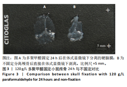
面所述的方法将硬脑膜与颅骨分离,将脑膜贴在载玻片上,大体可见硬脑膜舒展,形状完整,韧性较好;将颅骨取下后直接在体式显微镜下将硬脑膜剥离,此时硬脑膜较软,易破损,且易收缩,不利于后期进一步使用,见图3。"

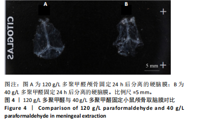
2.4 120 g/L多聚甲醛与40 g/L多聚甲醛取脑膜对比 将包含硬脑膜的颅骨分别在120 g/L的多聚甲醛与40 g/L的多聚甲醛中浸泡24 h,在体式显微镜下剥离脑膜,可见在120 g/L多聚甲醛浸泡后的脑膜形态较为完整,淋巴管结构明显;40 g/L的多聚甲醛浸泡下的硬脑膜在剥离时容易出现破损,质地较软,形态较120 g/L的多聚甲醛差,与120 g/L多聚甲醛对比无明显优势,见图4。"
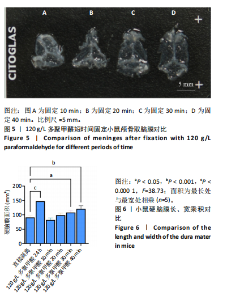
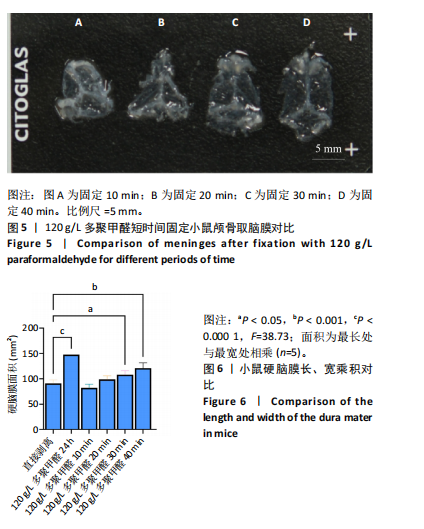
2.5 120 g/L多聚甲醛短时间取脑膜对比 对比了短时间泡120 g/L多聚甲醛取硬脑膜,测试了10,20,30, 40 min,将固定后的硬脑膜贴附于载玻片上,大体可见30-40 min的硬脑膜较为舒展,形态较好,测量多聚甲醛固定的硬脑膜长宽面积与直接剥离的硬脑膜予以对比,120 g/L多聚甲醛固定30 min有差异,40 min差异明显,24 h最佳;见图5,6。"
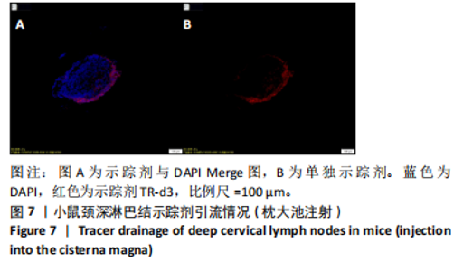
2.6 颈深颈深淋巴结示踪剂引流情况(枕大池注射) 经枕大池注射示踪剂30 min后,取颈深淋巴结,切成冠状冷冻切片,切片内可见明显的示踪剂,示踪剂通过枕大池进入淋巴管,淋巴结位置无误,见图7。"
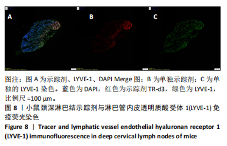
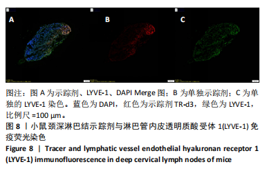
2.7 颈深淋巴结示踪剂与LYVE-1免疫荧光 淋巴结可见明显的示踪剂存在, LYVE-1免疫荧光染色阳性,淋巴结位置无误,颈深淋巴结内淋巴管存在,见图8。"

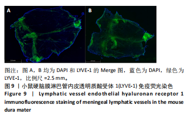
2.8 硬脑膜LYVE-1免疫荧光染色 结果显示,LYVE-1免疫荧光染色阳性,脑膜淋巴管形状明显存在,表明该方法免疫荧光染色结果良好,可作为后续实验使用,见图9。"

2.9 硬脑膜淋巴管示踪剂荧光与LYVE-1免疫荧光染色 脑膜淋巴管尾端可见明显的示踪剂,LYVE-1免疫荧光染色阳性,脑膜淋巴管形状明显存在,结果表明示踪剂可以通过枕大池进入硬脑膜淋巴管,可与免疫荧光染色同时进行,可作为后续实验使用,见图10。"

| [1] 何冠楠. 中国的老龄化趋势下阿尔兹海默症发病情况与预防[J]. 临床医药文献电子杂志,2016,3(40):8083-8084. [2] ILIFF JJ, WANG M, LIAO Y, et al. A Paravascular Pathway Facilitates CSF Flow Through the Brain Parenchyma and the Clearance of Interstitial Solutes, Including Amyloid β.Sci Transl Med. 2012;4(147):147ra111. [3] LOUVEAU A, SMIRNOV I, KEYES TJ, et al. Structural and functional features of central nervous system lymphatic vessels. Nature. 2015; 523(7560):337-341. [4] BOLAND B, YU WH, CORTI O, et al. Promoting the clearance of neurotoxic proteins in neurodegenerative disorders of ageing. Nat Rev Drug Discov. 2018;17(9):660-688. [5] 陈君媚, 凌云, 孙松娴, 等.中枢神经系统淋巴引流对β淀粉样蛋白清除的影响研究[J].中华神经医学杂志,2019,18(3):302-305. [6] TIAN Y, ZHAO M, CHEN Y, et al. The Underlying Role of the Glymphatic System and Meningeal Lymphatic Vessels in Cerebral Small Vessel Disease. Biomolecules. 2022;12(6):748. [7] MOGENSEN FL, DELLE C, NEDERGAARD M. The Glymphatic System (En)during Inflammation. Int J Mol Sci. 2021;22(14):7491. [8] AHN JH, CHO H, KIM JH, et al. Meningeal lymphatic vessels at the skull base drain cerebrospinal fluid. Nature. 2019;572(7767):62-66. [9] NEDERGAARD M, GOLDMAN SA. Glymphatic failure as a final common pathway to dementia. Science. 2020;370(6512):50-56. [10] CHOI D, PARK E, CHOI J, et al. Piezo1 regulates meningeal lymphatic vessel drainage and alleviates excessive CSF accumulation. Nat Neurosci. 2024;27(5):913-926. [11] DU T, RAGHUNANDAN A, MESTRE H, et al. Restoration of cervical lymphatic vessel function in aging rescues cerebrospinal fluid drainage. Nat Aging. 2024 Aug 15. doi: 10.1038/s43587-024-00691-3. [12] VAN DEN BROECK W, DERORE A, SIMOENS P. Anatomy and nomenclature of murine lymph nodes: Descriptive study and nomenclatory standardization in BALB/cAnNCrl mice.J Immunol Methods. 2006;312(1-2):12-19. [13] LI JX, HAO YW, WANG SF, et al. Yuanzhi powder facilitated Aβ clearance in APP/PS1 mice: Target to the drainage of glymphatic system and meningeal lymphatic vessels.J Ethnopharmacol. 2024;319(Pt 1): 117195. [14] WU Y, ZHANG TT, LI XQ, et al. Borneol-driven meningeal lymphatic drainage clears amyloid-β peptide to attenuate Alzheimer-like phenotype in mice. Theranostics. 2023;13(1):106-124. [15] JACOB L, DE BRITO NETO J, LENCK S, et al. Conserved meningeal lymphatic drainage circuits in mice and humans. J Exp Med. 2022;219(8):e20220035. [16] BRADSTREET JJ, RUGGIERO M, PACINI S. Commentary: Structural and functional features of central nervous system lymphatic vessels. Front Neurosci. 2015;9:485. [17] CHACHAJ A, GĄSIOROWSKI K, SZUBA A, et al. The Lymphatic System In The Brain Clearance Mechanisms - New Therapeutic Perspectives For Alzheimer’s Disease. Curr Neuropharmacol. 2023;21(2):380-391. [18] LI X, QI L, YANG D, et al. Meningeal lymphatic vessels mediate neurotropic viral drainage from the central nervous system. Nat Neurosci. 2022;25(5):577-587. [19] WANG M, YAN C, LI X, et al. Non-invasive modulation of meningeal lymphatics ameliorates ageing and Alzheimer’s disease-associated pathology and cognition in mice. Nat Commun. 2024;15(1):1453. [20] PATEL TK, HABIMANA-GRIFFIN L, GAO X, et al. Dural lymphatics regulate clearance of extracellular tau from the CNS. Mol Neurodegener. 2019;14(1):11. [21] DING XB, WANG XX, XIA DH, et al. Impaired meningeal lymphatic drainage in patients with idiopathic Parkinson’s disease. Nat Med. 2021;27(3):411-418. [22] YANKOVA G, BOGOMYAKOVA O, TULUPOV A. The glymphatic system and meningeal lymphatics of the brain: new understanding of brain clearance. Rev Neurosci. 2021;32(7):693-705. [23] 陈飞宇, 李庆新. 脑膜淋巴管功能及相关疾病的研究进展[J]. 西北国防医学杂志,2020,41(4):253-259. [24] HABLITZ LM, NEDERGAARD M. The Glymphatic System: A Novel Component of Fundamental Neurobiology. J Neurosci. 2021;41(37): 7698-7711. [25] LI G, CAO Y, TANG X, et al. The meningeal lymphatic vessels and the glymphatic system: Potential therapeutic targets in neurological disorders. J Cereb Blood Flow Metab. 2022;42(8):1364-1382. [26] 刘全磊. 增强MLVs功能缓解小鼠SAH后早期脑损伤的研究[D].天津:天津医科大学,2021. [27] DA MESQUITA S, LOUVEAU A, VACCARI A, et al. Functional aspects of meningeal lymphatics in ageing and Alzheimer’s disease. Nature. 2018;560(7717):185-191. [28] WEN YR, YANG JH, WANG X, et al.Induced dural lymphangiogenesis facilities soluble amyloid-beta clearance from brain in a transgenic mouse model of Alzheimer’s disease. Neural Regen Res. 2018;13(4):709-716. [29] ALDERFER L, RUSSO E, ARCHILLA A, et al. Matrix stiffness primes lymphatic tube formation directed by vascular endothelial growth factor‐C. FASEB J. 2021; 35(5):e21498. [30] MA W, GIL H J, LIU X, et al. Mitochondrial respiration controls the Prox1-Vegfr3 feedback loop during lymphatic endothelial cell fate specification and maintenance. Sci Adv. 2021;7(18):eabe7359. [31] 莫澜, 孟波, 袁桧, 等. 脑膜淋巴管在中枢神经系统疾病中作用和机制的研究进展[J]. 中国病理生理杂志,2022,38(1):162-166. [32] 田丽娜. FOXC2和LYVE-1在人胚淋巴管发生发育中的作用[D]. 长春:吉林大学,2007. [33] RINGSTAD G, EIDE PK. Cerebrospinal fluid tracer efflux to parasagittal dura in humans. Nat Commun. 2020;11(1):354. [34] 陈君媚. 苓桂术甘汤改善脑淋巴炎症保护神经元损伤作用机制研究[D].南京:南京中医药大学,2020. [35] 陈君媚. 当归芍药散改善脑膜淋巴管引流脑淋巴功能治疗sAD神经炎症小鼠认知障碍机制研究[D].广州:广州中医药大学,2023. [36] 孙睿, 高波, 郭传瑸. 裸小鼠淋巴结的解剖和组织学特点[J].北京大学学报(医学版),2017,49(5):893-898. [37] 师长宏,冯秀亮,张海. 基础动物实验技术与方法[M]. 西安:第四军医大学出版社,2011. [38] 谢雯, 董洪权, 侍崇龙, 等. 脑膜淋巴管转运功能障碍加重脂多糖诱导的小鼠中枢炎症[J]. 南京医科大学学报(自然科学版),2023,43(7):927-933. [39] LI D, LIN H, SUN S, et al. Photostimulation of lymphatic clearance of β-amyloid from mouse brain: a new strategy for the therapy of Alzheimer’s disease.Front Optoelectron. 2023;16(1):45. [40] 王临梅, 赵恬田, 邵水金. 一种小鼠硬脑膜淋巴管的分离与标记方法[C].昆明:中国解剖学会2019年年会论文文摘汇编会议论文集,2019. |
| [1] | Xie Liugang, Cui Shuke, Guo Nannan, Li Aoyu, Zhang Jingrui. Research hotspots and frontiers of stem cells for Alzheimer’s disease [J]. Chinese Journal of Tissue Engineering Research, 2025, 29(7): 1475-1485. |
| [2] |
Li Tian, Ren Yuhua, Gao Yanping, Su Qiang.
Mechanism of agomelatine alleviating anxiety- and depression-like behaviors in APP/PS1 transgenic mice #br#
#br#
[J]. Chinese Journal of Tissue Engineering Research, 2025, 29(6): 1176-1182.
|
| [3] | Chen Yilin, Jiang Xiaobo, Qu Honglin, Liu Ruilian. General pattern of GSK3/Nrf2-regulated biological rhythms in organismal aging [J]. Chinese Journal of Tissue Engineering Research, 2025, 29(6): 1257-1264. |
| [4] | Lu Xiuli, Xu Huazhen, Chen Yuxing, Yao Nan, Hu Zixuan, Huang Dane. Mechanism of Jiangu Formula in treating osteoporosis based on osteoclast-osteoblast coupling [J]. Chinese Journal of Tissue Engineering Research, 2025, 29(32): 6828-6835. |
| [5] | Yan Laijun, Ge Haiya, Wang Zhengming, Yang Zongrui, Niu Lifeng, Zhan Hongsheng. Mechanism by which Tongdu Huoxue Decoction inhibits macrophage inflammation to delay intervertebral disc degeneration in rats [J]. Chinese Journal of Tissue Engineering Research, 2025, 29(32): 6851-6857. |
| [6] | Nigeayi · Aihemaiti, Yilidanna · Dilixiati, An Wei, Maimaitituxun · Tuerdi. Expression of mitochondrial creatine kinase 2 in a rat model of temporomandibular joint osteoarthritis and its role in inflammation progression [J]. Chinese Journal of Tissue Engineering Research, 2025, 29(32): 6877-6884. |
| [7] | Wang Ziheng, Wu Shuang. Oxidative stress-related genes and molecular mechanisms after spinal cord injury: data analysis and verification based on GEO database [J]. Chinese Journal of Tissue Engineering Research, 2025, 29(32): 6893-6904. |
| [8] | Su Qin, Jia Siwei, Guo Minfang, Meng Tao, Li Yanbing, Mu Bingtao, Song Lijuan, Ma Cungen, Yu Jiezhong. Lycium barbarum polysaccharide intervenes in SH-SY5Y cell injury induced by beta-amyloid protein 1-42: protective effect of mitochondrial autophagy [J]. Chinese Journal of Tissue Engineering Research, 2025, 29(31): 6688-6696. |
| [9] | Yu Qinghe, Cai Ziming, Wu Jintao, Ma Pengfei, Zhang Xin, Zhou Longqian, Wang Yakun, Lin Xiaoqin, Lin Wenping. Vanillic acid inhibits inflammatory response and extracellular matrix degradation of endplate chondrocytes [J]. Chinese Journal of Tissue Engineering Research, 2025, 29(30): 6391-9397. |
| [10] | Fan Jiaxin, Jia Xiang, Xu Tianjie, Liu Kainan, Guo Xiaoling, Zhang Hui, Wang Qian . Metformin inhibits ferroptosis and improves cartilage damage in osteoarthritis model rats [J]. Chinese Journal of Tissue Engineering Research, 2025, 29(30): 6398-6408. |
| [11] | Zhou Ying, Tian Yong, Zhong Zhimei, Gu Yongxiang, Fang Hao. Inhibition of tumor necrosis factor receptor associated factor 6 regulates mTORC1/ULK1 signaling and promotes autophagy to improve myocardial injury in sepsis mice [J]. Chinese Journal of Tissue Engineering Research, 2025, 29(30): 6434-6440. |
| [12] | Wang Wanchun, , Yi Jun, Yan Zhangren, Yang Yue, Dong Degang, Li Yumei. 717 Jiedu Decoction remodels homeostasis of extracellular matrix and promotes repair of local injured tissues in rats after Agkistrodon halys bite [J]. Chinese Journal of Tissue Engineering Research, 2025, 29(30): 6457-6465. |
| [13] | Zhang Xin, Guo Baojuan, Xu Huixin, Shen Yuzhen, Yang Xiaofan, Yang Xufang, Chen Pei. Protective effects and mechanisms of 3-N-butylphthalide in Parkinson’s disease cell models [J]. Chinese Journal of Tissue Engineering Research, 2025, 29(30): 6466-6473. |
| [14] | Zhang Songjiang, Li Longyang, Zhou Chunguang, Gao Jianfeng. Central anti-inflammatory effect and mechanism of tea polyphenols in exercise fatigue model mice [J]. Chinese Journal of Tissue Engineering Research, 2025, 29(30): 6474-6481. |
| [15] | Hu Shujuan, Liu Dang, Ding Yiting, Liu Xuan, Xia Ruohan, Wang Xianwang. Ameliorative effect of walnut oil and peanut oil on atherosclerosis [J]. Chinese Journal of Tissue Engineering Research, 2025, 29(30): 6482-6488. |
| Viewed | ||||||
|
Full text |
|
|||||
|
Abstract |
|
|||||