Chinese Journal of Tissue Engineering Research ›› 2025, Vol. 29 ›› Issue (28): 6003-6011.doi: 10.12307/2025.460
Previous Articles Next Articles
Design of customized Gyroid condylar prosthesis and finite element analysis of articular disc
Jiang Tingting1, 2, Liu Danyu1, 2, Jiang Zhixiu1, 2, Ji Yuchen1, 2, Cao Yilin1, 2, Su Yucheng2, 3, 4, Wang Xinyu1, 2
- 1Jiamusi University, Jiamusi 154003, Heilongjiang Province, China; 2Key Laboratory of Oral Biomaterials and Clinical Applications of Heilongjiang Province, Stomatology Engineering Experimental Center of Jiamusi University, Stomatology College of Jiamusi University, Jiamusi 154007, Heilongjiang Province, China; 3Dental Implant Center, Peking Union Medica College Hospital, Chinese Academy of Medical Sciences, Beijing 100032, China; 4Beijing Implant Training College (BITC), Beijing Citident Stomatology Hospital, Beijing 100032, China
-
Received:2024-04-27Accepted:2024-06-26Online:2025-10-08Published:2024-12-07 -
Contact:Wang Xinyu, Associate chief physician, Master’s supervisor, Jiamusi University, Jiamusi 154003, Heilongjiang Province, China; Key Laboratory of Oral Biomaterials and Clinical Applications of Heilongjiang Province, Stomatology Engineering Experimental Center of Jiamusi University, Stomatology College of Jiamusi University, Jiamusi 154007, Heilongjiang Province, China -
About author:Jiang Tingting, Master candidate, Practicing physician, Jiamusi University, Jiamusi 154003, Heilongjiang Province, China; Key Laboratory of Oral Biomaterials and Clinical Applications of Heilongjiang Province, Stomatology Engineering Experimental Center of Jiamusi University, Stomatology College of Jiamusi University, Jiamusi 154007, Heilongjiang Province, China -
Supported by:Heilongjiang Natural Science Foundation Project, No. LH2022H089 (to WXY); Heilongjiang Provincial Health Commission Research Project, No. 2020-314 (to WXY)
CLC Number:
Cite this article
Jiang Tingting, Liu Danyu, Jiang Zhixiu, Ji Yuchen, Cao Yilin, Su Yucheng, Wang Xinyu. Design of customized Gyroid condylar prosthesis and finite element analysis of articular disc[J]. Chinese Journal of Tissue Engineering Research, 2025, 29(28): 6003-6011.
share this article
Add to citation manager EndNote|Reference Manager|ProCite|BibTeX|RefWorks
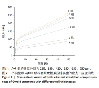
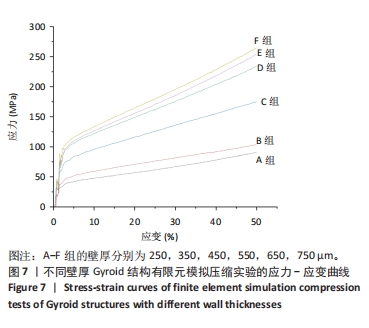
2.1 不同壁厚Gyroid结构力学性能分析 2.1.1 有限元模拟压缩实验结果 对不同壁厚Gyroid结构进行有限元模拟力学压缩实验,对实验数据进行处理后绘制应力-应变曲线(图7),通过拟合线性阶段的数据求出斜率,即为不同壁厚Gyroid结构对应的弹性模量,A-F组Gyroid结构弹性模量分别为1.47,1.95,2.80,3.69,3.89,4.39 GPa,其中B-E组Gyroid结构弹性模量在下颌骨松质骨弹性模量范围内。由于后续实验需细分组别且D-F组(孔径为800-1 000 μm)在成骨范围内,故筛选出D-F组进行后续实验。"
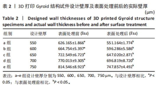
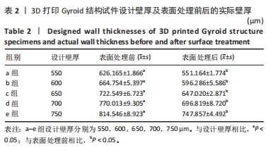
2.1.2 万能试验机力学压缩实验结果 壁厚分析结果表明,3D 打印Gyroid 结构试件的壁厚与设计壁厚相差较大,3D 打印Gyroid 结构试件的壁厚大于设计壁厚(P < 0.05),经大颗粒喷砂酸蚀表面处理后的试件壁厚可降低至与设计壁厚相近(P > 0.05),见表2,图8。"

"

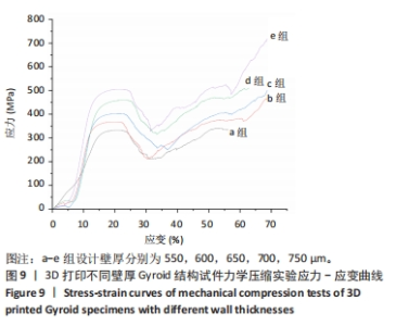
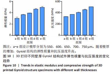
在室温下对不同壁厚Gyroid结构力学标准件进行压缩实验,对实验数据进行处理后绘制应力-应变曲线(图9),分别求出不同壁厚Gyroid结构的弹性模量和抗压强度,见表3。a-c组Gyroid结构弹性模量均在下颌骨松质骨弹性模量范围内,根据上述条件最终选取c组(壁厚为650 μm,孔径为900 μm)作为最终实验组。如图10所示,压缩实验结果表明,壁厚越大,Gyroid结构的弹性模量和抗压强度越大。"
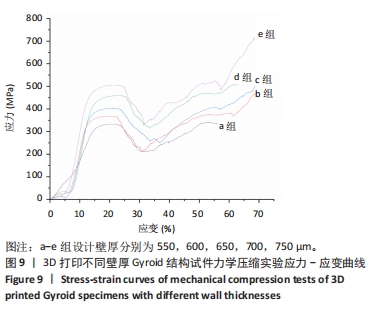

"
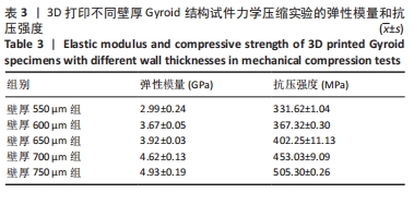

"
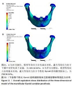
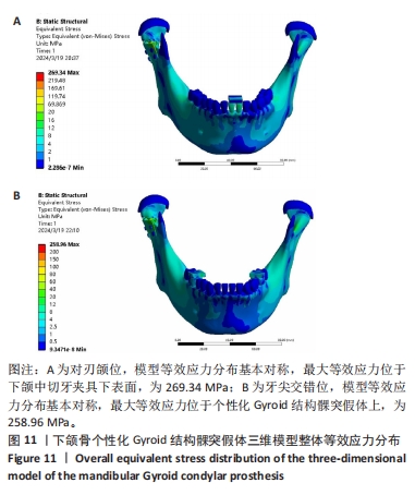
2.2 下颌骨个性化Gyroid结构髁突假体三维模型有限元分析 2.2.1 两种颌位下模型整体的等效应力分布 如图11所示,两种颌位在自然咬合状态下,模型等效应力分布基本对称且最大等效应力相近;对刃颌位时,最大等效应力位于下颌中切牙夹具下表面,为269.34 MPa;牙尖交错位时,最大等效应力位于个性化Gyroid结构髁突假体上,为258.96 MPa。"

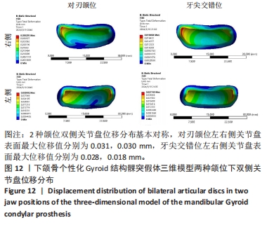
2.2.2 两种颌位下的关节盘位移及等效应力分布 如图12所示,在自然咬合状态下两种颌位双侧关节盘位移分布基本对称,对刃颌位左右侧关节盘表面最大位移值分别为0.031,0.030 mm,牙尖交错位左右侧关节盘表面最大位移值分别为0.028,0.018 mm。"

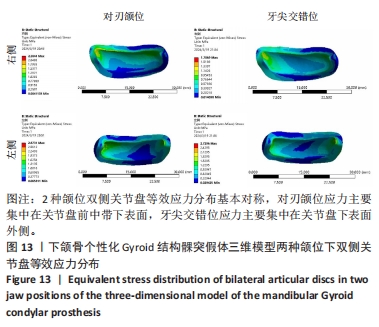
如图13所示,在自然咬合状态下两种颌位双侧关节盘等效应力分布基本对称,对刃颌位应力主要集中在关节盘前中带下表面,牙尖交错位应力主要集中在关节盘下表面外侧。两种颌位双侧关节盘表面最大等效应力相近,对刃颌位左右侧关节盘最大等效应力分别为2.87,2.30 MPa,牙尖交错位左右侧关节盘最大等效应力分别为2.73,1.71 MPa,满足植入条件。"
| [1] DEL CASTILLO PARDO DE VERA JL, CEBRIÁN CARRETERO JL, ARAGÓN NIÑO Í, et al. Virtual Surgical Planning for Temporomandibular Joint Reconstruction with Stock TMJ Prostheses: Pilot Study. Medicina (Kaunas). 2024;60(2):339. [2] AMARISTA FJ, PEREZ DE. Concomitant Temporomandibular Joint Replacement and Orthognathic Surgery. Diagnostics (Basel). 2023; 13(15):2486. [3] MERCURI LG. Alloplastic temporomandibular joint replacement - past, present, and future: “Learn from the past, prepare for the future, live in the present.” Thomas S. Monson. Br J Oral Maxillofac Surg. 2024;62(1):91-96. [4] BHARGAVA D. Hybrid total alloplastic temporomandibular joint replacement prosthesis: a pilot study to evaluate feasibility, functional performance and impact on post-operative quality of life. Oral Maxillofac Surg. 2024;28(2):767-777. [5] 黄相道,邵占英,王发生,等.人工髁突假体在颞下颌关节重建中的临床应用[J].中国修复重建外科杂志,2012,26(1):6-9. [6] 吴宝磊,张圃.异质性人工髁突修复重建颞下颌关节的相关临床决策因素[J].医学与哲学(B),2013,34(3):35-38. [7] PINTO L, LIMA BC, COUTINHO MA, et al. Temporomandibular joint patient specific implant as treatment for hemifacial microsomia. Natl J Maxillofac Surg. 2023;14(3):515-518. [8] GOKER F, RUSSILLO A, BAJ A, et al. Custom made/patient specific alloplastic total temporomandibular joint replacement in immature patient: a case report and short review of literature. Eur Rev Med Pharmacol Sci. 2022;26(3 Suppl):26-34. [9] MERCURI LG, NETO MQ, POURZAL R. Alloplastic temporomandibular joint replacement: present status and future perspectives of the elements of embodiment. Int J Oral Maxillofac Surg. 2022;51(12):1573-1578. [10] KIM D, ELIAS KL. Elastic, viscoelastic, and fracture properties of bone tissue measured by nanoindentation. Handbook of Nanomaterials Properties. 2014:1321-1341. [11] YANG X, WU L, LI C, et al. Synergistic Amelioration of Osseointegration and Osteoimmunomodulation with a Microarc Oxidation-Treated Three-Dimensionally Printed Ti-24Nb-4Zr-8Sn Scaffold via Surface Activity and Low Elastic Modulus. ACS Appl Mater Interfaces. 2024; 16(3):3171-3186. [12] 邹庆化.金属材料强度与硬度之间的相关关系[J].金属热处理, 1993(1):53-55. [13] ARCHARD JF. Surface topography and tribology. Tribology. 1974;7(5): 213-220. [14] CHMIELEWSKA A, DEAN D. The role of stiffness-matching in avoiding stress shielding-induced bone loss and stress concentration-induced skeletal reconstruction device failure. Acta Biomater. 2024;173:51-65. [15] SEEHANAM S, KHRUEADUANGKHAM S, SINTHUVANICH C, et al. Evaluating the effect of pore size for 3d-printed bone scaffolds. Heliyon. 2024;10(4):e26005. [16] MAEVSKAIA E, GUERRERO J, GHAYOR C, et al. Triply Periodic Minimal Surface-Based Scaffolds for Bone Tissue Engineering: A Mechanical, In Vitro and In Vivo Study. Tissue Eng Part A. 2023;29(19-20):507-517. [17] HAYASHI K, KISHIDA R, TSUCHIYA A, et al. Superiority of Triply Periodic Minimal Surface Gyroid Structure to Strut-Based Grid Structure in Both Strength and Bone Regeneration. ACS Appl Mater Interfaces. 2023;15(29):34570-34577. [18] GUO W, YANG Y, LIU C, et al. 3D printed TPMS structural PLA/GO scaffold: Process parameter optimization, porous structure, mechanical and biological properties. J Mech Behav Biomed Mater. 2023;142:105848. [19] PUGLIESE R, GRAZIOSI S. Biomimetic scaffolds using triply periodic minimal surface-based porous structures for biomedical applications. SLAS Technol. 2023;28(3):165-182. [20] TENG F, SUN Y, GUO S, et al. Topological and Mechanical Properties of Different Lattice Structures Based on Additive Manufacturing. Micromachines (Basel). 2022;13(7):1017. [21] ZHENG X, DUAN F, SONG Z, et al. A TMPS-designed personalized mandibular scaffolds with optimized SLA parameters and mechanical properties. Front Mater. 2022;9:966031. [22] 宋颐函,郑晓晓,孙子惠,等.均一与梯度孔隙率Gyroid结构力学性能研究及多孔种植体增材制造[J]. 中国口腔种植学杂志, 2023,28(3):204-209. [23] KARAGEORGIOU V, KAPLAN D. Porosity of 3D biomaterial scaffolds and osteogenesis. Biomaterials. 2005;26(27):5474-5491. [24] HARA D, NAKASHIMA Y, SATO T, et al. Bone bonding strength of diamond-structured porous titanium-alloy implants manufactured using the electron beam-melting technique. Mater Sci Eng C Mater Biol Appl. 2016;59:1047-1052. [25] ZHOU C, DENG C, CHEN X, et al. Mechanical and biological properties of the micro-/nano-grain functionally graded hydroxyapatite bioceramics for bone tissue engineering. J Mech Behav Biomed Mater. 2015;48: 1-11. [26] ZHOU C, YE X, FAN Y, et al. Biomimetic fabrication of a three-level hierarchical calcium phosphate/collagen/hydroxyapatite scaffold for bone tissue engineering. Biofabrication. 2014;6(3):35013. [27] NIEZEN ET, VAN MINNEN B, BOS R, et al. Temporomandibular joint prosthesis as treatment option for mandibular condyle fractures: a systematic review and meta-analysis. Int J Oral Maxillofac Surg. 2023;52(1):88-97. [28] 吕晶晶,米丛波.牙周膜的生物力学性能[J].国际口腔医学杂志, 2014,41(3):362-364. [29] DING X, LIAO S, ZHU X, et al. Effect of orthotropic material on finite element modeling of completely dentate mandible. Mater Design. 2015;84:144-153. [30] CHENG KJ, LIU YF, WANG JH, et al. 3D-printed porous condylar prosthesis for temporomandibular joint replacement: Design and biomechanical analysis. Technol Health Care. 2022;30(4):1017-1030. [31] 钱茹.种植体的应力应变分析及优化设计[J].科技信息,2011(24): 105-106. [32] 谢艳华.下颌运动及含种植体口腔生物力学分析[D].南京:东南大学,2017. [33] 戴雯婕.含种植体的下颌咬合生物力学分析[D].南京:东南大学, 2019. [34] BEEK M, KOOLSTRA JH, VAN RUIJVEN LJ, et al. Three-dimensional finite element analysis of the human temporomandibular joint disc. J Biomech. 2000;33(3):307-316. [35] FORSTER H, FISHER J. The influence of loading time and lubricant on the friction of articular cartilage. Proc Inst Mech Eng H. 1996;210(2): 109-119. [36] MABUCHI K, OBARA T, IKEGAMI K, et al. Molecular weight independence of the effect of additive hyaluronic acid on the lubricating characteristics in synovial joints with experimental deterioration. Clin Biomech (Bristol, Avon). 1999;14(5):352-356. [37] TANAKA E, KAWAI N, TANAKA M, et al. The frictional coefficient of the temporomandibular joint and its dependency on the magnitude and duration of joint loading. J Dent Res. 2004;83(5):404-407. [38] VAN EIJDEN TM, KORFAGE JA, BRUGMAN P. Architecture of the human jaw-closing and jaw-opening muscles. Anat Rec. 1997;248(3):464-474. [39] KORIOTH TW, HANNAM AG. Deformation of the human mandible during simulated tooth clenching. J Dent Res. 1994;73(1):56-66. [40] 赵珊笛,周昊.颞下颌关节重建的研究进展[J].北京口腔医学,2022, 30(1):65-68. [41] BAGHERI ZS, MELANCON D, LIU L, et al. Compensation strategy to reduce geometry and mechanics mismatches in porous biomaterials built with Selective Laser Melting. J Mech Behav Biomed Mater. 2017; 70:17-27. [42] ARABNEJAD S, BURNETT JR, PURA JA, et al. High-strength porous biomaterials for bone replacement: A strategy to assess the interplay between cell morphology, mechanical properties, bone ingrowth and manufacturing constraints. Acta Biomater. 2016;30:345-356. [43] PEI X, WU L, ZHOU C, et al. 3D printed titanium scaffolds with homogeneous diamond-like structures mimicking that of the osteocyte microenvironment and its bone regeneration study. Biofabrication. 2020;13(1).doi: 10.1088/1758-5090/abc060. [44] 刘伟洛.增材制造三周期极小曲面结构的力学性能研究[D].广州:广州大学,2021. [45] RAN Q, YANG W, HU Y, et al. Osteogenesis of 3D printed porous Ti6Al4V implants with different pore sizes. J Mech Behav Biomed Mater. 2018; 84:1-11. [46] XIE G, QIN F, ZHU S. Recent progress in Ti-based metallic glasses for application as biomaterials. Mater Trans. 2013;54(8):1314-1323. [47] ZOU L, ZHONG Y, XIONG Y, et al. A Novel Design of Temporomandibular Joint Prosthesis for Lateral Pterygoid Muscle Attachment: A Preliminary Study. Front Bioeng Biotechnol. 2020;8:630983. [48] ZHONG YQ, SUN Q, HE DM, et al. Study on the lateral pterygoid muscle status after artificial temporomandibular joint replacement. Int J Oral Maxillofac Surg. 2021;50(11):1496-1501. [49] 陈伟,房睿.微型钛板在内固定治疗下颌骨粉碎性骨折中的应用:21例临床分析[J].上海口腔医学,2020,29(3):333-336. [50] SHU J, MA H, JIA L, et al. Biomechanical behaviour of temporomandibular joints during opening and closing of the mouth: A 3D finite element analysis. Int J Numer Method Biomed Eng. 2020; 36(8):e3373. [51] LI A, SHAO B, CHONG D, et al. The influence of maxillary incisor angles on the stress distributions of temporomandibular joints under incisal biting. Int J Numer Method Biomed Eng. 2023;39(6):e3702. |
| [1] | Lai Pengyu, Liang Ran, Shen Shan. Tissue engineering technology for repairing temporomandibular joint: problems and challenges [J]. Chinese Journal of Tissue Engineering Research, 2025, 29(在线): 1-9. |
| [2] | Xu Hao, Ding Lu, Li Xiao. Investigating the effect of the mechanical wear on abutment screw in Morse taper connection implant implant system by using finite element analysis [J]. Chinese Journal of Tissue Engineering Research, 2025, 29(在线): 1-9. |
| [3] | Li Liangkui, Huang Yongcan, Wang Peng, Yu Binsheng. Effect of anterior controllable anteriodisplacement and fusion on vertebrae-ossification of posterior longitudinal ligament complex and implants: a finite element analysis [J]. Chinese Journal of Tissue Engineering Research, 2025, 29(9): 1761-1767. |
| [4] | Xu Biao, Lu Tan, Jiang Yaqiong, Yin Yujiao. Xu Biao, Lu Tan, Jiang Yaqiong, Yin Yujiao [J]. Chinese Journal of Tissue Engineering Research, 2025, 29(9): 1768-1774. |
| [5] | Zhou Jinhai, Li Jiangwei, Wang Xuquan, Zhuang Ying, Zhao Ying, Yang Yuyong, Wang Jiajia, Yang Yang, Zhou Shilian. Three-dimensional finite element analysis of anterior femoral notching during total knee arthroplasty at different bone strengths [J]. Chinese Journal of Tissue Engineering Research, 2025, 29(9): 1775-1782. |
| [6] | Chen Xi, Tang Tao, Chen Tongbing, Li Qing, Zhang Wen. Mechanical stability of intertrochanteric fracture of femur with different internal fixation systems [J]. Chinese Journal of Tissue Engineering Research, 2025, 29(9): 1783-1788. |
| [7] | Li Shuai, Liu Hua, Shang Yonghui, Liu Yicong, Zhao Qihang, Liu Wen. Stress distribution on the maxilla when wearing the Twin-block appliance for Class II malocclusion [J]. Chinese Journal of Tissue Engineering Research, 2025, 29(5): 881-887. |
| [8] | Zhou Zonghao, Luo Siyang, Chen Jiawen, Chen Guangneng, Feng Hongchao. Finite element analysis of bioabsorbable plates versus miniature titanium plates in mandibular fracture fixation in different bone qualities [J]. Chinese Journal of Tissue Engineering Research, 2025, 29(4): 818-826. |
| [9] | Chen Yilong, Zhang Xu, Li Hong. Mechanical analysis of fiber post combined with different crown restorations for endodontically treated non-carious cervical lesions [J]. Chinese Journal of Tissue Engineering Research, 2025, 29(4): 866-871. |
| [10] | Zhuang Yan, Wang Xinyu, Cao Yilin, Ding Yuanxin, Wang Jiaqi, Yu Miao, Luan Chunyang, Ding Yuansheng. Three-dimensional finite element analysis of personalized orthodontic devices for 3D printed maxillary single-rooted rotated tooth [J]. Chinese Journal of Tissue Engineering Research, 2025, 29(30): 6409-6415. |
| [11] | Akliya·Anwar, Nafisa·Gupur, Baibugafu·Yelisi, Zilalai·Gulaiti, Guzalnur·Emrayim, Nijat·Tursun. Dynamic stress analysis of maxillary sinus lifting without bone grafting and with immediate loading after bone grafting [J]. Chinese Journal of Tissue Engineering Research, 2025, 29(30): 6416-6425. |
| [12] | Su Dejun, Dong Wanpeng, Dong Yuefu, Zhang Jichao, Zhang Zhen. Design of asymmetric prosthesis and mechanical analysis of total knee arthroplasty [J]. Chinese Journal of Tissue Engineering Research, 2025, 29(3): 510-516. |
| [13] | Wang X, Wang Hm, Chen Sh, Feng Tx, Bu Hm, Zhu Lg, Chen Dd, Wei X. Stress and morphological characteristics of intervertebral foramen of cervical rotation-traction manipulation for treating cervical spondylotic radiculopathy: a three-dimensional finite element analysis [J]. Chinese Journal of Tissue Engineering Research, 2025, 29(3): 441-447. |
| [14] | Zhao Yuxin, Liang Liang, Jin Feng, Xu Yangyang, Kang Zhijie, Fang Yuan, He Yujie, Wang Xing, Wang Haiyan, Li Xiaohe. Establishment and stress analysis of a finite element model for adolescent cervical disc herniation [J]. Chinese Journal of Tissue Engineering Research, 2025, 29(3): 448-454. |
| [15] | Li Zhenggang, Shang Xuehong, Wu Zhang, Li Hong, Sun Chaojun, Chen Huadong, Sun Zhe, Yang Yi. Finite element analysis of three internal fixation modalities for treatment of Pauwels type III femoral neck fractures under different loading conditions [J]. Chinese Journal of Tissue Engineering Research, 2025, 29(3): 455-463. |
| Viewed | ||||||
|
Full text |
|
|||||
|
Abstract |
|
|||||