Chinese Journal of Tissue Engineering Research ›› 2025, Vol. 29 ›› Issue (7): 1321-1327.doi: 10.12307/2024.741
Mechanism by which nobiletin inhibits inflammatory response of BV2 microglia
Chi Wenxin1, 2, Zhang Cunxin2, Gao Kai3, Lyu Chaoliang2, Zhang Kefeng2
- 1School of Clinical Medicine, Jining Medical University, Jining 272067, Shandong Province, China; 2Department of Spine Surgery, 3Department of Joint Surgery, Jining No. 1 People’s Hospital, Jining 272000, Shandong Province, China
-
Received:2023-08-21Accepted:2023-12-14Online:2025-03-08Published:2024-06-27 -
Contact:Zhang Kefeng, Master, Associate chief physician, Department of Spine Surgery, Jining No. 1 People’s Hospital, Jining 272000, Shandong Province, China; Lyu Chaoliang, MD, Chief physician, Department of Spine Surgery, Jining No. 1 People’s Hospital, Jining 272000, Shandong Province, China -
About author:Chi Wenxin, Master candidate, School of Clinical Medicine, Jining Medical University, Jining 272067, Shandong Province, China; Department of Spine Surgery, Jining No. 1 People’s Hospital, Jining 272000, Shandong Province, China -
Supported by:Natural Science Foundation of Shandong Province, No. ZR2021LZY008 (to GK); Jining Key Research & Development Program Project, No. 2021YXNS137 (to GK); Jining Key Research & Development Program Project, No. 2021YXNS001 (to LCL)
CLC Number:
Cite this article
Chi Wenxin, Zhang Cunxin, Gao Kai, Lyu Chaoliang, Zhang Kefeng. Mechanism by which nobiletin inhibits inflammatory response of BV2 microglia[J]. Chinese Journal of Tissue Engineering Research, 2025, 29(7): 1321-1327.
share this article
Add to citation manager EndNote|Reference Manager|ProCite|BibTeX|RefWorks

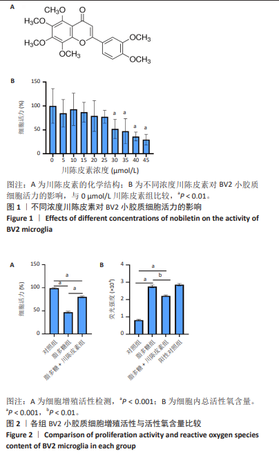
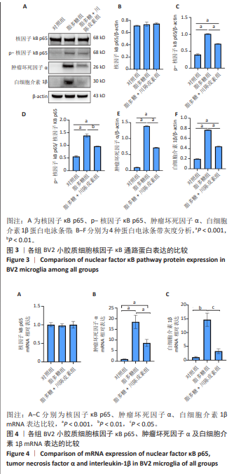
2.1 川陈皮素对BV2细胞存活率的影响 川陈皮素的化学结构见图1A。为了确定川陈皮素对BV2小胶质细胞的细胞毒性作用,首先采用CCK-8实验探究川陈皮素对BV2小胶质细胞的最佳药物浓度,结果显示:在0-25 μmol/L浓度下,BV2小胶质细胞活力无明显变化(P > 0.05),说明此浓度范围内的川陈皮素对BV2小胶质细胞没有明显的毒性作用;在30 μmol/L及以上浓度条件下,BV2小胶质细胞活力明显降低(P < 0.01),见图1B,表明该浓度范围的对川陈皮素BV2小胶质细胞有较为明显的细胞毒性作用。因此,选择20 μmol/L川陈皮素进行后续实验。 2.2 各组BV2小胶质细胞增殖活性检测 CCK-8实验结果显示,对照组、脂多糖组、脂多糖+川陈皮素组细胞活力分别为(99.19±3.73)%,(47.20±5.69)%,(80.26±5.17)%,组间两两比较差异有显著性意义(P < 0.001),见图2A。 2.3 各组BV2小胶质细胞活性氧含量检测 活性氧荧光探针检测结果显示,与对照组比较,脂多糖组细胞内活性氧含量升高(P < 0.001);与脂多糖组比较,脂多糖+川陈皮素组细胞内活性氧含量降低(P < 0.01),见图2B。"

2.4 各组BV2小胶质细胞核因子κB p65、肿瘤坏因子α和白细胞介素1β的表达 Western blot检测显示,与对照组比较,脂多糖组细胞中p-核因子κB p65、肿瘤坏死因子α、白细胞介素1β的蛋白表达水平均升高(P < 0.001);与脂多糖组比较,脂多糖+川陈皮素组细胞中p-核因子κB p65、肿瘤坏死因子α、白细胞介素1β的蛋白表达水平均降低(P < 0.001,P < 0.01);各组核因子κB p65蛋白表达水平比较差异无显著性意义(P > 0.05),见图3。 2.5 各组BV2小胶质细胞核因子κB p65、肿瘤坏因子α和白细胞介素1β的mRNA表达 qRT-PCR检测结果显示,各组细胞中核因子κB p65 mRNA表达比较差异无显著性意义(P > 0.05);与对照组比较,脂多糖组细胞中肿瘤坏死因子α、白细胞介素1β mRNA表达水平均升高(P < 0.001,P < 0.01);与脂多糖组比较,脂多糖+川陈皮素组细胞中肿瘤坏死因子α、白细胞介素1β mRNA表达水平均降低(P < 0.001,P < 0.05),见图4。 "
| [1] ANJUM A, DA’IN YAZID M, FAUZI DAUD M, et al. Spinal Cord Injury: Pathophysiology, Multimolecular Interactions, and Underlying Recovery Mechanisms. Int J Mol Sci. 2020;21(20):7533. [2] STERNER RC, STERNER RM. Immune response following traumatic spinal cord injury: Pathophysiology and therapies. Front Immunol. 2023;13:1084101. [3] KARSY M, HAWRYLUK G. Modern Medical Management of Spinal Cord Injury. Curr Neurol Neurosci Rep. 2019;19(9):65-72. [4] ALIZADEH A, DYCK SM, KARIMI-ABDOLREZAEE S. Traumatic Spinal Cord Injury: An Overview of Pathophysiology, Models and Acute Injury Mechanisms. Front Neurol. 2019;10:282. [5] ORR MB, GENSEL JC. Spinal Cord Injury Scarring and Inflammation: Therapies Targeting Glial and Inflammatory Responses. Neurotherapeutics. 2018;15(3): 541-553. [6] GRAEBER MB, LI W, RODRIGUEZ ML. Role of microglia in CNS inflammation. FEBS Lett. 2011;585(23):3798-3805. [7] YAN A, LIU Z, SONG L, et al. Idebenone Alleviates Neuroinflammation and Modulates Microglial Polarization in LPS-Stimulated BV2 Cells and MPTP-Induced Parkinson’s Disease Mice. Front Cell Neurosci. 2018;12:529. [8] DING Y, CHEN Q. The NF-κB Pathway: a Focus on Inflammatory Responses in Spinal Cord Injury. Mol Neurobiol. 2023;60(9): 5292-5308. [9] LIU HT, ZHANG JJ, XU XX, et al. SARM1 promotes neuroinflammation and inhibits neural regeneration after spinal cord injury through NF-κB signaling. Theranostics. 2021;11(9):4187-4206. [10] FAN L, DONG J, HE X, et al. Bone marrow mesenchymal stem cells-derived exosomes reduce apoptosis and inflammatory response during spinal cord injury by inhibiting the TLR4/MyD88/NF-κB signaling pathway. Hum Exp Toxicol. 2021;40(10):1612-1623. [11] GUAN B, JIANG C. Design and development of 1,3,5-triazine derivatives as protective agent against spinal cord injury in rat via inhibition of NF-ĸB. Bioorg Med Chem Lett. 2021;41:127964. [12] ZHOU MM, ZHANG WY, LI RJ, et al. Anti-inflammatory activity of Khayandirobilide A from Khaya senegalensis via NF-κB, AP-1 and p38 MAPK/Nrf2/HO-1 signaling pathways in lipopolysaccharide-stimulated RAW 264.7 and BV-2 cells. Phytomedicine. 2018;42: 152-163. [13] WU X, SONG M, RAKARIYATHAM K, et al. Anti-inflammatory effects of 4′-demethylnobiletin, a major metabolite of nobiletin. J Funct Foods. 2015;19(Pt A):278-287. [14] MILEYKOVSKAYA E, YOO SH, DOWHAN W, et al. Nobiletin: Targeting the Circadian Network to Promote Bioenergetics and Healthy Aging. Biochemistry (Moscow). 2020;85(12-13):1554-1559. [15] LIN Z, WU D, HUANG L, et al. Nobiletin Inhibits IL-1beta-Induced Inflammation in Chondrocytes via Suppression of NF-kappaB Signaling and Attenuates Osteoarthritis in Mice. Front Pharmacol. 2019;10:570. [16] QI G, MI Y, FAN R, et al. Nobiletin Protects against Systemic Inflammation-Stimulated Memory Impairment via MAPK and NF-κB Signaling Pathways. J Agric Food Chem. 2019; 67(18):5122-5134. [17] NAKAJIMA A, YAMAKUNI T, HARAGUCHI M, et al. Nobiletin, a Citrus Flavonoid That Improves Memory Impairment, Rescues Bulbectomy-Induced Cholinergic Neurodegeneration in Mice. J Pharmacol Sci. 2019;105(1):122-126. [18] DAI XJ, LI N, YU L, et al. Activation of BV2 microglia by lipopolysaccharide triggers an inflammatory reaction in PC12 cell apoptosis through a toll-like receptor 4-dependent pathway. Cell Stress Chaperones. 2015;20(2):321-331. [19] HE GB, HE YB, NI HW, et al. Dexmedetomidine attenuates neuroinflammation and microglia activation in LPS-stimulated BV2 microglia cells through targeting circ-Shank3/miR-140-3p/TLR4 axis. Eur J Histochem. 2023;67(3):3766. [20] XIAO DM, YANG R, GONG L, et al. Plantamajoside inhibits high glucose-induced oxidative stress, inflammation, and extracellular matrix accumulation in rat glomerular mesangial cells through the inactivation of Akt/NF-κB pathway. J Recept Signal Transduct Res. 2021;41(1):45-52. [21] YI LJ, LU Y, YU S, et al. Formononetin inhibits inflammation and promotes gastric mucosal angiogenesis in gastric ulcer rats through regulating NF-κB signaling pathway. J Recept Signal Transduct Res. 2022;42(1):16-22. [22] HE Y, RUGANZU JB, ZHENG Q, et al. Silencing of LRP1 Exacerbates Inflammatory Response Via TLR4/NF-κB/MAPKs Signaling Pathways in APP/PS1 Transgenic Mice. Mol Neurobiol. 2020;57(9):3727-3743. [23] CHEONG MH, LEE SR, YOO HS, et al. Anti-inflammatory effects of Polygala tenuifolia root through inhibition of NF-κB activation in lipopolysaccharide-induced BV2 microglial cells. J Ethnopharmacol. 2011;137(3):1402-1408. [24] HAN Q, YUAN Q, MENG X, et al. 6 Shogaol attenuates LPS induced inflammation in BV2 microglia cells by activatin. Oncotarget. 2017;8(26): 42001-42006. [25] TIMMERMAN R, BURM SM, BAJRAMOVIC JJ. Tissue-specific features of microglial innate immune responses. Neurochem Int. 2021;142: 104924. [26] YANG X, XU S, QIAN Y, et al. Resveratrol regulates microglia M1/M2 polarization via PGC-1α in conditions of neuroinflammatory injury. Brain Behav Immun. 2017;64:162-172. [27] YOU ZJ, YANG ZZ, CAO S, et al. The Novel KLF4/BIG1 Regulates LPS-mediated Neuro-inflammation and Migration in BV2 Cells via PI3K/Akt/NF-kB Signaling Pathway. Neuroscience. 2022;488:102-111. [28] WALSH CM, WYCHOWANIEC JK, BROUGHAM DF, et al. Functional hydrogels as therapeutic tools for spinal cord injury: New perspectives on immunopharmacological interventions. Pharmacol Ther. 2022; 234:108043. [29] JENDELOVA P. Therapeutic Strategies for Spinal Cord Injury. Int J Mol Sci. 2018; 19(10):3200-3202. [30] XU S, WANG J, ZHONG J, et al. CD73 alleviates GSDMD‐mediated microglia pyroptosis in spinal cord injury through PI3K/AKT/Foxo1 signaling. Clin Transl Med. 2021;11(1):e269. [31] ZHENG Y, QI S, WU F, et al. Chinese Herbal Medicine in Treatment of Spinal Cord Injury: A Systematic Review and Meta-Analysis of Randomized Controlled Trials. Am J Chin Med. 2020;48(7):1593-1616. [32] JIANG S, BABA K, OKUNO T, et al. Go-sha-jinki-Gan Alleviates Inflammation in Neurological Disorders via p38-TNF Signaling in the Central Nervous System. Neurotherapeutics. 2020;18(1):460-473. [33] MARK PETRASH J, SHIEH B, AMMAR DA, et al. Diabetes-Independent Retinal Phenotypes in an Aldose Reductase Transgenic Mouse Model. Metabolites. 2021;11(7):450-460. [34] WU A, YANG Z, HUANG Y, et al. Natural phenylethanoid glycosides isolated from Callicarpa kwangtungensis suppressed lipopolysaccharide-mediated inflammatory response via activating Keap1/Nrf2/HO-1 pathway in RAW 264.7 macrophages cell. J Ethnopharmacol. 2020;258:112857. [35] JIA R, LI Y, CAO L, et al. Antioxidative, anti-inflammatory and hepatoprotective effects of resveratrol on oxidative stress-induced liver damage in tilapia (Oreochromis niloticus). Comp Biochem Physiol C Toxicol Pharmacol. 2019;215: 56-66. [36] LI BJ, WANG MM, CHEN S, et al. Baicalin Mitigates the Neuroinflammation through the TLR4/MyD88/NF-κB and MAPK Pathways in LPS-Stimulated BV-2 Microglia. Biomed Res Int. 2022: 2022:3263446. [37] YANG GL, LI SM, YUAN L, et al. Effect of nobiletin on the MAPK/NF-κB signaling pathway in the synovial membrane of rats with arthritis induced by collagen. Food Funct. 2017;8(12):4668-4674. [38] LI SY, LI X, CHEN FY, et al. Nobiletin mitigates hepatocytes death, liver inflammation, and fibrosis in a murine model of NASH through modulating hepatic oxidative stress and mitochondrial dysfunction. J Nutr Biochem. 2022; 100:108888. |
| [1] | Zhang Yibo, Lu Jianqi, Mao Meiling, Pang Yan, Dong Li, Yang Shangbing, Xiao Xiang. Exploring the causal relationship between rheumatoid arthritis and coronary atherosclerosis: a Mendel randomized study involving serum metabolites and inflammatory factors [J]. Chinese Journal of Tissue Engineering Research, 2025, 29(在线): 1-9. |
| [2] | Hu Taotao, Liu Bing, Chen Cheng, Yin Zongyin, Kan Daohong, Ni Jie, Ye Lingxiao, Zheng Xiangbing, Yan Min, Zou Yong. Human amniotic mesenchymal stem cells overexpressing neuregulin-1 promote skin wound healing in mice [J]. Chinese Journal of Tissue Engineering Research, 2025, 29(7): 1343-1349. |
| [3] | Liu Qi, Li Linzhen, Li Yusheng, Jiao Hongzhuo, Yang Cheng, Zhang Juntao. Icariin-containing serum promotes chondrocyte proliferation and chondrogenic differentiation of stem cells in the co-culture system of three kinds of cells [J]. Chinese Journal of Tissue Engineering Research, 2025, 29(7): 1371-1379. |
| [4] | He Longcai, Song Wenxue, Ming Jiang, Chen Guangtang, Wang Junhao, Liao Yidong, Cui Junshuan, Xu Kaya. An experimental method for simultaneous extraction and culture of primary cortical neurons and microglial cells from SD rats [J]. Chinese Journal of Tissue Engineering Research, 2025, 29(7): 1395-1400. |
| [5] | Yu Ting, Lyu Dongmei, Deng Hao, Sun Tao, Cheng Qian. Icariin pretreatment enhances effect of human periodontal stem cells on M1-type macrophages [J]. Chinese Journal of Tissue Engineering Research, 2025, 29(7): 1328-1335. |
| [6] | Zhao Ruihua, Chen Sixian, Guo Yang, Shi Lei, Wu Chengjie, Wu Mao, Yang Guanglu, Zhang Haoheng, Ma Yong. Wen-Shen-Tong-Du Decoction promoting spinal cord injury repair in mice [J]. Chinese Journal of Tissue Engineering Research, 2025, 29(6): 1118-1126. |
| [7] | Ji Huihui, Jiang Xu, Zhang Zhimin, Xing Yunhong, Wang Liangliang, Li Na, Song Yuting, Luo Xuguang, Cui Huilin, Cao Ximei. SR9009 combined with indolepropionic acid alleviates inflammation in C2C12 myoblasts through the nuclear factor-kappa B signaling pathway [J]. Chinese Journal of Tissue Engineering Research, 2025, 29(6): 1220-1229. |
| [8] | Lang Mecuo, Zhang Yilin, Wang Li. MiR-338-3p affects proliferation and apoptosis of alveolar bone osteoblasts by targeting receptor activator of nuclear factor-kappaB ligand [J]. Chinese Journal of Tissue Engineering Research, 2025, 29(5): 899-907. |
| [9] | Bai Jing, Zhang Xue, Ren Yan, Li Yuehui, Tian Xiaoyu. Effect of lncRNA-TNFRSF13C on hypoxia-inducible factor 1alpha in periodontal cells by modulation of #br# miR-1246 #br# [J]. Chinese Journal of Tissue Engineering Research, 2025, 29(5): 928-935. |
| [10] | Wang Rongrong, Huang Yushan, Li Xiangmiao, Bai Jinzhu. Prostaglandin E1 regulates vascular-related factors and protects microcirculatory function during the acute phase of traumatic spinal cord injury [J]. Chinese Journal of Tissue Engineering Research, 2025, 29(5): 958-967. |
| [11] | Zhao Zengbo, Li Chenxi, Dou Chenlei, Ma Na, Zhou Guanjun. Anti-inflammatory and osteogenic effects of chitosan/sodium glycerophosphate/sodium alginate/leonurine hydrogel [J]. Chinese Journal of Tissue Engineering Research, 2025, 29(4): 678-685. |
| [12] | Yang Bin, Tao Guangyi, Yang Shun, Xu Junjie, Huang Junqing . Visualization analysis of research hotspots of artificial intelligence in field of spinal cord nerve injury and repair [J]. Chinese Journal of Tissue Engineering Research, 2025, 29(4): 761-770. |
| [13] | Guo Jia, Ren Yafeng, Li Bing, Huang Jing, Shang Wenya, Yang Yike, Liu Huiyao. Action mechanism of mesenchymal stem cell-derived exosomes carrying miRNAs in improving spinal cord injury [J]. Chinese Journal of Tissue Engineering Research, 2025, 29(36): 7827-7838. |
| [14] | Zheng Yitong, Wang Yongxin, Liu Wen, Amujite, Qin Hu. Action mechanism of intrathecal transplantation of human umbilical cord mesenchymal stem cell-derived exosomes for repair of spinal cord injury under neuroendoscopy [J]. Chinese Journal of Tissue Engineering Research, 2025, 29(36): 7743-7751. |
| [15] | Liu Chengyuan, Guo Qianping. Differential effects of kartogenin on chondrogenic and osteogenic differentiation of rat and rabbit bone marrow mesenchymal stem cells [J]. Chinese Journal of Tissue Engineering Research, 2025, 29(35): 7490-7498. |
| Viewed | ||||||
|
Full text |
|
|||||
|
Abstract |
|
|||||