Chinese Journal of Tissue Engineering Research ›› 2023, Vol. 27 ›› Issue (9): 1462-1468.doi: 10.12307/2022.999
Previous Articles Next Articles
Application of trunk surface morphometry in vertebral column defects
Hu Xijian, Zhao Bin, Chai Huo, Liu Haifeng
- Department of Orthopedics, Second Hospital of Shanxi Medical University, Taiyuan 030001, Shanxi Province, China
-
Received:2022-01-25Accepted:2022-02-25Online:2023-03-28Published:2022-07-02 -
Contact:Zhao Bin, MD, Chief physician, Department of Orthopedics, Second Hospital of Shanxi Medical University, Taiyuan 030001, Shanxi Province, China -
About author:Hu Xijian, Master candidate, Department of Orthopedics, Second Hospital of Shanxi Medical University, Taiyuan 030001, Shanxi Province, China -
Supported by:a grant from Shanxi Provincial Health Commission, No. 2020073 (to ZB)
CLC Number:
Cite this article
Hu Xijian, Zhao Bin, Chai Huo, Liu Haifeng. Application of trunk surface morphometry in vertebral column defects[J]. Chinese Journal of Tissue Engineering Research, 2023, 27(9): 1462-1468.
share this article
Add to citation manager EndNote|Reference Manager|ProCite|BibTeX|RefWorks
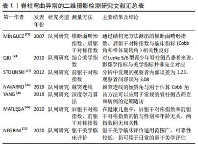
2.1 脊柱弯曲异常的体格检查 躯体的形态异常是脊柱弯曲异常最重要也是最早出现的临床表现,体表形态度量在该疾病中的应用主要通过对躯干对称性的评估和测量以对脊柱弯曲异常进行评估与筛查,使用历史已有一百余年。中国卫计委于2014年发布,2015年开始实施的《儿童青少年脊柱弯曲异常的筛查》(GB/T 16133-2014)中规定[16],脊柱侧凸的检查包括:①一般检查:包括对双肩、肩胛骨、腰凹和棘突连线的对称性进行评估;②前屈试验;③脊柱运动试验;④俯卧试验;⑤脊柱侧凸测量仪检查。脊柱前后弯曲异常的检查分为以下2步:首先侧向评估受检者的胸腰椎前、后凸程度,其次采用俯卧试验。 中华医学会骨科学分会2020年发布的《中国青少年脊柱侧凸筛查临床实践指南及路径指引》则建议选择目测法[17]、Adams前屈试验、躯干旋转角测量(包括脊柱侧凸测量尺或便携式电子脊柱侧凸筛查工具等)联合使用进行青少年脊柱侧凸筛查。该指南中目测法定义为观察双肩外观、腰线外观及骨盆水平度,此方法高度依赖检测者的主观判断,不可单独作为判定标准。除外观检查外,前屈试验是另一个非常重要的体格检查手段,亦无需借助任何工具,是临床上最简单易行的检测方法。研究显示Cobb角大于20°时前屈试验的检测结果如下:胸椎侧凸敏感度为92%,特异度为60%;腰椎侧凸敏感度为73%,特异度为68%[18]。前屈试验虽然被认为是一项非常敏感的临床检查,然而灵敏度和特异度取决于检查人员的技术水平、弯曲的位置和弯曲的程度[19]。该方法假阴性率高,易导致漏诊,因此指南同样不建议其单独作为判定标准使用。而SENKOYLU等[20]对前屈试验的改良方案得到了很高的特异性和敏感度,ROC曲线分析显示曲线下面积范围为0.7-0.8,但样本量较少,有待进一步进行大样本量研究。 脊柱侧凸测量仪通过测量躯干旋转角度,可将前屈试验中的躯干不对称的程度量化,在躯干旋转角> 5°时高度怀疑脊柱侧凸。研究显示Cobb角大于20°时ATR的特异度和敏感度如下:胸椎侧凸敏感度为71%,特异度为83%;腰椎侧凸敏感度为51%,特异度为83%[18]。在近年来已有基于手机重力感应的脊柱侧凸检测软件出现如Scoliogauge,其原理与脊柱侧凸测量仪相同,极大方便了居家的早期筛查[21]。目测法和前屈试验经过多年使用及研究其可靠性极高,直到现在仍是脊柱侧凸初筛最主要的方法,但由于其过分依赖主观性和无法量化等缺陷,仅可对脊柱弯曲异常进行初步判断,促使人们设计更加准确、定量的体表形态度量方法。最著名的定量测量便是躯干旋转角度,其作为脊柱侧凸初筛方法使用至今,并且随着科技进步,更多设备能够作为躯干旋转角度测量的载体,如智能手机等,显著降低了其使用限制。 2.2 脊柱弯曲异常的光学技术检测 2.2.1 二维摄影 背部及躯干侧方的二维照片用于脊柱弯曲异常的评估已应用多年,既有定性评估,也有定量测量。对于仅需定性观察的指标如躯干美学临床评价,观察患者本人与观察照片的效果是相同的[22]。而对于定量测量的体表指标来说,需要对身体的特征点进行标记,再从照片中测量所需的距离、角度及其他指标如棘突连线等[23]。也有无需对患者体表进行标记即可测量的指标,如综合美学指数、后躯干对称指数和前躯干对称指数等,其中后躯干对称指数是最常用的评价指标[24-27]。随着科学技术的进步,人工智能技术已被引入到一些疾病的筛查中,并发挥了重要作用,如视网膜病变和遗传性疾病[28-29]。 近年来,人工智能也应用于脊柱侧凸的筛查中,为脊柱侧凸的早期诊断提供了便捷的,可操作性强的方法。YANG等[30]于2019年开发的计算机深度学习算法在对背部图像进行多重算法训练后,能够判断脊柱侧凸的存在和严重程度,准确度优于人类专家的平均水平。该研究是人工智能技术在脊柱侧凸检测方面的一次大胆尝试,检测脊柱侧凸比人类专家准确性更高,对脊柱侧凸严重程度的判断正确率为55.5%,与相应对比的4位人类专家中的最高分值(56.8%)相当且优于平均水平(46.9%),但该算法仍存在局限性,如缺少具体的测量方法、在阴性和轻度脊柱侧凸病例中检测准确性欠佳等,有待进一步完善。但该方法方便快捷的检测方式(仅需背部正位照片)及其有利于在脊柱侧凸大规模筛查中使用。二维测量文献汇总详见表1。 "
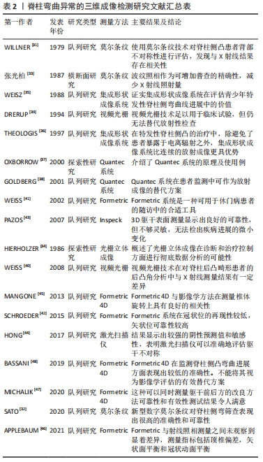
2.2.2 三维成像 由于脊柱形态畸形是三维形态改变,诸多研究也致力于观察并记录躯干畸形的三维变化,利用光学技术研发的各类非侵入性成像系统在脊柱弯曲异常的筛查及评估工作中已经投入使用多年。有研究利用相机、光源和光栅评估人体表面身体两侧不同区域的条纹对称性以评估脊柱侧凸程度,称之为莫尔条纹技术或波纹照相技术 [31-32]。张光铂等[33]在北京市城郊区开展青少年脊柱侧凸筛查时,同时使用前屈试验、波纹照相及X射线摄片3种检测方法,极大减少了前屈试验导致的假阳性结果,也很大程度减少了青少年X射线检测的辐射暴露量。该方法由于分析速度慢而妨碍了其在临床中的应用,随着计算机技术的进步,这种技术的分析能力也在不断改进,并衍生出新型的光栅立体成像技术,该方法类似于传统的立体摄影,但2台摄像机中的其中一台被具有更强反光性的投影仪所取代,另外在摄影时添加等距光栅线条,系统将光栅线投射到待测表面上,观察投影中的光栅线扭曲程度以反映三维表面形状的信息[34]。莫尔条纹与光栅立体摄影属于使用二维图像表现背部三维特征的方法,与人工测量方法相比速度更快,可用于脊柱侧凸的大规模普查。 结构光方法则直接对背部轮廓进行三维重建。集成形状成像系统(Integrated Shape Imaging System,ISIS)使用投影仪与相机从绕测量对象轴向移动,提取其轮廓,产生三维表面形状[35-36]。Quantec系统又称QSIS(Quantec Spinal Image System),与集成形状成像系统原理类似,使用一台摄像机和一台投射特殊设计阴影图案的投影仪,在背部标记T1-L5棘突及双侧髂后上棘,使用特殊软件生成12个参数并对图像进行分析[37-38]。以上2种结构光方法直接进行了躯干的三维重建,测量精度更高,但由于需要手工标记解剖标志和仪器的复杂性,测量效率较低。而近年来,随着计算机运算速度和摄像机精度的提升,出现了新型的视频光栅技术应用于表面形貌测量[39-40],如Formetric系统无需手动标记即可利用光栅技术重建背部三维结构,结合计算机处理,较集成形状成像系统精度更高、更加自动化,是目前表面形貌测量的主流技术[41-42]。近年来随着信息技术的迅速发展,新型三维重建技术如Inspeck系统、激光扫描仪等也在尝试应用于脊柱侧凸的检测 [43-44]。 近年最多被报道的体表成像技术为Diers Formetric 4D,是一种无辐射、非接触式扫描仪,可根据表面形貌对脊柱进行3D重建,也是Formetric系统的最新版本。诸多研究者对该系统的准确性进行了研究。MANGONE等[45]验证了该技术与躯干旋转角具有很高的相关性,提出这种方法可用于青少年特发性脊柱侧凸患者畸形程度的评估。APPLEBAUM等[46]将X射线参数与Formetric系统测量的参数作差异性分析,结果显示两种方法参数信度良好(0.90)。多年的研究结果表明该系统在临床工作中的表现出色。随着不断探索及科学进步,目前体表成像技术较其研发之初更加成熟,如MICHALIK等[47]专家在Formetric 4D基础上采用前后位两个视角,这种系统的改良进一步增加了检测的可信度并极大地提高了效能。但值得注意的是,亦有文献报道Formetric 4D在测量脊柱侧弯程度方面表现出中等的准确性,而在监测曲线进展方面表现出较低的准确性[48]。从早期的波纹照相法、光栅立体摄影法到ISIS-ISIS2和Diers Formical 4D测量仪,三维测量的可靠性很高。但是与EOS、超声测量或全脊柱X射线检查相比,体表形态测量的最大缺陷是由于体表畸形与脊柱畸形程度不一致导致体表形态无法完全反映脊柱畸形情况。尽管目前表面形貌的三维测量技术的精度已经很高,但仍不能将其视为影像学评估的替代方案。三维测量文献汇总详见表2。 "
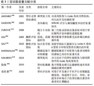
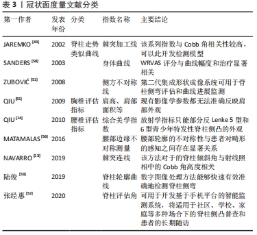
2.3 体表形态度量在各平面上的应用 2.3.1 冠状面 作为诊断脊柱侧凸的金标准,Cobb角是在全脊柱正位X射线片上测量的,对应人体的冠状位,故冠状位是脊柱弯曲异常评价指标中使用率最高的平面。许多研究尝试从背部体表特征中提取出与实际脊柱走向类似的曲线,如棘突加工线(Spinous process line)、沃尔特·里德视觉评估量表(Walter Reed Visual Assessment Scale,WRVAS)的身体曲线(Body curve)、第二代集成形状成像系统中的侧方不对称线(Lateral asymmetry,LA)等[49-51]。近年来,有研究尝试从2D图像中提取类似曲线,并检验其弯曲角度与Cobb角的相似性。无论提取方法是通过在体表进行解剖标志点的标记还是在照片中对背部沟壑评估,棘突连线都是最常用来描述脊柱侧凸的整体性畸形的曲线。NAVARRO等[23]在2019年报道背部照片中的棘突连线与Cobb角的相关系数在0.75-0.88,标准误差为5°-11°,具有较高的相关性。张经惠等[52]利用“单相机,多视角”的方式构造立体视觉的脊柱形态测量方法,推算出用于评估脊柱形态的脊柱评估角,结果表明与 Cobb角相关性达 0.871。陆俊等[53]开发了一种脊柱侧凸快速检测系统,采用了背部感兴趣区域提取,得到背部脊柱特征点,拟合成脊柱轮廓曲线,计算得到脊柱侧弯Cobb角大小,误差小于20%,但该系统的缺点是不能有效地检测出背部脊柱轮廓不明显的患者。此类的脊柱类似曲线具有一定辅助诊断价值,均存在临床研究样本量较少的问题,仍需进一步进行大样本量的实验来验证其有效性。 另一方面,肩部、肩胛骨、腰部和胸廓等结构的不对称性也被用于躯干表面测量。国际脊柱侧弯矫形与康复治疗学会建议的体表解剖标志点包括C7棘突、L4棘突、双侧肩峰、双侧肩胛上角、双侧肩胛下角、双侧髂后上棘[54]。除此之外髂嵴最高点或髂后上棘也可用于定位。除解剖标志点外,躯体轮廓边缘组成的曲线在进行双侧差异对比时也可被用于评估躯干不对称所致躯干畸形,如《中国青少年脊柱侧凸筛查临床实践指南及路径指引》中提到的脊柱侧凸筛查目测法便包括对双肩不等高和一侧腰部出现皱褶的观察。肩部与腰部的边缘是最常用于体表形态评估的线性指标。躯干美学临床评价工具对肩部、肩胛骨、腰部以及半胸4个位置的不对称性进行评估,分为轻度、中度和重度3个等级,该工具由测试者通过肉眼观察背部给出主观定性评价。集成形状成像使用不平衡、侧向不对称和体积不对称指数作为冠状面的评价指标。后躯干对称指数是由6个子指数相加组成,每个子指数都由背部各类解剖标志之间的直线距离经数学处理后计算为比值。前躯干对称指数与后躯干对称指数原理相近,但由于其在需要受试者裸露上半身的情况下在躯干前方辨识解剖标志并测量,应用受到一定限制。 近年来,利用各类角度及距离等指标体现躯干不对称性的评估方法层出不穷,不仅是对体表指标的补充,也对这些体表参数与相关影像学参数的相关性做出一定的验证。Qiu等[55]于2009年测量双胸弯患者的数个肩部体表指标,与影像学指标进行相关性分析,发现体表参数与影像学参数并非完全相关。其团队于2010年设计的综合美学指数由5个腰骶部子指数组成,并将其应用于Lenke5,6型青少年特发性脊柱侧凸患者,与腰骶部影像学参数相关性较好[24]。MATAMALAS等[56]也发现部分腰部边缘体表参数如腰高角(waist height angle,WHA)、腰围距离比(waistline distance ratio,WLDR)等与影像参数有一定的相关性。需要注意的是,正常青少年也可能存在轻微躯干不对称的情况[57]。而这些研究中体表参数与影像学参数相关系数均不超过0.8,表明体表参数变化并不能完整反映实际的脊柱畸形。冠状面评估指标内容庞杂,其分类详见表3。 "

从目前的技术水平讲,单一的冠状面评估虽然检测速度较快,但与实际脊柱畸形相关性欠佳,无法实际反映脊柱在冠状面的畸形情况。虽然精度欠佳,但冠状面的形态评估仍有运用于脊柱畸形筛查方面的潜力,有待研究者进一步发掘。 2.3.2 横断面 主要测量棘突两侧躯干高低不等的程度,可通过测量两侧躯干高低差或躯干旋转的角度实现。需要将这种差异量化时,最常使用的指标是躯干旋转角度,这也是脊柱侧凸测量仪的工作原理。与躯干旋转角度相似的是第二代集成形状成像系统(ISIS2)中的Transverse index,将背部分为19个等间距的截面进行计算,得出冠状面体表畸形程度[58]。JAREMKO等[49]所研究的部分指数与通过计算机确立躯干旋转角度和横断面躯干中心,并通过复杂的计算得出躯干不对称指数。利用表面形貌技术测定的SHS (Suzuki Hump Sum)指数[59-60],测量3组高度差,并将相关的子指数相加而得出其结果。横断面畸形指数中也包含类似的冠状面测量指数 [61]。 2.3.3 矢状面 矢状面的体表形态测量方法在3个平面中最少提及。除脊柱前后弯曲异常的目测筛查法外,定量指标一般取自侧位X射线片中的胸椎后凸角和腰椎前凸角,但此类角度以胸腰椎终板作为测量标志,体表无对应位置可供测量,不方便开展人群普查。NAULT等[1]提及枕骨隆突和眉弓的中点可用于确立头颅位置。集成形状成像及Quantec系统等结构光技术及其他三维重建技术可计算各生理曲度的最大前后凸位置和大小并粗略判断脊柱前后弯曲的情况。 "

| [1] NAULT ML, ALLARD P, HINSE S, et al. Relations between standing stability and body posture parameters in adolescent idiopathic scoliosis. Spine (Phila Pa 1976). 2002;27(17):1911-1917. [2] EL HAWARY R, CHUKWUNYERENWA C. Update on evaluation and treatment of scoliosis. Pediatr Clin North Am. 2014;61(6):1223-1241. [3] 陈绍丰,李博,周潇逸,等.青少年特发性脊柱侧凸相关生活质量评价量表现状[J].第二军医大学学报,2020,41(11):1188-1192. [4] WEISS HR, MORAMARCO M. Indication for surgical treatment in patients with adolescent Idiopathic Scoliosis-a critical appraisal. Patient Saf Surg. 2013;7(1):17. [5] LUAN FJ, WAN Y, MAK KC, et al. Cancer and mortality risks of patients with scoliosis from radiation exposure: a systematic review and meta-analysis. Eur Spine J. 2020;29(12):3123-3134. [6] LEVY AR, GOLDBERG MS, MAYO NE, et al. Reducing the lifetime risk of cancer from spinal radiographs among people with adolescent idiopathic scoliosis. Spine (Phila Pa 1976). 1996;21(13):1540-1547. [7] RONCKERS CM, LAND CE, MILLER JS, et al. Cancer mortality among women frequently exposed to radiographic examinations for spinal disorders. Radiat Res. 2010;174(1):83-90. [8] RONCKERS CM, DOODY MM, LONSTEIN JE. Multiple diagnostic X-rays for spine deformities and risk of breast cancer. Cancer Epidemiol Biomarkers Prev. 2008;17(3):605-613. [9] HOFFMAN DA, LONSTEIN JE, MORIN MM, et al. Breast cancer in women with scoliosis exposed to multiple diagnostic X rays. J Natl Cancer Inst. 1989;81(17):1307-1312. [10] OAKLEY PA, EHSANI NN, HARRISON DE. The scoliosis quandary: are radiation exposures from repeated X-rays harmful? Dose Response. 2019;17(2):1559325819852810. [11] DESCHÊNES S, CHARRON G, BEAUDOIN G, et al. Diagnostic imaging of spinal deformities: reducing patients radiation dose with a new slot-scanning X-ray imager. Spine (Phila Pa 1976). 2010;35(9):989-994. [12] BRANCHINI M, DEL VECCHIO A, GIGLIOTTI CR. Organ doses and lifetime attributable risk evaluations for scoliosis examinations of adolescent patients with the EOS imaging system. Radiol Med. 2018;123(4):305-313. [13] SUZUKI S, YAMAMOTO T, SHIKATA J, et al. Ultrasound measurement of vertebral rotation in idiopathic scoliosis. J Bone Joint Surg Br. 1989; 71:252-255. [14] KWOK G, YIP J, YICK KL, et al. postural screening for adolescent idiopathic scoliosis with infrared thermography. Sci Rep. 2017;7(1):14431. [15] VASILIADIS E, GRIVAS TB. Quality of life after conservative treatment of adolescent idiopathic scoliosis. Stud Health Technol Inform. 2008; 135:409-413. [16] 中华人民共和国国家卫生和计划生育委员会、中国国家标准化管理委员会.儿童青少年脊柱弯曲异常的筛查:GB/T 16133-2014[S]. 2014. [17] 中国青少年脊柱侧凸筛查临床实践指南及路径指引[J].中华骨科杂志,2020,40(23):1574-1582. [18] CÔTÉ P, KREITZ BG, CASSIDY JD, et al. A study of the diagnostic accuracy and reliability of the Scoliometer and Adam’s forward bend test. Spine (Phila Pa 1976). 1998;23(7):796-803. [19] VIVIANI GR, BUDGELL L, DOK C, et al. Assessment of accuracy of the scoliosis school screening examination. Am J Public Health. 1984;74(5): 497-498. [20] SENKOYLU A, ILHAN MN, ALTUN N, et al. A simple method for assessing rotational flexibility in adolescent idiopathic scoliosis: modified Adam’s forward bending test. Spine Deform. 2021;9(2):333-339. [21] FRANKO OI, BRAY C, NEWTON PO. Validation of a scoliometer smartphone app to assess scoliosis. J Pediatr Orthop. 2012;32(8):72-75. [22] Negrini S, Donzelli S, Di Felice F, et al. Construct validity of the trunk aesthetic clinical evaluation (TRACE) in young people with idiopathic scoliosis. Ann Phys Rehabil Med. 2020;63(3):216-221. [23] NAVARRO IJ, CANDOTTI CT, FURLANETTO TS, et al. Validation of a mathematical procedure for the cobb angle assessment based on photogrammetry. J Chiropr Med. 2019;18(4):270-277. [24] QIU Y, QIU XS, MA WW, et al. How well do radiological measurements correlate with cosmetic indices in adolescent idiopathic scoliosis with Lenke 5, 6 curve types? Spine. 2010;35(18):882-888. [25] MÍNGUEZ MF, BUENDÍA M, CIBRIÁN RM, et al. Quantifier variables of the back surface deformity obtained with a noninvasive structured light method: evaluation of their usefulness in idiopathic scoliosis diagnosis. Eur Spine J. 2007;16(1):73-82. [26] MATLĘGA A, STĘPOWSKA J, WIŚNIEWSKI A, et al. Assessment of the coronal plane trunk symmetry in children. Phys Theory Pract. 2020; 36(12):1502-1508. [27] STOLINSKI L, KOTWICKI T, CZAPROWSKI D, et al. Analysis of the Anterior Trunk Symmetry Index (ATSI). Preliminary report. Stud Health Technol Inform. 2012;176:242-246. [28] ZHANG Y, SHI J, PENG Y, et al. Artificial intelligence-enabled screening for diabetic retinopathy: a real-world, multicenter and prospective study. BMJ Open Diabetes Res Care. 2020;8(1):e001596. [29] GUROVICH Y, HANANI Y, BAR OY, et al. Identifying facial phenotypes of genetic disorders using deep learning. Nat Med. 2019;25(1):60-64. [30] YANG J, ZHANG K, FAN H, et al. Development and validation of deep learning algorithms for scoliosis screening using back images. Commun Biol. 2019;25(2):390. [31] WILLNER S. Moiré topography for the diagnosis and documentation of scoliosis. Acta Orthop Scand. 1979;50(3):295-302. [32] SATO T, YONEZAWA I, AKIMOTO T, et al. Novel hump measurement system with a 3D camera for early diagnosis of patients with adolescent idiopathic scoliosis: a study of accuracy and reliability. Cureus. 2020; 12(5):e8229. [33] 张光铂,李子荣,魏新荣,等.波纹照相在脊柱侧凸普查中的应用[J].中华外科杂志,1987,25(7):387-389. [34] HIERHOLZER E, SCHIER F. Rasterstereography in the measurement and postoperative follow-up of anterior chest wall deformities. Z Kinderchir. 1986;41(5):267-271. [35] WEISZ I, JEFFERSON RJ, TURNER-SMITH AR, et al. ISIS scanning: a useful assessment technique in the management of scoliosis. Spine. 1988; 13:405-408. [36] THEOLOGIS T, FAIRBANK JCT, TURNER-SMITH A, et al. Early detection of progression in adolescent idiopathic scoliosis by measurement of changes in back shape with the integrated shape imaging system scanner. Spine. 1997;22(11):1223-1227. [37] OXBORROW N. Assessing the child with scoliosis: the role of surface topography. Arch Dis Child. 2000;83:453-455. [38] GOLDBERG CJ, KALISZER M, MOORE DP, et al. Surface topography, Cobb angles, and cosmetic change in scoliosis. Spine (Phila Pa 1976). 2001;26(4):E55-E63. [39] DRERUP B, HIERHOLZER E. Back shape measurement using video rasterstereography and three-dimensional reconstruction of spinal shape. Clin Biomech. 1994;9(1):28-36. [40] WEISS HR, ELOBEIDI N. Comparison of the kyphosis angle evaluated by video rasterstereography (VRS) with x-ray measurements. Stud Health Technol Inform. 2008;140:137-139. [41] WEISS HR, DIECKMANN J, GERNER HJ. Outcome of in-patient rehabilitation in patients with M. Scheuermann evaluated by surface topography. Stud Health Technol Inform. 2002;88:246-249. [42] SCHROEDER J, REER R, BRAUMANN KM. Video raster stereography back shape reconstruction: a reliability study for sagittal, frontal, and transversal plane parameters. Eur Spine J. 2015;24(2):262-269. [43] PAZOS V, CHERIET F, DANSERAU J, et al. Reliability of trunk shape measurements based on 3-D surface reconstructions. Eur Spine J. 2007;16(11):1882-1891. [44] HONG A, JASWAL N, WESTOVER L, et al. surface topography classification trees for assessing severity and monitoring progression in adolescent idiopathic scoliosis. Spine (Phila Pa 1976). 2017;42(13): E781-E787. [45] MANGONE M, RAIMONDI P, PAOLONI M, et al. Vertebral rotation in adolescent idiopathic scoliosis calculated by radiograph and back surface analysis-based methods: correlation between the Raimondi method and rasterstereography. Eur Spine J. 2013;22(2):367-371. [46] APPLEBAUM A, CHO W, NESSIM A, et al. Establishing the validity of surface topography for assessment of scoliosis: a prospective study. Spine Deform. 2021;9(3):685-689. [47] MICHALIK R, KNOD M, SIEBERS H, et al. Introduction and evaluation of a novel multi-camera surface topography system.Gait Posture. 2020; 80:367-373. [48] BASSANI T, STUCOVITZ E, GALBUSERA F, et al. Is rasterstereography a valid noninvasive method for the screening of juvenile and adolescent idiopathic scoliosis? Eur Spine J. 2019;28(3):526-535. [49] JAREMKO JL, PONCET P, RONSKY J, et al. Indices of torso asymmetry related to spinal deformity in scoliosis. Clin Biomech. 2002;17:559-568. [50] SANDERS JO, POLLY DW JR, CATS-BARIL W, et al. Analysis of patient and parent assessment of deformity in idiopathic scoliosis using the Walter Reed Visual Assessment Scale. Spine (Phila Pa 1976). 2003;28(18): 2158-2163. [51] ZUBOVIĆ A, DAVIES N, BERRYMAN F, et al. New method of Scoliosis Deformity Assessment: ISIS2 System. Stud Health Technol Inform. 2008; 140:157-160. [52] 张经惠,沈林勇,宋薇,等.用于日常体表监测的三维脊柱形态测量技术[J].生物医学工程学杂志,2020,37(5):809-817. [53] 陆俊,邢丽冬,钱志余,等.基于数字图像处理的脊柱侧弯快速检测系统[J].中国医疗器械杂志,2019,43(4):259-262. [54] KOTWICKI T, NEGRINI S, GRIVAS TH, et al. Methodology of evaluation of morphology of the spine and the trunk in idiopathic scoliosis and other spinal deformities -6th SOSORT consensus paper. Scoliosis. 2009;4:26. [55] QIU XS, MA WW, LI WG, et al. Discrepancy between radiographic shoulder balance and cosmetic shoulder balance in adolescent idiopathic scoliosis patients with double thoracic curve. Eur Spine J. 2009;18(1):45-51. [56] MATAMALAS A, BAGÓ J, AGATA E, et al. Validity and reliability of photographic measures to evaluate waistline asymmetry in idiopathic scoliosis. Eur Spine J. 2016;25(10):3170-3179. [57] GARDNER A, BERRYMAN F, PYNSENT P. What is the variability in shoulder, axillae and waist position in a group of adolescents? J Anat. 2017;231(2):221-228. [58] BERRYMAN F, PYNSENT P, FAIRBANK J, et al. A new system for measuring 3D back shape in scoliosis. Eur Spine J. 2008;17:663-672. [59] ASHER M, LAI SM, BURTON D, et al. The influence of spine and trunk deformity on preoperative idiopathic scoliosis patients’ health-related quality of life questionnaire responses. Spine (Phila Pa 1976). 2004; 29(8):861-868. [60] PARENT EC, DAMARAJU S, HILL D, et al. Identifying the best surface topography parameters for detecting idiopathic scoliosis curve progression. Stud Health Technol Inform. 2010;158:78-82. [61] MINGUEZ MF, BUENDÍA M, CIBRIÁN RM, et al. Quantifier variables of the back surface deformity obtained with a noninvasive structured light method: evaluation of their usefulness in idiopathic scoliosis diagnosis. Eur Spine J. 2007;16(1):73-82. [62] FAIRBANK J. Historical perspective: william adams, the forward bending test, and the spine of gideon algernon mantell. Spine (Phila Pa 1976). 2004;29(17):1953-1955. [63] BUNNELL WP. An objective criterion for scoliosis screening. J Bone Joint Surg Am. 1984;66(9):1381-1387. [64] SUZUKI N, INAMI K, ONO T, et al. In: Research into Spinal Deformities 2. Stokes IAF, editor. Amsterdam: IOS Press; 1999. Analysis of posterior trunk symmetry index (POTSI) in Scoliosis: Part 1: 81-84. [65] ZAINA F, NEGRINI S, ATANASIO S. TRACE (Trunk Aesthetic Clinical Evaluation), a routine clinical tool to evaluate aesthetics in scoliosis patients: development from the Aesthetic Index (AI) and repeatability. Scoliosis. 2009;4:3. [66] 徐锡明,魏显招,等.青少年特发性脊柱侧凸学校筛查的进展及意义[J].脊柱外科杂志,2013,11(6):360-364. [67] DRERUP B, HIERHOLZER E. Evaluation of frontal radiographs of scoliotic spines--Part I. Measurement of position and orientation of vertebrae and assessment of clinical shape parameters. J Biomech. 1992;25(11):1357-1362. [68] DRERUP B, HIERHOLZER E. Evaluation of frontal radiographs of scoliotic spines--Part II. Relations between lateral deviation, lateral tilt and axial rotation of vertebrae. J Biomech. 1992;25(12):1443-1450. [69] DRERUP B, HIERHOLZER E. Assessment of scoliotic deformity from back shape asymmetry using an improved mathematical model. Clin Biomech (Bristol, Avon). 1996;11(7):376-383. [70] 牛升波,杨桓,杨明园,等.脊柱畸形的影像学参数测量及其研究进展[J].第二军医大学学报,2020,41(11):1177-1182. [71] 蒋志成,徐慧琼,万宇辉,等.儿童青少年脊柱弯曲异常筛查研究进展[J].中国学校卫生,2021,42(2):312-315, 320. [72] 吕东辉,顼超静,孙九爱.图像处理技术在脊柱侧弯检查中的应用[J].生物医学工程学杂志,2012,29(4):663-668. |
| [1] | Zhang Yufang, Lü Meng, Mei Zhao. Construction and verification of a full spine biomechanical model of adolescent scoliosis [J]. Chinese Journal of Tissue Engineering Research, 2022, 26(9): 1351-1356. |
| [2] | Wang Shuai, Wang Liancheng, Zhang Shuhao, Li Fuli, Dong Jiaxing, Zhang Yajie. Correlation of the electromyography ratio of the paraspinal muscles on the convex and concave sides with Cobb angle, apical vertebra translation, and coronal balance distance in adolescent idiopathic scoliosis patients [J]. Chinese Journal of Tissue Engineering Research, 2022, 26(9): 1402-1406. |
| [3] | Song Yuxin, Zhang Tongtong, Niu Jianxiong, Wang Zengping, Wen Jie, Zhang Qunli, Xue Wen, Liu Lin. Precise screw placement of 3D printing model and orthopedic robot in spinal deformity [J]. Chinese Journal of Tissue Engineering Research, 2022, 26(6): 904-907. |
| [4] | Xin Daqi, Wang Guoqiang, Han Di, Xing Wenhua, Fu Yu, Zhu Yong, Zhou Yang, Bai Xianming, He Chenyang, Zhao Yan. Finite element simulation surgical modeling of Lenke 3 adult idiopathic scoliosis: modeling evaluation twice in 5 years [J]. Chinese Journal of Tissue Engineering Research, 2022, 26(36): 5755-5763. |
| [5] | Lu Yangyang, Xu Chunxin, Chen Cen, Ren Jie, Zhai Xinyi, Zhu Shenyu, Shen Min. Establishment of thoracic and abdominal volume models of adolescent idiopathic scoliosis based on three-dimensional motion capture system [J]. Chinese Journal of Tissue Engineering Research, 2022, 26(36): 5807-5811. |
| [6] | Sun Jinpeng, Liu Jun, Bai Yunfeng, Hua Feng, Wang Haoran, Zheng Hongrui, Wu Tao. Melatonin effect on the activity of mouse ATDC5 cells [J]. Chinese Journal of Tissue Engineering Research, 2022, 26(35): 5665-5668. |
| [7] | Pan Xian, Liang Simin, Liu Bingxia, Song Tianzeng, Ma He, Wu Peng, Ge Zhaohui. Correlation between pelvic incidence-lumbar lordosis and clinical outcome in patients with degenerative lumbar scoliosis [J]. Chinese Journal of Tissue Engineering Research, 2022, 26(33): 5370-5375. |
| [8] | Zhang Jian, Lin Jianping, Zhou Gang, Fang Yehan, Wang Benchao, Wu Yongchang. Semi-quantitative MRI evaluation of cartilage degeneration in early knee osteoarthritis [J]. Chinese Journal of Tissue Engineering Research, 2022, 26(3): 425-429. |
| [9] | Sun Jinpeng, Liu Jun, Bai Yunfeng, Hua Feng, Wang Haoran, Zheng Hongrui, Wu Tao. Effects of suppressor of cytokine signaling 3 on osteogenic activity in the cartilage of adolescent idiopathic scoliosis [J]. Chinese Journal of Tissue Engineering Research, 2022, 26(26): 4160-4165. |
| [10] | Li Xiuya, Sheng Yang, Wang Shengyi, Li Yue, Zhang Ruzhi, Pan Yan, Sun Yixin, Deng Linhong, Mark Bradley, Zhang Rong. Preparation of protein chips for screening matrix for Muse cell culture based on fibrinogen and gelatin [J]. Chinese Journal of Tissue Engineering Research, 2022, 26(21): 3312-3318. |
| [11] | Ren Dong, Zhu Ye, Lei Lei, Wang Yuren. Orthopedic force applied to the rib influences the displacement and rotation angle of thoracic vertebrae: a finite element analysis [J]. Chinese Journal of Tissue Engineering Research, 2022, 26(18): 2812-2816. |
| [12] | Yuan Haoxiang, Xu Jing, Zeng Jinshu, Chen Hao, Yan Yelei, Chen Jiahao, Liu Qingshan, Xu Fei. Sequence of prevention for anterior cruciate ligament injury: screening, intervention and assessment [J]. Chinese Journal of Tissue Engineering Research, 2022, 26(17): 2775-2781. |
| [13] | Lu Dezhi, Mei Zhao, Li Xianglei, Wang Caiping, Sun Xin, Wang Xiaowen, Wang Jinwu. Digital design and effect evaluation of three-dimensional printing scoliosis orthosis [J]. Chinese Journal of Tissue Engineering Research, 2021, 25(9): 1329-1334. |
| [14] | Liang Yan, Zhao Yongfei, Xu Shuai, Zhu Zhenqi, Wang Kaifeng, Liu Haiying, Mao Keya. Imaging evaluation of short-segment fixation and fusion for degenerative lumbar scoliosis assisted by highly selective nerve root block [J]. Chinese Journal of Tissue Engineering Research, 2021, 25(9): 1423-1427. |
| [15] | Wu Chao, Gao Mingjie, Wang Jianzhong, Zhang Yunfeng, Yu Jinghong, Cai Yongqiang, Wang Haiyan, He Yujie, Tong Ling, Li Jiawei, Gao Shang, Wang Xing, Wu Min, Li Zhijun, Li Xiaohe. Finite element analysis of stress and displacement of Lenke 3 adolescent idiopathic scoliosis thoracolumbar spine [J]. Chinese Journal of Tissue Engineering Research, 2021, 25(33): 5273-5280. |
| Viewed | ||||||
|
Full text |
|
|||||
|
Abstract |
|
|||||