Chinese Journal of Tissue Engineering Research
Previous Articles Next Articles
Labial and lingual alveolar bone thickness of adult tooth root
Ding Ji-qun1, Fang Jian-qiang2, Yuan Chang-qing1, Chen Jie1
- 1 Department of Stomatology, Affiliated Hospital of Qingdao University Medical College, Qingdao 266003, Shandong Province, China
2 Department of Orthodontics, Hangzhou Stomatological Hospital, Hangzhou 310006, Zhejiang Province, China
-
Received:2012-11-04Revised:2012-11-05Online:2013-04-09Published:2013-04-09 -
Contact:Yuan Chang-qing, Master, Associate chief physician, Department of Stomatology, Affiliated Hospital of Qingdao University Medical College, Qingdao 266003, Shandong Province, China chenfjq2010@163.com -
About author:Ding Ji-qun★, Master, Department of Stomatology, Affiliated Hospital of Qingdao University Medical College, Qingdao 266003, Shandong Province, China chenfjq2010@163.com
CLC Number:
Cite this article
Ding Ji-qun, Fang Jian-qiang, Yuan Chang-qing, Chen Jie . Labial and lingual alveolar bone thickness of adult tooth root[J]. Chinese Journal of Tissue Engineering Research, doi: 10.3969/j.issn.2095-4344.2013.15.008.
share this article

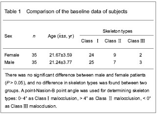
Quantitative analysis of subjects A total of 70 patients meeting the inclusion criteria were enrolled in the result analysis with no loss. Baseline analysis of subjects Age has a great influence on the bone and tooth root; therefore, selected subjects were all young adults. No difference was found in the baseline analysis between different genders (P > 0.05; Table 1)."
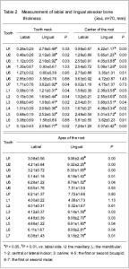
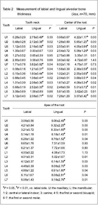
Measurement results of the labial and lingual alveolar bone thickness of the tooth root (Table 2)"
| [1] Guo QY, Zhang SJ, Liu H, et al. Three-dimensional evaluation of upper anterior alveolar bone dehiscence after incisor retraction and intrusion in adult patients with bimaxillary protrusion malocclusion. J Zhejiang Univ Sci B. 2011;12(12):990-997. [2] Nahm KY, Kang JH, Moon SC, et al. Alveolar bone loss around incisors in Class I bidentoalveolar protrusion patients: a retrospective three-dimensional cone beam CT study. Dentomaxillofac Radiol. 2012;41(6): 481-488. [3] Evangelista K, Vasconcelos Kde F, Bumann A, et al. Dehiscence and fenestration in patients with Class I and Class Ⅱ Division 1 malocclusion assessed with cone-beam computed tomography. Am J Orthod Dentofacial Orthop. 2010;138(2):e1-e7.[4] Fuh LJ, Huang HL, Chen CS, et al. Variations in bone density at dental implant sites in different regions of the jawbone. J Oral Rehabil. 2010;37(5):346-351. [5] Choi JH, Park CH, Yi SW, et al. Bone density measurement in interdental areas with simulated placement of orthodontic miniscrew implants. Am J Orthod Dentofacial Orthop. 2009; 136(6):e1-e12.[6] Farnsworth D, Rossouw PE, Ceen RF, et al. Cortical bone thickness at common miniscrew implant placement sites. Am J Orthod Dentofacial Orthop. 2011;139(4): 495-503. [7] Park J, Cho HJ. Three-dimensional evaluation of interradicular spaces and cortical bone thickness for the placement and initial stability of microimplants in adults. Am J Orthod Dentofacial Orthop. 2009;136(3): e1-e12.[8] Vasak C, Watzak G, Gahleitner A, et al. Computed tomography-based evaluation of template (NobelGuide™)-guided implant positions: a prospective radiological study. Clin Oral Implants Res. 2011;22(10): 1157-1163.[9] Blok Y, Gravesteijn FA, van Ruijven LJ, et al. Micro-architecture and mineralization of the human alveolar bone obtained with microCT. Arch Oral Biol. 2012.[10] Kapila S, Conley RS, Harrell WE Jr. The current status of cone beam computed tomography imaging in orthodontics. Dentomaxillofac Radiol. 2011;40(1):24-34. [11] State Council of the People’s Republic of China. Administrative Regulations on Medical Institution. 1994-09-01.[12] Lee SL, Kim HJ, Son MK, et al. Anthropometric analysis of maxillary anterior buccal bone of Korean adults using cone-beam CT. J Adv Prosthodont. 2010;2(3):92-96.[13] Kumar V, Ludlow J, Soares Cevidanes LH, et al. In vivo comparison of conventional and cone beam CT synthesized cephalograms. Angle Orthod. 2008;78(5): 873-879.[14] Jeffrey CK, Kwong J. Image quality produced by different cone beam cephalometry tomography settings. Am J Orthod Dentofacial Orthop. 2008;133(2):317-327.[15] Suomalainen A, Vehmas T, Kortesniemi M, et al. Accuracy of linear measurements using dental cone beam and conventional multislice computed tomography. Dentomaxillofac Radiol. 2008;37(1):10-17. [16] Suomalainen A, Kiljunen T, Käser Y, et al. Dosimetry and image quality of four dental cone beam computed tomography scanners compared with multislice computed tomography scanners. Dentomaxillofac Radiol. 2009;38(6): 367-378.[17] Loubele M, Bogaerts R, Van Dijck E, et al. Comparison between effective radiation dose of CBCT and MSCT scanners for dentomaxillofacial applications. Eur J Radiol. 2009;71(3):461-468. [18] Roberts JA, Drage NA, Davies J, et al. Effective dose from cone beam CT examinations in dentistry. Br J Radiol. 2009;82(973):35-40.[19] Huang QH, Wang QB, Luo MY, et al. Xiangnan Xueyuan Xuebao. 2009;11(2):4-6.[20] Tyndall DA, Rathore S. Cone-beam CT diagnostic applications: caries, periodontal bone assessment, and endodontic applications. Dent Clin N Am. 2008;52(4):825-841.[21] Kau CH, Richmond S, Palomo JM, et al. Three-dimensional cone beam computerized tomography in orthodontics. J Orthod. 2005;32(4):282-293.[22] Ludlow JB, Ivanovic M. Comparative dosimetry of dental CBCT devices and 64-slice CT for oral and maxillofacial radiology. Oral Surg Oral Med Oral Pathol Oral Radiol Endod. 2008;106(1):106-114.[23] Zhao BD, Li NY, Zhou YG, et al. A study of rebuild of a three-dimensional anatomic model of mandibles. Huaxi Kouqiang Yixue Zazhi. 2002;20(1):21-23.[24] Januário AL, Duarte WR, Barriviera M, et al. Dimension of the facial bone wall in the anterior maxilla: a cone-beam computed tomography study. Clin Oral Implants Res. 2011;22(10):1168-1171.[25] Ghassemian M, Nowzari H, Lajolo C, et al. The thickness of facial alveolar bone overlying healthy maxillary anterior teeth. J Periodontol. 2012;83(2):187-197. [26] Henneman S, Von den Hoff JW, Maltha JC. Mechanobiology of tooth movement. Eur J Orthod. 2008; 30(3):299-306.[27] Anwar N, Fida M. Compensation for vertical dysplasia and its clinical application. Eur J Orthod. 2009;31(5):516-522.[28] Uysal T, Yagci A, Ozer T, et al. Mandibular anterior bony support and incisor crowding: Is there a relationship? Am J Orthod Dentofacial Orthop. 2012;142(5):645-653. [29] Poggio PM, Incorvati C, Velo S, et al. “Safe Zones”: A Guide for miniscrew positioning in the maxillary and mandibular Arch. Angle Orthod. 2006;76(2):191-197.[30] Kim SH, Yoon HG, Choi YS, et al. Evaluation of interdental space of the maxillary posterior area for orthodontic mini-implants with cone-beam computed tomography. Am J Orthod Dentofacial Orthop. 2009;135(5):635-641.[31] Fayed MM, Pazera P, Katsaros C. Optimal sites for orthodontic mini-implant placement assessed by cone beam computed tomography. Angle Orthod. 2010;80(5): 939-951. [32] Liou EI, Chen PH, Wang YC, et al. A computed tomographic image study on the thickness of the infrazygomatic crest of the maxilla and its clinical implications for mini screw insertion. Am J Orthod Dentofacial Orthop. 2007;131(3):352-356. [33] Lin JR, Chen S. Treatment of severe class Ⅲ with buccal shelf mini-screws. Zhonghua Kouqiang Zhengjixue Zazhi. 2010;17(3):121-126.[34] Lin JR. The new method of IZC mini-screws placement. Zhonghua Kouqiang Zhengjixue Zazhi. 2009;16(1):38-44.[35] Kwak HH, Park HD, Yoon HR, et al. Topographic anatomy of the inferior wall of the maxillary sinus in Koreans. Int J Oral Maxillofac Surg. 2004;33(4):382-388. [36] Bodner L, Brennan PA, McLeod NM. Characteristics of iatrogenic mandibular fractures associated with tooth removal: review and analysis of 189 cases. Br J Oral Maxillofac Surg. 2011;49(7):567-572. |
| [1] | Fan Jiabing, Zhang Junmei. Morphological measurement and analysis of the mandible in adult females with different vertical skeletal types [J]. Chinese Journal of Tissue Engineering Research, 2021, 25(8): 1177-1183. |
| [2] | Li Yuanyuan, Lu Yingjuan, Ye Yushan, Mustafa M.M Weldali, Chang Shaohai. Constructing finite element models of three maxillary arch forms [J]. Chinese Journal of Tissue Engineering Research, 2021, 25(20): 3125-3129. |
| [3] | Nie Jing, Shi Xiaoyu. Cone-beam CT measurement of alveolar bone thickness of the maxillary anterior area at implant anchorage site in different sexes [J]. Chinese Journal of Tissue Engineering Research, 2021, 25(14): 2133-2136. |
| [4] | Luo Yicai, Li Hao. Effect of enhanced aryl hydrocarbon receptor expression on inflammatory response and healing of alveolar bone defects in diabetic rats [J]. Chinese Journal of Tissue Engineering Research, 2021, 25(14): 2166-2171. |
| [5] | Zhang Yicen, Wang Peixin, Liu Zhicheng. Ultrasound-guided injection of hyaluronic acid and corticosteroid for treating plantar fasciitis: evaluation of pain, fascia thickness and ankle-foot function [J]. Chinese Journal of Tissue Engineering Research, 2021, 25(11): 1670-1674. |
| [6] | Xu Guofeng, Li Xuebin, Tang Yifan, Zhao Yin, Zhou Shengyuan, Chen Xiongsheng, Jia Lianshun. The role of autophagy in ossification of the human ligamentum flavum [J]. Chinese Journal of Tissue Engineering Research, 2020, 24(8): 1174-1181. |
| [7] | Zhang Bin, Sun Lihua, Zhang Junhua, Liu Tongbin, Liu Yusan, Cui Caiyun, Li Jun. Short-term effect comparison of a modified socket shield technique and conventional flapless immediate implant and immediate restoration in maxillary aesthetic area [J]. Chinese Journal of Tissue Engineering Research, 2020, 24(34): 5514-5519. |
| [8] |
Zhang Xuan, Li Yunpeng, Zhang Xuejian, Yin Chuanrong, Deng Yue.
Guided bone regeneration using preformed titanium mesh combined with bioabsorbable membranes in aesthetic area [J]. Chinese Journal of Tissue Engineering Research, 2020, 24(26): 4112-4117. |
| [9] | Qin Haikuo, Luo Shixing. Correlation of cortical bone thickness and X-ray gray value in different planes of proximal femur with brittle fracture of female hip [J]. Chinese Journal of Tissue Engineering Research, 2020, 24(18): 2867-2872. |
| [10] |
Cheng Yuting, Wu Chao, Huang Xiaolin, Li Fang, Shi Qianhui, Zhou Qian, Hong Wei, Wang Yong, Liao Jian.
Low-dose zoledronic acid regulates osteoclasts and
osteoblasts in the extraction socket of ovariectomized rats |
| [11] |
Zhang Yujing, Peng Yuzhi, Liu Baozhen, Jing Fang.
Effect of Xianling Gubao Capsule
on alveolar bone mass in postmenopausal women with periodontitis: a cone-beam
CT evaluation
|
| [12] | Chen Zhouyan, Zhou Rong, He Song, Wu Dingdan, Wu Xi, Bai Rui, Huang Yue. Effects of the changes of thickness and placement position of rectangular attachment on canine tooth torsion correction [J]. Chinese Journal of Tissue Engineering Research, 2020, 24(16): 2513-2519. |
| [13] | Song Xudong, He Yunwu, Li Yonglin, Chen Jing, Hu Junlan. Ultrasound-guided paravertebral nerve block for zoster-associated pain: a Meta-analysis [J]. Chinese Journal of Tissue Engineering Research, 2020, 24(11): 1797-1804. |
| [14] | Wang Hailong, Lü Ying, An Meiwen, Hou Chunsheng. Mechanic property evaluation of medical Kirschner wire’s large deflection bending [J]. Chinese Journal of Tissue Engineering Research, 2019, 23(4): 567-572. |
| [15] | Chen Xingjie, Yi Honglei, Chen Xuqiong, Wu Zenghui, Ma Xiangyang, Ai Fuzhi, Wang Jianhua, Zhang Kai, Xia Hong. Radiological analysis of oropharyngeal airway space after posterior internal fixation for the atlantoaxial dislocation [J]. Chinese Journal of Tissue Engineering Research, 2019, 23(36): 5856-5860. |
| Viewed | ||||||
|
Full text |
|
|||||
|
Abstract |
|
|||||