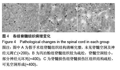
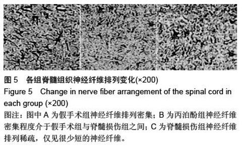
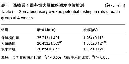
| [1] Fitch MT, Doller C, Combs CK,et al.Cellular and molecular mechanisms of glial scarring and progressive cavitation: in vivo and in vitro analysis of inflammation-induced secondary injury after CNS trauma.J Neurosci. 1999;19(19):8182-8198.[2] Young W.Spinal cord contusion models.Prog Brain Res. 2002; 137:231-255.[3] 张海明,张映.神经生长因子对神经元作用的研究进展动物医学进展[J].动物医学进展,2006,27(9):39-41.[4] Haku T, Okuda S, Kanematsu F,et al.Repair of cervical esophageal perforation using longus colli muscle flap: a case report of a patient with cervical spinal cord injury.Spine J. 2008; 8(5):831-835.[5] Pearse DD, Sanchez AR, Pereira FC,et al.Transplantation of Schwann cells and/or olfactory ensheathing glia into the contused spinal cord: Survival, migration, axon association, and functional recovery.Glia. 2007;55(9):976-1000.[6] Papastefanaki F, Chen J, Lavdas AA,et al.Grafts of Schwann cells engineered to express PSA-NCAM promote functional recovery after spinal cord injury.Brain. 2007;130(Pt 8): 2159-2174.[7] Finch AM, Wong AK, Paczkowski NJ,et al.Low-molecular- weight peptidic and cyclic antagonists of the receptor for the complement factor C5a.J Med Chem. 1999; 42(11):1965- 1974.[8] Pallini R, Vitiani LR, Bez A,et al.Homologous transplantation of neural stem cells to the injured spinal cord of mice. Neurosurgery. 2005;57(5):1014-1025.[9] Albin RL, Mink JW.Recent advances in Tourette syndrome research.Trends Neurosci. 2006;29(3):175-182.[10] 陈雪扉,莫万斌.注射用七叶皂苷钠联合脐带间充质干细胞移植改善脑梗死大鼠神经功能[J].中国组织工程研究与临床康复, 2011,15(27):5080-5084.[11] Amar AP, Levy ML.Pathogenesis and pharmacological strategies for mitigating secondary damage in acute spinal cord injury.Neurosurgery. 1999;44(5):1027-1039.[12] Crowe MJ, Bresnahan JC, Shuman SL,et al.Apoptosis and delayed degeneration after spinal cord injury in rats and monkeys.Nat Med. 1997;3(1):73-76.[13] Ohta S, Iwashita Y, Takada H,et al.Neuroprotection and enhanced recovery with edaravone after acute spinal cord injury in rats.Spine (Phila Pa 1976). 2005;30(10):1154-1158.[14] Banno M, Mizuno T, Kato H,et al.The radical scavenger edaravone prevents oxidative neurotoxicity induced by peroxynitrite and activated microglia.Neuropharmacology. 200548(2):283-290.[15] Björklund E, Lindberg E, Rundgren M,et al.Ischaemic brain damage after cardiac arrest and induced hypothermia--a systematic description of selective eosinophilic neuronal death. A neuropathologic study of 23 patients. Resuscitation. 2014;85(4):527-532.[16] Ariake K, Ohtsuka H, Motoi F,et al.GCF2/LRRFIP1 promotes colorectal cancer metastasis and liver invasion through integrin-dependent RhoA activation.Cancer Lett. 2012;325(1): 99-107.[17] Pearse DD, Sanchez AR, Pereira FC,et al.Transplantation of Schwann cells and/or olfactory ensheathing glia into the contused spinal cord: Survival, migration, axon association, and functional recovery.Glia. 2007;55(9):976-1000.[18] Pallini R, Vitiani LR, Bez A,et al.Homologous transplantation of neural stem cells to the injured spinal cord of mice. Neurosurgery. 2005;57(5):1014-1025.[19] Lepore AC, Neuhuber B, Connors TM, et al.Long-term fate of neural precursor cells following transplantation into developing and adult CNS.Neuroscience. 2006;139(2): 513-530.[20] Chuang SK.Limited evidence to demonstrate that the use of hyperbaric oxygen (HBO) therapy reduces the incidence of osteoradionecrosis in irradiated patients requiring tooth extraction.J Evid Based Dent Pract. 2011;11(3):129-131.[21] 丁进京.丙泊酚治疗对急性期脑梗死患者血清ACA水平的影响[J]. 中国实用神经疾病杂志,2012,15(3):34-36. |