Chinese Journal of Tissue Engineering Research
Previous Articles Next Articles
Biological characteristics of endothelial progenitor cells from the peripheral blood versus umbilical cord blood
Wu Hong-tao1, Ma Yan2, Bi Xiao-juan2, Jiang Ming2, Yang Kai1, Wang Ning1
- 1Department of No.1 Cadre Ward Medicine, First Affiliated Hospital, Xinjiang Medical University, Urumqi 830000, Xinjiang Uygur Autonomous Region, China; 2Stem Cell Laboratory, Medical Research Center, First Affiliated Hospital, Xinjiang Medical University, Urumqi 830000, Xinjiang Uygur Autonomous Region, China
-
Revised:2013-09-02Online:2013-11-05Published:2013-11-05 -
Contact:Wang Ning, M.D., Chief physician, Master’s supervisor, Department of No.1 Cadre Ward Medicine, First Affiliated Hospital, Xinjiang Medical University, Urumqi 830000, Xinjiang Uygur Autonomous Region, China 13999994126@163.com -
About author:Wu Hong-tao★, Studying for master’s degree, Department of No.1 Cadre Ward Medicine, First Affiliated Hospital, Xinjiang Medical University, Urumqi 830000, Xinjiang Uygur Autonomous Region, China whtbmd_2008@126.com -
Supported by:the Natural Science Foundation of Xinjiang Uygur Autonomous Region, No. 2011211A075*
CLC Number:
Cite this article
Wu Hong-tao, Ma Yan, Bi Xiao-juan, Jiang Ming, Yang Kai, Wang Ning. Biological characteristics of endothelial progenitor cells from the peripheral blood versus umbilical cord blood[J]. Chinese Journal of Tissue Engineering Research, doi: 10.3969/j.issn.2095-4344.2013.45.014.
share this article

2.1 人外周血与脐血单个核细胞形态变化 外周血与脐血分离的单个核细胞在形态上均呈圆形,体积较小,诱导培养2 d后可见大量的单个核细胞黏附于培养皿底,培养4天后均见细胞体积明显变大,形态不规则,可见少量梭形细胞,见图1A、B;诱导培养7 d长梭形内皮祖细胞明显增多,外周血源组的有细胞集落形成,集落中心聚集为圆形细胞,见图1C;而脐血源组可见梭形细胞自行排列为典型的线样阵列。见图1D。"

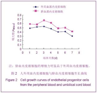
2.2 人外周血与脐血内皮祖细胞的扩增能力 锥虫蓝染色结果显示,实验获得单个核细胞数量在外周血源为(1.00±0.39)×106 L-1,脐血源为(2.00±0.66)×106 L-1;二者相比差异有显著性意义(P < 0.05)。以相同细胞浓度接种单个核细胞,培养诱导7 d收获外周血源内皮祖细胞为(107.63±8.22)×104个,脐血源内皮祖细胞为(423.5±34.91)×104个,二者差异有显著性意义(P < 0.05)。 内皮祖细胞的活率:收获时外周血和脐血内皮祖细胞活率分别为(97.6±0.9)%和(99.3±0.3)%,两者比较差异有显著性意义(P < 0.05)。 内皮祖细胞增殖特性测定:绘制诱导后内皮祖细胞的生长曲线显示,外周血源与脐血源内皮祖细胞的增殖在接种后3 d达到峰值,并且脐血内皮祖细胞的增殖力明显高于外周血,随后呈逐渐衰减态。见图2。"

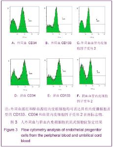
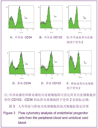
2.3 人外周血与脐血内皮祖细胞的增殖能力 流式细胞检测结果显示,培养7 d,流式细胞仪检测分析表明,人外周血/脐血来源的内皮祖细胞分别表达CD34:59.8%/60.8%;CD133:54.2%/53.5%;血管内皮细胞因子受体2:56.0%/47.4%,见图3。"

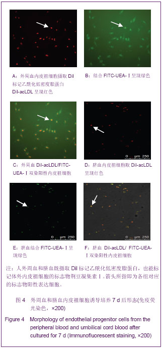
免疫荧光染色结果见图4,诱导培养7 d的人外周血和脐血内皮祖细胞经激光共聚焦显微镜观察,细胞既摄取DiI-acLDL呈现红色,见图4A,D;也结合FITC-UEA-1呈现绿色,见图4B,E;双染色具有黄绿色阳性细胞特征,见图4C,F,即为正在分化的内皮祖细胞。"
| [1] AsallaraT, MuroharaT, Sullivan A, et al. Isolation of putative endothelial cells for angiogenesis. Science. 1997;275(5302): 964-967. [2] Peichev M, Naiyer AJ, Pereira D, et al. Expression of VEGFR-2 and AC133 by circulating human CD34(+)cells identifies a population of functional endothelial precrsors. Blood. 2000;95(3):952-958. [3] Gill M, Dias S, Hattori K, et al. Vascular trauma induces rapid but transient mobilization of VEGF2(+)AC133(+)endothelial precursor cells. Circ Res. 2001;88(2):167-174. [4] Hill JM, Zalos G, Halcox JP, et al. Circulating endothelial progenitor cells,vascular function,and cardiovascular risk. N EngI J Med. 2003;348(7):593-600. [5] Walter DH, Rittig K, Bahlmann FH, et al. Statin therapy accelerates reendothelialization: a novel effect involving mobilization and incorporation of bone marrow-derived endothelial progenitor cells. Circulation. 2002;105(25): 3017-3024. [6] Kocher AA, Schuster MD, Szabolcs MJ, et al. Neovascularization of ischemic myocardium by human bone-marrow-derived angioblasts prevents cardiomyocyte apoptosis, reduce remodeling and improves cardiac function. Nat Med. 2001;7(4):430-436. [7] Takahashi T, Kalka C, Masnda H, et al. Ischemia-and cytokine- induced mobilization of bone marrow-derived endothelial progenitor cells for neovascularization. Nat Med. 1999;5(4): 434-438. [8] Asahara T, Masuda H, Takahashi T, et al. Bone marrow origin of endothelial progenitor cells responsible for postnatal vasculogenesis in physiological and pathological neovascularization. Circ Res. 1999;85(3):221-228. [9] Murayama T, Tepper OM, Silver M, et al. Determination of bone marrow-derived endothelial progenitor cell significance in angiogenic growth factor-induced neovascularization in vivo. Exp Hematol. 2002;30(8):967-972. [10] Suzuki T, Nishida M, Futami S, et al. Neoendothelialization after peripheral blood stem cell transplantation in humans:a case report of a Tokaimura nuclear accident victim. Cardiovasc Res. 2003;58(2):487-492. [11] 彭艳,程培,徐勇,等.脐血内皮祖细胞尾静脉与局部注射治疗糖尿病下肢缺血[J].中国组织工程研究与临床康复,2011,15(19): 3499-3502. [12] Giannotti G, Doerries C, Mocharla PS, et al. Impaired endothelial repair capacity of early endothelial progenitor cells in prehypertension: relation to endothelial dysfunction. Hypertension. 2010;55(6):1389-1397. [13] Chen LL, Liao YF, Zeng TS, et al. Number and function of circulating endothelial progenitor cell in diabetics with different vascular complications. Zhonghua Yi Xue Za Zhi. 2009;89(18): 1234-1239. [14] Maitra B, Szekely E, Gjini K, et al. Human mesenchymal stem cells support unrelated donor hematopoietic stem cells and suppress T-cell activation. Bone Marrow Transplant. 2004;33: 597-604. [15] Rocha V, Wagner JE Jr, Sobocinski KA, et al. Graft-versus-host disease in children who have received a cord-blood or bone marrow transplant from an HLA-identical sibling. Eurocord and International Bone Marrow Transplant Registry Working Committee on Alternative Donor and Stem Cell Sources. N Engl J Med. 2000;342(25):1846-1854. [16] Teleron AA, Carlson B, Young PP. Blood donor white blood cell reduction filter as a source of human peripheral blood-derived endohelial progenitor cell. Transfusion. 2005; 45(1):21-25. [17] Eggermann J, Kliche S, Hoffmann K, et al. Endothelial progenitor cell culture and differentiation in vitro: a methodological comparison using human umbilical cord blood.Cardiovasc Res. 2003;58(2):478-486. [18] Fadini GP, Albiero M, Cignarella A, et al. Effects of androgens on endothelial progenitor cells in vitro and in vivo.Clin Sci (Lond). 2009;117(10):355-364. [19] Hur J, Yoon CH, Kim HS, et al. Characterization of two types of endothelial progenitor cells and their different contributions to neovasculogenesis. Arterioscler Thromb Vasc Biol. 2004; 24(2):288-293. [20] Chivu M, Diaconu CC, Bleotu C, et al. The comparison of different protocols forexpansion of umbilical-cord hematopoietic stem cells. J Cell Mol Med. 2004;8(2): 223-231. [21] Reyftmann L, Dechaud H, Hamamah S, et al. Fetal and umbilical blood cord stem cells:a room for the obstetrician and gynaecologist. Part two, Gynecol Obstel Fertil. 2004;3(11): 969-975. [22] 方立建,宋鄂,栾瑛,等.人脐血内皮祖细胞体外培养和鉴定[J].中国组织工程研究与临床康复,2010,45(14):8450-8454. [23] 徐燕,孟恒星,于珍,等.不同来源内皮祖细胞培养及其生物学特性比较[J].中国组织工程研究与临床康复,2010,23(14): 4309-4316. [24] He T, Peterson TE, Holmuhamedov EL, et al. Human endothelial progenitor cells tolerate oxidative stress due to intrinsically high expression of manganese superoxide dismutase. Arterioscler Thromb vasc Biol. 2004;24(11): 2021-2027. [25] Maitra B, Szekely E, Gjini K, et al. Human mesenchymal stem cells support unrelated donor hematopoietic stem cells and suppress T-cell activation. Bone Marrow Transplant. 2004; 33(6): 597-604. [26] 周建良,邹明晖,陈义初,等.人脐血内皮祖细胞与血管内皮生长因子和碱性成纤维细胞生长因子共培养及其增殖和迁移能力[J].中国组织工程研究与临床康复,2011,15(6):1000-1004. [27] Kovacic JC, Moore J, Herbert A, et al. Endothelial progenitor cells, angioblasts, and angiogenesis--old terms reconsidered from acurrent perspective. Trends Cardiovasc Med. 2008; 18(2): 45-51. [28] Prater DN, Case J, Ingram DA, et al. Working hypothesis to redefine endothelial progenitor cells. Leukemia. 2007;21(6): 1141-1149. [29] Hirschi KK, Ingram DA, Yoder MC. Assessing identity, phenotype,and fate of endothelial progenitor cells. Arterioscler Thromb VascBiol. 2008;28(9):1584-1595. [30] 宋晓红,张罗,韩德民,等.鼠尾胶原为底物的人鼻腔纤毛上皮细胞培养模式的建立[J].中国耳鼻咽喉头颈外科,2007,2(14):107- 111. [31] 章必成,王俊,赵勇,等.以鼠尾胶为贴黏剂的小鼠淋巴管内皮细胞培养体系的建立[J].第四军医大学学报,2007,28(5):390-393. |
| [1] | Kong Desheng, He Jingjing, Feng Baofeng, Guo Ruiyun, Asiamah Ernest Amponsah, Lü Fei, Zhang Shuhan, Zhang Xiaolin, Ma Jun, Cui Huixian. Efficacy of mesenchymal stem cells in the spinal cord injury of large animal models: a meta-analysis [J]. Chinese Journal of Tissue Engineering Research, 2020, 24(在线): 3-. |
| [2] | Zhao Weibiao, He Ziwei, Li Ji, Li Yi. Application value of 3D printing guide plate in SuperPATH technology for elderly hip arthroplasty: retrospective study and evidence analysis of literature retrieval [J]. Chinese Journal of Tissue Engineering Research, 2020, 24(9): 1324-1330. |
| [3] | Gao Yang, Bai Shengchao, Chen Shengju, Wang Ruiyuan, Li Junping. Effect of exercise-induced muscle damage on desmin aggregates [J]. Chinese Journal of Tissue Engineering Research, 2020, 24(8): 1243-1248. |
| [4] | Chen Juan, Zhang Ting, Wu Yidan, Lu Yan, Ouyang Zhaolian. China’s strengths in basic research in the main subfields of tissue engineering [J]. Chinese Journal of Tissue Engineering Research, 2020, 24(8): 1267-1271. |
| [5] | Chen Jinsong, Wang Zhonghan, Chang Fei, Liu He. Tissue engineering methods for repair of articular cartilage defect under special conditions [J]. Chinese Journal of Tissue Engineering Research, 2020, 24(8): 1272-1279. |
| [6] | Wang Gang, Li Donghui, Bai Zhiming. Evolution and progress of replacement therapy, materials and reconstruction of long ureteral injuries [J]. Chinese Journal of Tissue Engineering Research, 2020, 24(8): 1299-1305. |
| [7] | Liu Chundong, Shen Xiaoqing, Zhang Yanli, Zhang Xiaogen, Wu Buling. Effects of strontium-modified titanium surfaces on adhesion, migration and proliferation of bone marrow mesenchymal stem cells and expression of bone formation-related genes [J]. Chinese Journal of Tissue Engineering Research, 2020, 24(7): 1009-1015. |
| [8] | Lin Ming, Pan Jinyong, Zhang Huirong. Knockout of NIPBL gene down-regulates the abilities of proliferation and osteogenic differentiation in mouse bone marrow mesenchymal stem cells [J]. Chinese Journal of Tissue Engineering Research, 2020, 24(7): 1002-1008. |
| [9] | Zhang Wen, Lei Kun, Gao Lei, Li Kuanxin. Neuronal differentiation of rat bone marrow mesenchymal stem cells via lentivirus-mediated bone morphogenetic protein 7 transfection [J]. Chinese Journal of Tissue Engineering Research, 2020, 24(7): 985-990. |
| [10] | Wu Zhifeng, Luo Min. Biomechanical analysis of chemical acellular nerve allograft combined with bone marrow mesenchymal stem cell transplantation for repairing sciatic nerve injury [J]. Chinese Journal of Tissue Engineering Research, 2020, 24(7): 991-995. |
| [11] | Huang Yongming, Huang Qiming, Liu Yanjie, Wang Jun, Cao Zhenwu, Tian Zhenjiang, Chen Bojian, Mai Xiujun, Feng Enhui. Proliferation and apoptosis of chondrocytes co-cultured with TDP43 lentivirus transfected-human umbilical cord mesenchymal stem cells [J]. Chinese Journal of Tissue Engineering Research, 2020, 24(7): 1016-1022. |
| [12] | Qin Xinyu, Zhang Yan, Zhang Ningkun, Gao Lianru, Cheng Tao, Wang Ze, Tong Shanshan, Chen Yu. Elabela promotes differentiation of Wharton’s jelly-derived mesenchymal stem cells into cardiomyocyte-like cells [J]. Chinese Journal of Tissue Engineering Research, 2020, 24(7): 1046-1051. |
| [13] | Liu Mengting, Rao Wei, Han Bing, Xiao Cuihong, Wu Dongcheng. Immunomodulatory characteristics of human umbilical cord mesenchymal stem cells in vitro [J]. Chinese Journal of Tissue Engineering Research, 2020, 24(7): 1063-1068. |
| [14] |
Cen Yanhui, Xia Meng, Jia Wei, Luo Weisheng, Lin Jiang, Chen Songlin, Chen Wei, Liu Peng, Li Mingxing, Li Jingyun, Li Manli, Ai Dingding, Jiang Yunxia.
Baicalein inhibits the biological behavior of hepatocellular
carcinoma stem cells by downregulation of Decoy receptor 3 expression |
| [15] | Zhang Peigen, Heng Xiaolai, Xie Di, Wang Jin, Ma Jinglin, Kang Xuewen. Electrical stimulation combined with neurotrophin 3 promotes proliferation and differentiation of endogenous neural stem cells after spinal cord injury in rats [J]. Chinese Journal of Tissue Engineering Research, 2020, 24(7): 1076-1082. |
| Viewed | ||||||
|
Full text |
|
|||||
|
Abstract |
|
|||||