Chinese Journal of Tissue Engineering Research ›› 2013, Vol. 17 ›› Issue (10): 1761-1765.doi: 10.3969/j.issn.2095-4344.2013.10.009

Previous Articles     Next Articles

Cardiac function of myocardial infarction rats after bone marrow mesenchymal stem cell transplantation

Li Xiao-lin1, Fan Zhong-cai1, Bai Xue2   

  1. 1 Department of Cardiology and Neurology, Affiliated Hospital of Traditional Chinese Medicine of Luzhou Medical College, Luzhou 646000, Sichuan Province, China
    2 Affiliated Hospital of Luzhou Medical College, Luzhou 646000, Sichuan Province, China
  • Received:2012-06-26 Revised:2012-07-27 Online:2013-03-05 Published:2013-03-05
  • About author:Li Xiao-lin★, Master, Physician, Department of Cardiology and Neurology, Affiliated Hospital of Traditional Chinese Medicine of Luzhou Medical College, Luzhou 646000, Sichuan Province, China lxlgft@126.com

Abstract:

BACKGROUND: Previous studies have shown that the transplanted bone marrow mesenchymal stem cells can survive and differentiate in the damaged myocardial tissue.
OBJECTIVE: To observe the effect of local injection and transplantation of allogeneic bone marrow mesenchymal stem cells on cardiac function in rat model of myocardial infarction.
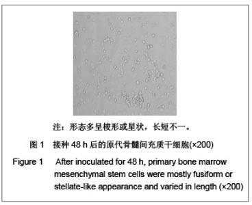
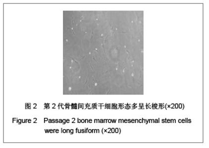
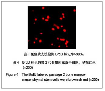
METHODS: The allogeneic bone marrow mesenchymal stem cells were cultured in vitro and then labeled with BrdU after the number of the cells reached to a certain amount (106). A total of 24 Sprague-Dawley rats were randomly divided into three groups. Rats in the normal control group did not undergo the anterior descending coronary artery ligation, and then 1 mL bone marrow mesenchymal stem cells were injected into the myocardium surrounding the anterior descending coronary artery; in the sham-transplantation group, 1 mL dulbecco’s modified eagle’s medium was injected into the surrounding infarcted myocardium at 7 days after anterior descending coronary artery ligation; in the transplantation group, bone marrow msenchymal stem cells in the same dose were injected into the surrounding infarcted myocardium at 7 days after anterior descending coronary artery ligation. The cardiac function of the rats was measured before transplantation and 5 days after transplantation.
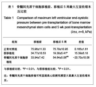
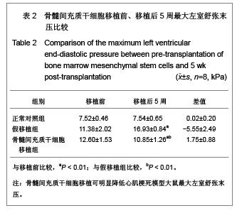
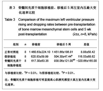
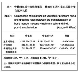
RESULTS AND CONCLUSION: The maximum left ventricular end-systolic pressure and maximum and minimum left ventricular pressure rising and dropping rates in the sham-transplantation group at 5 weeks after transplantation were lower than those before transplantation (P < 0.01), while the left ventricular end-diastolic pressure was higher than that before transplantation (P < 0.01); the maximum left ventricular end-systolic pressure and maximum and minimum left ventricular pressure rising and dropping rates in the transplantation group at 5 weeks after transplantation were higher than those before transplantation (P < 0.01), and the left ventricular end-diastolic pressure was lower than that before transplantation (P < 0.01). At 5 weeks after transplantation, the maximum left ventricular end-systolic pressure and maximum and minimum left ventricular pressure rising and dropping rates in the transplantation group were higher than those in the sham-transplantation group (P < 0.01), while the left ventricular end-diastolic pressure in the transplantation group was lower than that in the sham-transplantation group (P < 0.01). The results showed that bone marrow mesenchymal stem cell transplantation can significantly improve the cardiac function of a rat model of myocardial infarction.

Key words: stem cells, stem cell transplantation, bone marrow mesenchymal stem cells, transplantation, myocardial infarction, maximum left ventricular end-systolic pressure, maximum and minimum left ventricular pressure rising and dropping rate, cardiac function, injection transplantation, myocardial tissue, stem cell survival and differentiation, stem cell photographs-containing paper

CLC Number: