Chinese Journal of Tissue Engineering Research ›› 2024, Vol. 28 ›› Issue (11): 1762-1766.doi: 10.12307/2024.231
Previous Articles Next Articles
Delayed onset muscle soreness and exercise-induced skeletal muscle memory
Bai Zhengrong1, 2, Sun Yu3, Zhang Zhenxian1, Pan Shinong2
- 1Department of CT Diagnosis, Yan’an People’s Hospital, Yan’an 716000, Shaanxi Province, China; 2Department of Radiology, Shengjing Hospital, China Medical University, Shenyang 110000, Liaoning Province, China; 3The First Hospital of Jinzhou Medical University, Jinzhou 121000, Liaoning Province, China
-
Received:2022-12-21Accepted:2023-04-08Online:2024-04-18Published:2023-07-27 -
Contact:Pan Shinong, Professor, Department of Radiology, Shengjing Hospital, China Medical University, Shenyang 110000, Liaoning Province, China -
About author:Bai Zhengrong, MD candidate, Associate chief physician, Department of CT Diagnosis, Yan’an People’s Hospital, Yan’an 716000, Shaanxi Province, China; Department of Radiology, Shengjing Hospital, China Medical University, Shenyang 110000, Liaoning Province, China -
Supported by:Key Research & Development Project of Xi’an City (General Project), No. 2021YF-08 (to BZR)
CLC Number:
Cite this article
Bai Zhengrong, Sun Yu, Zhang Zhenxian, Pan Shinong. Delayed onset muscle soreness and exercise-induced skeletal muscle memory[J]. Chinese Journal of Tissue Engineering Research, 2024, 28(11): 1762-1766.
share this article
Add to citation manager EndNote|Reference Manager|ProCite|BibTeX|RefWorks
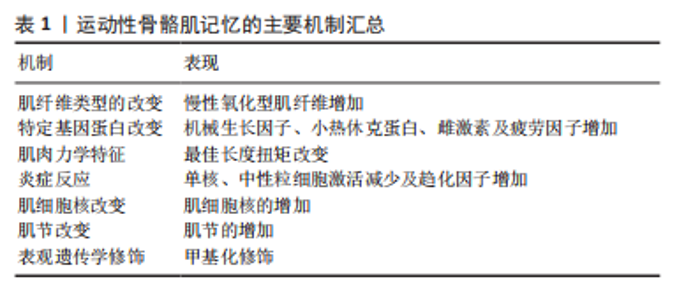
2.1 运动延迟性肌肉损伤中的骨骼肌记忆 骨骼肌损伤是运动中最常见损伤,离心运动导致骨骼肌损伤和迟发性肌肉酸痛多见于大强度、不习惯的剧烈离心运动、耐力及力量训练后,如马拉松及运动员的短期训练中,影响人群生活、工作、健身锻炼及专业运动人群运动训练、竞技比赛[1-5]。运动医学上主要是指运动后肌肉酸痛并伴有疲倦、乏力,甚至出现肌肉僵硬、痉挛,12-24 h出现、24-72 h达高峰,然后在运动后5-7 d逐渐消退和消失。损伤的机制目前国际主流观点倾向于机械损伤学说、代谢紊乱学说及炎症学说等多种因素的共同作用。对运动性肌肉损伤评价通常是在运动后的数天至数周内通过肌肉结构、功能、肌肉酸痛的间接标记进行检测。有趣的是,迟发性肌肉酸痛强度与其他运动性肌肉损伤间接标记物相关性很低,因此似乎不能反映肌肉损伤的程度[6-7]。关于迟发性肌肉酸痛的发生人们提出了几种假说来解释其发病机制,包括乳酸释放、痉挛、结缔组织损伤、肌肉损伤、炎症和氧化应激[8-9]。多年来炎症反应相关的生化、热和机械变化使肌肉传入神经纤维(Ⅲ型和Ⅳ型)敏感得到了较为广泛的支持[10]。直到2010年,MURASE和他的同事们[11-13]提出了神经营养因子(GDNF)和神经生长因子(NGF)在离心性收缩后迟发性肌肉酸痛的发展中起重要作用。 延迟性肌肉酸痛在日常生活中较为普遍,目前尚没有一种方法可以完全消除症状,因此寻找有效的防治方法将是未来的研究方向。运动本身是机体经受超负荷运动所致的由多个因素参与的对抗机体生理功能的反应,具有防御性是一种保护性反应,运动性骨骼肌记忆早在1983年已经为人所知了[14],有证据表明,第一次离心运动可以在随后的类似运动中产生预防运动性肌肉损伤效果[15-16],它被称为重复回合效应或者骨骼肌记忆 [17-20],其特点是减弱肌肉肿胀及肌肉损伤的标志[21-22],减轻迟发性肌肉酸痛,使肌肉力量和活动范围恢复更快[23]。运动性骨骼肌记忆被分为3类:神经性[24],外周机械性[25],肌肉本身肌细胞性[26]。神经性适应是由于高强度锻炼后细胞膜渗透压改变,细胞外液进入细胞内,促进炎症的产生,同时挤压神经痛觉感受器并释放化学物质刺激神经,引起脑部产生疼痛。神经适应不能完全解释骨骼肌的记忆,在第二次离心运动中,第一次对侧肢体的离心运动可以预防运动性肌肉损伤,然而对侧肢体的保护程度低于同侧肢体[27]。同样在电刺激引起的偏心收缩实验中,表明重复回合效应可独立于神经适应而发生,它涉及外周或肌肉适应[28]。SHARPLES等[29]提出长时间运动及短暂运动都存在肌肉记忆,通过训练-停止训练回到基线水平-再训练的运动干预过程是研究骨骼肌记忆的合适模型。在骨骼肌一次离心运动模型中发现这种保护作用[19,30-31],但这种保护效果有限[32],而通过几次的离心运动后这种适应效果更明显[33-34]。又有学者研究运动干预小鼠骨骼肌记忆,通过对腓肠肌、比目鱼肌及足底肌的研究发现,足底肌和腓肠肌的适应在6个月内完全消失[35],同时提出保护作用的大小不一定取决于初始肌肉损伤的严重程度[36-37]。有研究者提出事先下坡跑可对运动性肌肉损伤产生保护作用,但时间有限,通过下坡跑训练来提高跑步成绩至关重要[38]。BYRNES等[22]研究表明30 min下坡跑获得的重复回合效应在9周后消失,最大的运动性肌肉损伤导致最高的重复回合效应。因此骨骼肌在离心运动后的快速记忆提高了其临床应用价值,阐明离心运动骨骼肌记忆的机制能够为设计治疗干预措施及确定潜在治疗靶点提供宝贵意见。 2.2 目前有关运动性骨骼肌记忆的机制 2.2.1 纤维类型的改变 股四头肌纤维类型分为快速收缩糖酵解纤维和慢型氧化肌纤维,离心运动后小鼠骨骼肌纤维类型的比例与适应性有关,雄性SD大鼠后肢主要以快速收缩糖酵解纤维类型为主,离心训练后慢速氧化肌纤维的比例增加[37]。在离心运动后,氧化型肌纤维参与了对运动性肌肉损伤的保护作用[38]。 2.2.2 基因、蛋白表达的改变 是骨骼肌适应耐力运动的基础,离心运动明显上调了肌细胞活性和合成代谢信号通路[39],增加对肝脏型胰岛素样生长因子1[40]、机械生长因子(肌肉生长的正调节因子)[41]、微管相关蛋白或MAP蛋白(肌细胞机械状态敏感的蛋白质)的表达[42-43]。HSPB1 (Hsp27)和α-结晶蛋白的小热休克蛋白(sHSPs)在细胞适应中发挥重要作用,它涉及到未折叠蛋白的伴随、细胞骨架的稳定以及细胞氧化还原状态的调节和细胞凋亡的抑制[44],由于高敏感蛋白参与了对几种压力耐受性的发展,很可能由最初的运动回合引起的高敏感蛋白反应赋予了对第二次潜在破坏性运动的抵抗,有学者发现2次间隔3周的最大离心运动导致细胞骨架部分的热休克蛋白(HSPs)水平增加[26],尽管第二次运动造成的损伤比第一次运动要小,这些热休克蛋白更有效的移位可能是重复回合效应背后的机制。对人体受试者进行了2次下坡跑实验证明了当肌肉功能和迟发性肌肉酸痛评分恢复到运动前值时,活性氧达到峰值,这表明在离心收缩后的几天内,吞噬细胞产生的活性氧可能在调节恢复中发挥生理作用[45]。重复回合效应影响了循环血中的疲劳相关元素(K、Ca、Cl)、炎症反应和葡萄糖代谢(Zn)的浓度[46]。女性比男性更快地恢复到基线[29],雌激素水平升高可对运动性肌肉损伤产生保护作用[30],可能减少由局部炎症引起的继发性改变[31],与减少血浆肌酸激酶升高相关[32]。 2.2.3 肌肉力学特性 离心运动能够促进特定肌肉的慢性功能适应,这意味着离心训练能够将最佳长度-张力关系转移到较长的肌肉[47]。在偏心训练后,肌肉-肌腱复合体提高了承受应力的能力[48],归因于肌内结缔组织的增加,中间纤维系统的增强,Desmin是肌肉中间纤维的主要蛋白成分,在保护未来的肌节破坏中发挥作用[49-50]。从肌肉力学特性角度解释离心运动的训练效果,10名受试者腘绳肌训练效果可以通过肌肉最佳收缩长度的长期变化来证明,训练效果是肌肉纤维中肌节数量增加,角度-扭矩曲线变得更宽,收缩的最佳长度出现在更长的骨骼肌长度上,在一次偏心运动后,肌肉的扭矩曲线持续转移;较长肌肉的劳损会引起最佳角度向较长肌肉长度的转移[51],这种移位表明了一种训练效果,为肌肉提供了保护,防止离心运动造成进一步的损伤[52]。 2.2.4 肌节的增加 由于最初的偏心性发作导致的应力敏感纤维或肌节的去除,肌肉对运动性肌肉损伤的抵抗力更强[53-54],离心运动促进骨骼肌肌节的增加[55-56],这种肌节的纵向增加被认为有助于保护作用,可以避免肌节过度伸展超过重叠,从而导致肌节破坏。肌膜和肌浆网也被认为在最初的一轮离心运动后会变得更强[57],它限制钙稳态的破坏,从而防止钙蛋白酶激活和细胞骨架蛋白的降解。出现在平行肌原纤维中肌节数量增加代表肌节形成的不同阶段,受到偏心收缩的肌肉纤维通过增加新的肌节来适应不习惯的活动[58]。 2.2.5 炎症反应的改变 最初的一轮离心运动后,免疫反应会更有效地促进肌肉组织的再生,特别是通过增强炎症细胞浸润到肌肉和成肌细胞增殖[59],如单核细胞和中性粒细胞的激活降低与重复回合效应有关[60]。适应依赖于单核细胞趋化蛋白1 (MCP-1),通过巨噬细胞和卫星细胞之间的信号通路,单核细胞趋化蛋白1已被证明在单次离心运动后在转录水平上显著过表达,在第二次运动后表现得更大[61]。在反复的偏心运动后,趋化因子CCL2的上调和NF-kB活性的降低,可能有助于运动诱导的肌肉损伤的保护性适应。 2.2.6 肌细胞胞核的增加 离心运动促进了肌核的增加[55],同时肌膜和肌浆也在最初的离心运动后增加。肌纤维大小和肌核含量之间存在线性关系,在肌纤维增大和萎缩期间,通过细胞核增加(由肌肉卫星细胞供应)和细胞核丢失(由细胞凋亡)来保持肌核区域(相对)不变。长时间规律运动后存在骨骼肌记忆,增加的肌细胞核的数量有助于在随后的再次运动中更快的恢复,对肌肉在训练期间生长的能力产生长期的或永久的影响[29]。LEEDER 等[32]及MCHUGH 等[62]由阻力运动训练诱导的Sprague-Dawley大鼠实验中发现,运动干预后骨骼肌纤维中各种类型肌纤维含量、肌纤维大小和肌核数量发生改变,在随后的脱训练过程中肌纤维大小恢复,但肌核数量未恢复到基线水平,再次重复之前的运动后,肌纤维的大小恢复,肌核含量不变,这些增加的肌核有助于肌肉更快的恢复。相反,在肌肉萎缩模型中,肌核并未丢失[63-64]。这种肌核的永久性被认为与肌纤维在再训练中更有效地生长有关[65]。 又有学者研究运动干预小鼠骨骼肌记忆,通过对小鼠腓肠肌、比目鱼肌及足底肌的研究发现,肥大过程中获得的肌核随着肌肉的脱训练而丢失,足底肌和腓肠肌的肌肉适应在6个月内完全恢复,比目鱼肌肌核的增多发生在运动早期且与肌肉肥大分离,位于中心位置的肌核其形状可随训练及脱训练而改变[29]。早期参与阻力训练者,肌萎缩中观察到肌核凋亡来源于肌肉卫星细胞。在肌肉肥大过程中获得的肌核随着肌肉的脱训练而丢失[66]。 2.2.7 表观遗传学修饰的关系 运动适应和运动干预的表观遗传学可作为研究骨骼肌记忆的合适模型[23],骨骼肌记忆的表观遗传适应性,主要体现在终末分化的肌纤维和具有有丝分裂潜能的卫星细胞中。肌纤维是有丝分裂后的,表观遗传修饰短暂,对急性的适应很重要。卫星细胞可以增殖、自我更新,并将表观遗传修饰传递给子代群体[67],与骨骼肌的长期记忆有关。 利用小鼠运动训练模型——递进加权轮跑(PoWeR),在腓肠肌中观察到miRNA在训练时水平较低,在训练后仍较低,证明了miRNA水平能反映堆先前适应的记忆[29] 。SEABORNE 等[68]通过人类全基因组DNA甲基化在肌肉肥大、恢复至控制水平、随后再肥大后的分析中发现,与首次肥大相比,再肥大后整个基因组的低甲基化水平增加,即使在恢复过程中也保持低甲基化状态。提出在急性运动中GRIK2,TRAF1,BICC1,STAGI表现出表观遗传敏感性,在单次抵抗运动后表现出低甲基化,维持22周后,基因表达和肌肉质量在重新负荷后增加更大[69]。终身从事体育活动的健康老年男性骨骼肌的整体低甲基化水平,与同年龄组的相比氧化应激抵抗明显增强[70];反之由于缺乏运动造成骨骼肌组织中不同DNA甲基化模式的“记忆”也被报告。急性运动和慢性运动均会影响收缩肌肉中DNA甲基化的状态。在一场剧烈的自行车运动后,全基因组CpG甲基化下降显著[71],同时由慢性肥大训练引起的不同水平甲基化中,提出几个基因在随后的几轮运动中被保留[72-78]。 表1为目前运动性骨骼肌记忆的主要机制汇总。"
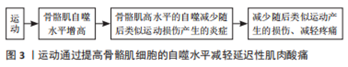
2.3 提出骨骼肌自噬参与了运动性骨骼肌记忆 在不习惯的离心运动迟发性肌肉酸痛时骨骼肌收缩后,骨骼肌的形态学发生改变,肌小节呈痉挛状态,部分肌丝断裂或消失,相对应处出现散在的小颗粒或减小颗粒有串成细的现状结构的趋势,并可见线粒体肿胀,溶酶体增多、增大。自噬是真核生物体内存在的溶酶体依懒性降解途径,在多种应激状态下激活,使受损的细胞器被包裹吞噬并转运至溶酶体降解回收,这也是适应训练的必要过程,它将有利于维持内环境的稳态,并具有组织及细胞保护和抗炎作用[79]。 运动引起的骨骼肌适应性反应,需要细胞凋亡和自噬的过程的参与[80]。 在运动过程中,细胞凋亡和自噬被激活以限制组织损伤,恢复组织完整性,是终止炎症反应或诱导适应的直接信号。自噬激活后被改变的蛋白质/细胞器,为肌肉可塑性提供条件、动员干细胞系统凋亡来进行适应。同时骨骼肌自噬在其质量控制中也起着至关重要的作用,能维持骨骼肌的内稳态和完整性,适当水平的运动诱导的自噬或自噬的改变还被认为是一种应激刺激,可以调节细胞信号并促进代谢适应[81],改善线粒体质量和静止卫星细胞的数量[82]。运动训练诱导骨骼肌适应和提高体能需要自噬,病理和生理刺激如急性运动可激活自噬。耐力运动训练诱导的骨骼肌适应和体能提高是通过增加骨骼肌细胞的基础自噬水平来实现的[83],自噬在骨骼肌中特别活跃,近年来已有研究证实当运动强度提高、超耐力跑步比赛、禁食状态下锻炼及急性严重缺氧会增加人体骨骼肌中的自噬体含量[84-85],有研究提出,长期适度运动能通过增强自噬进而恢复自噬受损类大鼠骨骼肌肌肉炎症。一些研究通过适当的刺激细胞自噬,预防由于久坐不动导致的衰老与疾病[86-87]。Levine 的研究团队在 2012 年提出,运动训练后细胞自噬水平通过Bcl2-Beclin1影响骨骼肌能量代谢[88]。短时间高强度运动和耐力运动同样可以提高骨骼肌细胞的自噬水平[86]。小鼠急性跑步机训练后,骨骼肌线粒体氧化应激和线粒体自噬增强,从而去除受损及功能失调的线粒体。 2.3.1 自噬和炎症的关系 研究表明,延迟性肌肉损伤的产生伴随着炎症反应的发生,出现局部的红、肿、热、痛等炎性表现,白细胞含量与白细胞介素1、白细胞介素6及趋化因子增多[89-90]。炎症是一种机体对组织损伤和病原体入侵的应激性反应,应激的不平衡会导致身体受伤。许多疾病都伴随着炎症反应,在多种病理状态下,自噬可抑制不同组织的炎症反应,起到组织保护作用。 在2型糖尿病中,通过细胞自噬可缓解2型糖尿病炎症,为2型糖尿病治疗提供新的途径[91]。用脂多糖刺激RAW264.7细胞后,槲皮素抑制炎症反应是通过激活IL-6/STAT3/FOXO3a通路增强自噬来实现的[92]。Atg5介导的自噬在肾小管中靶向NF-kB信号从而保护肾脏免受炎症[93]。二甲双胍通过下调Akt信号通路介导的自噬来保护心肌细胞免受缺血再灌注诱导的凋亡和炎症[94]。肿瘤相关炎症可预测预后,自噬可抑制癌症中的炎症,并通过多种机制影响细胞因子的产生和分泌,这与肿瘤的免疫和炎症微环境有关[95]。BCL2/腺病毒E1B 19 kD蛋白相互作用蛋白3 (BNIP3)通过激活自噬,降低脂多糖诱导的软骨细胞炎症和凋亡[96]。 2.3.2 运动-自噬与组织的适应性 运动诱导的自噬增强在多种组织器官均能发挥保护作用[97],运动可以激活心肌细胞自噬,使心脏在随后的高强度运动中得到保护。高强度运动与自噬中高LC311和低p62水平有关,这依赖于运动预处理心肌保护过程中的细胞效应[98]。运动训练介导的自噬在神经系统疾病中介导了神经系统疾病过程中的多种细胞病理状态[99]。LU等[100]研究表明,跑步机运动可明显缓解链霉素所致大鼠海马神经退行性障碍。TANG等[101]踏车运动可通过提高缺血周围脑组织微血管中MT1-MMP的表达,改善神经功能,促进血管生成。有氧运动可通过调节结肠癌的自噬来抵消恶病质[102]。根据以上分析,推测运动可以通过提高骨骼肌细胞的自噬水平,减轻二次运动中骨骼肌损伤后的炎症水平,进而减轻延迟性肌肉酸痛,见图3。"
| [1] ARMSTRONG RB, WARREN GL, WARREN JA. Mechanisms of exercise-induced muscle fibre injury. 1991;12(3):184-207. [2] VEQAR Z, IMTIYAZ S. Vibration Therapy in Management of Delayed Onset Muscle Soreness (DOMS). J Clin Diagn Res. 2014;8(6):LE01-4. [3] DEYHLE M, GIER A, EVANS K, et al. Skeletal Muscle Inflammation Following Repeated Bouts of Lengthening Contractions in Humans. 2015;6:424. [4] ABOODARDA SJ, GEORGE J, MOKHTAR AH, et al. Muscle strength and damage following two modes of variable resistance training. J Sports Sci Med. 2011;10(4):635-642. [5] HOTFIEL T, FREIWALD J, HOPPE MW, et al. Advances in Delayed-Onset Muscle Soreness (DOMS): Part I: Pathogenesis and Diagnostics. Sportverletz Sportschaden. 2018;32(4):243-250. [6] MORGAN DL. New insights into the behavior of muscle during active lengthening. Biophys J. 1990;57(2):209-221. [7] NOSAKA K, NEWTON M, SACCO P. Delayed-onset muscle soreness does not reflect the magnitude of eccentric exercise-induced muscle damage. Scand J Med Sci Sports. 2002;12(6):337-346. [8] ISNER-HOROBETI ME, DUFOUR SP, VAUTRAVERS P, et al. Eccentric exercise training: modalities, applications and perspectives. Sports Med. 2013;43(6):483-512. [9] HYLDAHL RD, HUBAL MJ. Lengthening our perspective: morphological, cellular, and molecular responses to eccentric exercise. Muscle Nerve. 2014;49(2):155-170. [10] FRIDÉN J, LIEBER RL. Structural and mechanical basis of exercise-induced muscle injury. Med Sci Sports Exerc. 1992;24(5):521-530. [11] MURASE S, TERAZAWA E, HIRATE K, et al. Upregulated glial cell line-derived neurotrophic factor through cyclooxygenase-2 activation in the muscle is required for mechanical hyperalgesia after exercise in rats. J Physiol. 20135;591(12):3035-3048. [12] MIZUMURA K, TAGUCHI T. Delayed onset muscle soreness: Involvement of neurotrophic factors. J Physiol Sci. 2016;66(1):43-52. [13] LIEBER RL, FRIDÉN J. Muscle damage is not a function of muscle force but active muscle strain. J Appl Physiol (1985). 1993;74(2):520-526. [14] SCHWANE JA, ARMSTRONG RB. Effect of training on skeletal muscle injury from downhill running in rats. J Appl Physiol Respir Environ Exerc Physiol. 1983;55(3):969-975. [15] BROWN M, HOWATSON G, KEANE K, et al. Adaptation to Damaging Dance and Repeated-Sprint Activity in Women. J Strength Cond Res. 2016;30(9):2574-2581. [16] BARSS T, MAGNUS C, CLARKE N, et al. Velocity-specific strength recovery after a second bout of eccentric exercise. J Strength Cond Res. 2014;28(2):339-349. [17] MCHUGH M. Can exercise induced muscle damage be avoided?. Br J Sports Med. 1999; 33(6):377. [18] MCHUGH MP, TETRO DT. Changes in the relationship between joint angle and torque production associated with the repeated bout effect. J Sports Sci. 2003;21(11):927-932. [19] CLARKSON PM, NOSAKA K, BRAUN B. Muscle function after exercise-induced muscle damage and rapid adaptation. Med Sci Sports Exerc. 1992;24(5):512-520. [20] SNIJDERS T, AUSSIEKER T, HOLWERDA A, et al. The concept of skeletal muscle memory: Evidence from animal and human studies. Acta Physiol (Oxf). 2020;229(3):e13465. [21] PIERRYNOWSKI MR, TÜDUS PM, PLYLEY MJ. Effects of downhill or uphill training prior to a downhill run. Eur J Appl Physiol Occup Physiol. 1987;56(6):668-672. [22] BYRNES W, CLARKSON P, WHITE J, et al. Delayed onset muscle soreness following repeated bouts of downhill running. J Appl Physiol (1985). 1985;59(3):710-715. [23] Nosaka K, Clarkson PM. Variability in serum creatine kinase response after eccentric exercise of the elbow flexors. Int J Sports Med. 1996;17(2):120-127. [24] LINDSTEDT SL, LASTAYO PC, REICH TE. l. When active muscles lengthen: properties and consequences of eccentric contractions. News Physiol Sci. 2001;16:256-261. [25] ABBOTT BC, WILKIE DR. The relation between velocity of shortening and the tension-length curve of skeletal muscle. J Physiol. 1952;117(3):26 P. [26] MCHUGH M, CONNOLLY D, ESTON R, et al. Exercise-induced muscle damage and potential mechanisms for the repeated bout effect. Sports Med. 1999;27(3):157-170. [27] HOWATSON G, VAN SOMEREN KA. Evidence of a contralateral repeated bout effect after maximal eccentric contractions. Eur J Appl Physiol. 2007;101(2):207-214. [28] BLACK CD, MCCULLY KK. Muscle injury after repeated bouts of voluntary and electrically stimulated exercise. Med Sci Sports Exerc. 2008;40(9):1605-1615. [29] SHARPLES AP, STEWART CE, SEABORNE RA. Does skeletal muscle have an ‘epi’-memory? The role of epigenetics in nutritional programming, metabolic disease, aging and exercise. Aging Cell. 2016;15(4):603-616. [30] VINCENT HK, VINCENT KR. The effect of training status on the serum creatine kinase response, soreness and muscle function following resistance exercise. Int J Sports Med. 1997;18(6):431-437. [31] NOSAKA K, CLARKSON PM. Muscle damage following repeated bouts of high force eccentric exercise. Med Sci Sports Exerc. 1995;27(9):1263-1269. [32] LEEDER J DC, VAN SOMEREN KA, GAZE D, et al. Recovery and adaptation from repeated intermittent-sprint exercise. Int J Sports Physiol Perform. 2014;9(3):489-496. [33] NICOL C, AVELA J, KOMI PV. The stretch-shortening cycle : a model to study naturally occurring neuromuscular fatigue. Sports Med. 2006;36(11):977-999. [34] HODY S, LEPRINCE P, SERGEANT K, et al. Human muscle proteome modifications after acute or repeated eccentric exercises. Med Sci Sports Exerc. 2011;43(12):2281-2296. [35] MURACH KA, MOBLEY CB, ZDUNEK CJ, et al. Muscle memory: myonuclear accretion, maintenance, morphology, and miRNA levels with training and detraining in adult mice. J Cachexia Sarcopenia Muscle. 2020;11(6):1705-1722. [36] BARNETT A. Using recovery modalities between training sessions in elite athletes: does it help? .Sports Med. 2006;36(9):781-796. [37] CHEN T, TSENG W, HUANG G, et al. Low-intensity eccentric contractions attenuate muscle damage induced by subsequent maximal eccentric exercise of the knee extensors in the elderly. Eur J Appl Physiol. 2013;113(4):1005-1015. [38] BONTEMPS B, VERCRUYSSEN F, GRUET M, et al. Downhill Running: What Are The Effects and How Can We Adapt? A Narrative Review. Sports Med. 2020;50(12):2083-2110. [39] HYLDAHL R, CHEN T, NOSAKA K. Mechanisms and Mediators of the Skeletal Muscle Repeated Bout Effect. Exerc Sport Sci Rev. 2017;45(1):24-33. [40] HODY S, LACROSSE Z, LEPRINCE P, et al. Effects of eccentrically and concentrically biased training on mouse muscle phenotype. Med Sci Sports Exerc. 2013;45(8):1460-1468. [41] DU C, ZHANG T, XIAO X, et al. Protease-activated receptor-2 promotes kidney tubular epithelial inflammation by inhibiting autophagy via the PI3K/Akt/mTOR signalling pathway. Biochem J. 2017;474(16):2733-2747. [42] NEUBAUER O, SABAPATHY S, ASHTON KJ, et al. Time course-dependent changes in the transcriptome of human skeletal muscle during recovery from endurance exercise: from inflammation to adaptive remodeling. J Appl Physiol (1985). 2014;116(3):274-287. [43] TEE JC, BOSCH AN, LAMBERT MI. Metabolic consequences of exercise-induced muscle damage. Sports Med. 2007;37(10):827-836. [44] BARASH I, MATHEW L, RYAN A, et al. Rapid muscle-specific gene expression changes after a single bout of eccentric contractions in the mouse. Am J Physiol Cell Physiol. 2004;286(2):C355-364. [45] HENTZEN E, LAHEY M, PETERS D, et al. Stress-dependent and -independent expression of the myogenic regulatory factors and the MARP genes after eccentric contractions in rats. J Physiol. 2006;570(Pt 1):157-167. [46] YU JG, MALM C, THORNELL LE. Eccentric contractions leading to DOMS do not cause loss of desmin nor fibre necrosis in human muscle. Histochem Cell Biol. 2002;118(1):29-34. [47] OREJUELA D, JORQUERA R, BERGERON A, et al. Hepatic stress in hereditary tyrosinemia type 1 (HT1) activates the AKT survival pathway in the fah-/- knockout mice model. J Hepatol. 2008;48(2):308-317. [48] CLOSE G, ASHTON T, CABLE T, et al. Effects of dietary carbohydrate on delayed onset muscle soreness and reactive oxygen species after contraction induced muscle damage. Br J Sports Med. 2005;39(12):948-953. [49] DIAS S, WEBER M, PADOIN S, et al. Circulating Concentration of Chemical Elements During Exercise-Induced Muscle Damage and the Repeated Bout Effect. Biol Trace Elem Res. 2022;200(3):1060-1070. [50] OOSTHUYSE T, BOSCH AN. The Effect of Gender and Menstrual Phase on Serum Creatine Kinase Activity and Muscle Soreness Following Downhill Running. Antioxidants (Basel). 2017;6(1):16. [51] CARTER A, DOBRIDGE J, HACKNEY AC. Influence of estrogen on markers of muscle tissue damage following eccentric exercise. Fiziol Cheloveka. 2001;27(5):133-137. [52] WELSH MC, ALLEN DL, BYRNES WC. Plasma matrix metalloproteinase-9 response to downhill running in humans. Int J Sports Med. 2014;35(5):363-370. [53] WESTERLIND K, BYRNES W, HARRIS C, et al. Alterations in oxygen consumption during and between bouts of level and downhill running. Med Sci Sports Exerc. 1994;26(9):1144-1152. [54] HOPPELER H. Moderate Load Eccentric Exercise; A Distinct Novel Training Modality. Front Physiol. 2016;7:483.. [55] PETTITT R, SYMONS D, EISENMAN P, et al. Eccentric strain at long muscle length evokes the repeated bout effect. J Strength Cond Res. 2005;19(4):918-924. [56] BROCKETT CL, MORGAN DL, PROSKE U. Human hamstring muscles adapt to eccentric exercise by changing optimum length. Med Sci Sports Exerc. 2001;33(5):783-790. [57] ARMSTRONG RB. Mechanisms of exercise-induced delayed onset muscular soreness: a brief review. Med Sci Sports Exerc. 1984;16(6):529-538. [58] NEWHAM DJ, JONES DA, CLARKSON PM. Repeated high-force eccentric exercise: effects on muscle pain and damage. J Appl Physiol (1985). 1987;63(4):1381-1386. [59] HOWELL JN, CHLEBOUN G, CONATSER R. Muscle stiffness, strength loss, swelling and soreness following exercise-induced injury in humans. J Physiol. 1993;464:183-196. [60] FÉASSON L, STOCKHOLM D, FREYSSENET D, et al. Molecular adaptations of neuromuscular disease-associated proteins in response to eccentric exercise in human skeletal muscle. J Physiol. 2002;543(Pt 1):297-306. [61] LEHTI TM, KALLIOKOSKI R, KOMULAINEN J. Repeated bout effect on the cytoskeletal proteins titin, desmin, and dystrophin in rat skeletal muscle. J Muscle Res Cell Motil. 2007;28(1):39-47. [62] MCHUGH MP. Recent advances in the understanding of the repeated bout effect: the protective effect against muscle damage from a single bout of eccentric exercise. Scand J Med Sci Sports. 2003;13(2):88-97. [63] LYNN R, MORGAN D L. Decline running produces more sarcomeres in rat vastus intermedius muscle fibers than does incline running. J Appl Physiol (1985). 1994;77(3):1439-1444. [64] YU JG, CARLSSON L, THORNELL LE. Evidence for myofibril remodeling as opposed to myofibril damage in human muscles with DOMS: an ultrastructural and immunoelectron microscopic study. Histochem Cell Biol. 2004;121(3):219-227. [65] YU JG, FÜRST DO, THORNELL LE. The mode of myofibril remodelling in human skeletal muscle affected by DOMS induced by eccentric contractions. Histochem Cell Biol. 2003;119(5):383-393. [66] PEAKE J, NOSAKA K, SUZUKI K. Characterization of inflammatory responses to eccentric exercise in humans. Exerc Immunol Rev. 2005;11:64-85. [67] PIZZA FX, DAVIS BH, HENRICKSON SD, et al. Adaptation to eccentric exercise: effect on CD64 and CD11b/CD18 expression. J Appl Physiol (1985). 1996;80(1):47-55. [68] SEABORNE RA, STRAUSS J, COCKS M, et al. Human Skeletal Muscle Possesses an Epigenetic Memory of Hypertrophy. Sci Rep. 2018;8(1):1898. [69] LEE H, KIM K, KIM B, et al. A cellular mechanism of muscle memory facilitates mitochondrial remodelling following resistance training. J Physiol. 2018;596(18):4413-4426. [70] BRUUSGAARD JC, EGNER IM, LARSEN TK, et al. No change in myonuclear number during muscle unloading and reloading. J Appl Physiol (1985). 2012;113(2):290-296. [71] EGNER I, BRUUSGAARD J, EFTESTøL E, et al. A cellular memory mechanism aids overload hypertrophy in muscle long after an episodic exposure to anabolic steroids. J Physiol. 2013;591(24):6221-6230. [72] GUNDERSEN K, BRUUSGAARD JC, EGNER IM, et al. Muscle memory: virtues of your youth?. J Physiol. 2018;596(18):4289-4290. [73] MURASE S, TERAZAWA E, QUEME F, et al. Bradykinin and nerve growth factor play pivotal roles in muscular mechanical hyperalgesia after exercise (delayed-onset muscle soreness). J Neurosci. 2010;30(10):3752-3761. [74] SHARPLES AP, HUGHES DC, DEANE CS, et al. Longevity and skeletal muscle mass: the role of IGF signalling, the sirtuins, dietary restriction and protein intake. Aging Cell. 2015;14(4):511-523. [75] LASTAYO P, PIEROTTI D, PIFER J, et al. Eccentric ergometry: increases in locomotor muscle size and strength at low training intensities. Am J Physiol Regul Integr Comp Physiol. 2000;278(5):R1282-1288. [76] NITERT MD, DAYEH TD, VOLKOV P, et al. Impact of an exercise intervention on DNA methylation in skeletal muscle from first-degree relatives of patients with type 2 diabetes. Diabetes. 2012;61(12):3322-3332. [77] SAILANI MR, HALLING JF, MøLLER HD, et al. Lifelong physical activity is associated with promoter hypomethylation of genes involved in metabolism, myogenesis, contractile properties and oxidative stress resistance in aged human skeletal muscle. Sci Rep. 2019;9(1):3272. [78] BARRÈS R, YAN J, EGAN B, et al. Acute exercise remodels promoter methylation in human skeletal muscle. Cell Metab. 2012;15(3):405-411. [79] DERETIC V, LEVINE B. Autophagy balances inflammation in innate immunity. Autophagy. 2018;14(2):243-251. [80] LAKER RC, DRAKE JC, WILSON RJ, et al. Ampk phosphorylation of Ulk1 is required for targeting of mitochondria to lysosomes in exercise-induced mitophagy. Nat Commun. 2017;8(1):548. [81] MOOREN F C, KRUGER K. Exercise, Autophagy, and Apoptosis. Prog Mol Biol Transl Sci. 2015;135:407-422. [82] LIANG J, ZENG Z, ZHANG Y, et al. Regulatory role of exercise-induced autophagy for sarcopenia. Exp Gerontol. 2020;130:110789. [83] LIRA VA, OKUTSU M, ZHANG M, et al. Autophagy is required for exercise training-induced skeletal muscle adaptation and improvement of physical performance. FASEB J. 2013;27(10):4184-4193. [84] YUAN JQ, YUAN Y, PAN SS, et al. Altered expression levels of autophagy-associated proteins during exercise preconditioning indicate the involvement of autophagy in cardioprotection against exercise-induced myocardial injury. J Physiol Sci. 2020;70(1):10. [85] MOREIRA O, ESTÉBANEZ B, MARTÍNEZ-FLOREZ S, et al. Mitochondrial Function and Mitophagy in the Elderly: Effects of Exercise. Oxid Med Cell Longev. 2017;2017:2012798. [86] JAMART C, BENOIT N, RAYMACKERS J, et al. Autophagy-related and autophagy-regulatory genes are induced in human muscle after ultraendurance exercise. Eur J Appl Physiol. 2012;112(8):3173-3177. [87] JAMART C, FRANCAUX M, MILLET G, et al. Modulation of autophagy and ubiquitin- proteasome pathways during ultra-endurance running. 2012;112(9):1529-1537. [88] HE C, BASSIK M, MORESI V, et al. Exercise-induced BCL2-regulated autophagy is required for muscle glucose homeostasis. Nature. 2012;481(7382):511-515. [89] JAFARIYAN S, MONAZZAMI A, NIKOUSEFAT Z, et al. Inflammatory and immune responses to a 3-day period of downhill running in active females. Cell Mol Biol (Noisy-le-grand). 2017;63(7):76-83. [90] LIU X, CHEN P, ZHAO L, et al. Macrophages depletion impairs skeletal muscle regeneration by regulating inflammation and oxidative stress levels. Sheng Li Xue Bao. 2018;70(1):23-32. [91] HE Q, WANG L, ZHAO R, et al. Mesenchymal stem cell-derived exosomes exert ameliorative effects in type 2 diabetes by improving hepatic glucose and lipid metabolism via enhancing autophagy. Stem Cell Res Ther. 2020;11:223. [92] RONG X, XU J, JIANG Y, et al. Citrus peel flavonoid nobiletinalleviates lipopolysaccharide-induced inflammation by activating IL-6/STAT3/FOXO3a-mediated autophagy. Food Funct. 2021;12(3):1305-1317. [93] PENG X, WANG Y, LI H, et al. ATG5-mediated autophagy suppresses NF-kappaB signaling to limit epithelial inflammatory response to kidney injury. Cell Death Dis. 2019;10(4):253. [94] HUANG KY, QUE JQ, HU ZS, et al. Metformin suppresses inflammation and apoptosis of myocardiocytes by inhibiting autophagy in a model of ischemia-reperfusion injury. Int J Biol Sci. 2020;16(14):2559-2579. [95] MONKKONEN T, DEBNATH J. Inflammatory signaling cascades and autophagy in cancer. Autophagy. 2018;14(2):190-198. [96] MA Z, WANG D, WENG J, et al. BNIP3 decreases the LPS-induced inflammation and apoptosis of chondrocytes by promoting the development of autophagy. J Orthop Surg Res. 2020;15(1):284. [97] ZHANG Y, CHEN N. Autophagy Is a Promoter for Aerobic Exercise Performance during High Altitude Training. Oxid Med Cell Longev. 2018;2018:3617508. [98] WANG L, WANG J, CRETOIU D, et al. Exercise-mediated regulation of autophagy in the cardiovascular system. J Sport Health Sci. 2020;9(3):203-210. [99] XING Y, YANG SD, WANG MM, et al. The beneficial roles of exercise training via autophagy in neurological diseases and possible mechanisms. Life Sci. 2019;221:130-134. [100] LU Y, DONG Y, TUCKER D, et al. Treadmill Exercise Exerts Neuroprotection and Regulates Microglial Polarization and Oxidative Stress in a Streptozotocin-Induced Rat Model of Sporadic Alzheimer’s Disease. J Alzheimers Dis. 2017;56(4):1469-1484. [101] TANG Y, ZHANG Y, ZHENG M, et al. Effects of treadmill exercise on cerebral angiogenesis and MT1-MMP expression after cerebral ischemia in rats. Brain Behav. 2018;8(8):e01079. [102] PIGNA E, BERARDI E, AULINO P, et al. Aerobic Exercisand Pharmacological Treatments Counteract Cachexia by Modulating Autophagy in Colon Cancer. Sci Rep. 2016;6:26991. |
| [1] | Geng Zhizhong, Pei Ziwen, Yan Gongli, Chen Jian. A meta-analysis of kinesio taping in the treatment of delayed onset muscle soreness [J]. Chinese Journal of Tissue Engineering Research, 2020, 24(35): 5733-5740. |
| Viewed | ||||||
|
Full text |
|
|||||
|
Abstract |
|
|||||