Chinese Journal of Tissue Engineering Research ›› 2019, Vol. 23 ›› Issue (1): 132-138.doi: 10.3969/j.issn.2095-4344.0693
Previous Articles Next Articles
Interactions between bone marrow stromal stem cells and nano-hydroxyapatite
Li Lu1, Shu Jingyuan1, Zheng Lixia2, Cui Yingying1, Niu Yueyue1, Wang Qian1, Wang Qingshan1
- 1Department of Pediatric Stomatology, Affiliated Hospital of Binzhou Medical University, Binzhou 256603, Shandong Province, China; 2Department of Stomatology, Liaocheng Retired Soldier Hospital, Liaocheng 252000, Shandong Province, China
-
Revised:2018-09-06Online:2019-01-08Published:2018-11-28 -
Contact:Wang Qingshan, Master, Professor, Department of Pediatric Stomatology, Affiliated Hospital of Binzhou Medical University, Binzhou 256603, Shandong Province, China -
About author:Li Lu, Master candidate, Department of Pediatric Stomatology, Affiliated Hospital of Binzhou Medical University, Binzhou 256603, Shandong Province, China -
Supported by:the Natural Science Foundation of Shandong Province, No. ZR2018LH010 (to WQS); the Medical and Health Technological Development Plan of Shandong Province, No. 2013WS0308 (to WQS)
CLC Number:
Cite this article
Li Lu, Shu Jingyuan, Zheng Lixia, Cui Yingying, Niu Yueyue, Wang Qian, Wang Qingshan. Interactions between bone marrow stromal stem cells and nano-hydroxyapatite[J]. Chinese Journal of Tissue Engineering Research, 2019, 23(1): 132-138.
share this article
Add to citation manager EndNote|Reference Manager|ProCite|BibTeX|RefWorks
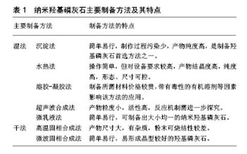
2.1 nHA结构、性质及制备方法 天然骨组织为无机相(主要是羟基磷灰石)和有机相(主要是Ⅰ型胶原纤维)的矿化复合物,在骨组织矿化过程中,纳米尺寸的羟基磷灰石晶体以层状形式周期性沉积于胶原纤维间隙间,这种层次结构使天然骨组织具有优异强度及韧性[14]。人工合成的nHA(Ca10(PO4)6(OH)2)晶体结构与骨组织中的无机成分类似,晶体属六方晶系,结构为六棱柱体,其晶胞特征为a1=a2=0.943 2 nm,c=0.688 1 nm,a轴互成120°夹角,a轴与c轴垂直[15]。从羟基磷灰石晶体结构来看,Ca2+容易与阳离子发生交换,Mg2+、Mn2+、Zn2+、Sr2+,OH-易与阴离子交换并被吸附,除此之外还可与蛋白质、有机酸等发生反应。这种离子交换反应可改变羟基磷灰石晶体生长习性,从而改变羟基磷灰石机械性能[16]。因此,大量学者对nHA及复合物进行研究。大量的实验研究表明nHA有优良的生物学活性及骨诱导性、骨传导性,是一种较为理想的骨缺损修复材料,能够广泛应用于骨缺损的修复、种植体表面改性、研制生物型人工韧带、引导骨组织再生膜材料和治疗股骨头坏死等方面。 nHA的制备方法主要有沉淀法、水热法、溶胶-凝胶法、超声合成法、微乳液法、固相反应法等[17],见表1。"

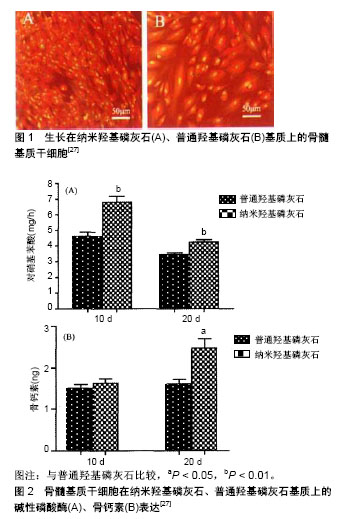
尹美林等[18]采用化学沉淀方法制备nHA,并对其进行表征,扫描电镜下见羟基磷灰石为针状晶体,结晶程度好,粒径分布均匀。 2.2 BMSCs生物学特性 20世纪60年代Friedenstein等在骨髓细胞培养中发现一群成纤维样、贴壁生长与造血干细胞不同的细胞,其主要来源于中胚层,在骨膜、肌肉、外周血以及结缔组织中均有存在,以骨髓和脐血中含量最为丰富。研究发现其具有较强的自我更新、多向分化潜能,可在不同的诱导条件下分化为成骨细胞、软骨细胞、脂肪细胞、肌细胞等多种细胞。徐俊昌等[19]在体外分离培养BMSCs并对其表面标志物、多向分化特性进行鉴定,结果显示BMSCs表面表达CD105、CD166、CD29,成骨诱导分化后,胞体变宽,钙结节茜素红染色、碱性磷酸酶染色为阳性,成软骨分化3周后阿利辛染色阳性,成脂肪诱导后细胞胞浆内出现脂肪小体。 2.3 nHA及其复合材料对BMSCs的影响 2.3.1 nHA微粒浓度对BMSCs的影响 Liu等[20]研究了不同浓度nHA微粒对BMSCs增殖分化的影响,研究结果显示,当nHA微粒浓度<20 μg/104个细胞时,nHA颗粒可促进BMSCs的增殖,但差异无显著性意义,nHA微粒浓度为20 μg/104个细胞时,可显著促进BMSCs的增殖,nHA微粒浓度大于20 μg/104个细胞时,可显著抑制BMSCs的增殖;在无血清培养条件下,nHA浓度为20 μg/104个细胞时,没有促进BMSCs增殖的作用,而浓度大于20 μg/104个细胞时,nHA微粒显著抑制BMSCs的增殖;在成骨诱导培养液中,nHA微粒浓度< 20 μg/104个细胞时,对BMSCs矿化无显著促进作用,随着微粒浓度的增加,其对BMSCs成骨分化的促进作用逐渐增强,呈浓度依从性关系,但当浓度大于20 μg/104个细胞时,其对BMSCs矿化促进作用随微粒浓度的增加而逐渐减弱。研究发现,羟基磷灰石生物毒性受颗粒直径、接触剂量和接触方式的影响[21]。Remya等[22]将不同质量浓度nHA(10,50,100,500,1 000 mg/L)与BMSCs相互作用,结果显示,暴露在高质量浓度(1 000 mg/L)nHA溶液中悬浮细胞出现纺锤形改变和颗粒状细胞质的变化,随着nHA质量浓度的增加,细胞形态出现极其不规则状,细胞扩散模式受限制;通过检测线粒体活动对细胞生存能力进行评估,结果表明,低质量浓度中的细胞生存能力与对照组无显著差异,高质量浓度中的细胞存活率约60%;对细胞氧化应激进行检测发现,高质量浓度nHA溶液中细胞皱褶变化高,而早期细胞凋亡则出现在nHA较低的质量浓度中。nHA微粒甚至更高的质量浓度只改变了细胞形态,并不诱导细胞凋亡。 nHA在复合材料中所占比例同样对BMSCs有明显影响。Zhu等[23]以含体积分数为10%胎牛血清的DMEM完全培养基为阴性实验对照组,以0.64%质量浓度苯酚为阳性实验对照组,将BMSCs与材料浸提液共同培养为实验组,观察第3,5,7天培养的BMSCs相对生长情况。结果显示,实验组细胞吸光度值显著增加,BMSCs的相对生长高于对照组和阴性组。在纳米羟基磷灰石/聚乙交酯-丙交酯(nanograde hydroxyapatite/poly lactic-co-glycolic acid,nHA/PLGA)复合支架材料中,当nHA/PLGA复合体积比为50/50时,BMSCs在黏附性、增殖率、成骨分化等方面均优于其他比例,表明合适比例的nHA在生物活性及骨传导方面扮演者重要的角色[24]。谭羽莹等[25]观察改性PLGA/HA复合材料与BMSCs体外培养后的细胞活性及生物相容性,MTT比色法检测在不同浓度PLGA/HA浸提液(10%,30%,50%,80%)中BMSCs的增殖情况,观察材料表面细胞的黏附性及细胞形态,结果显示,高浓度材料浸提液(50%,80%)对BMSCs的体外增殖呈负影响,但CTS均在1级,符合安全评价标准;实验还表明,BMSCs能够黏附并伸展形成伪足固定于支架材料表面,部分细胞已融合成细胞层,可见细胞外基质。 2.3.2 nHA支架材料表面微形貌对BMSCs的影响 生物材料表面状态及物理结构对生物性能有较大的影响,天然骨组织中的羟基磷灰石以数十纳米的晶体形式存在,而正是这些数十纳米晶体的有序排列构成了骨组织的力学属性。nHA表面与细胞相互作用,其化学成分和表面形貌强烈影响着细胞的生存、附着、迁移分化和增殖[26]。周琰春等[27]采用十六烷基三甲基溴化铵(cetyltrimethylammonium bromide,CTAB)为添加剂,在水相体系中制备了形貌均匀的nHA粉末,以无CTAB添加剂的普通羟基磷灰石粉末为对照,分别制备成羟基磷灰石膜并对其物相进行表征,nHA粉末外貌呈球形,为多晶状态,分散好,大小均匀,具有较好的浸润性及亲水性。普通羟基磷灰石粉末外貌呈条棒状,单晶态,浸润性及亲水性不及nHA。将分离培养后的BMSCs在两种基质上复合,观察BMSCs增殖、分化状况,结果证明,nHA基质表面细胞数量显著高于普通羟基磷灰石基质,而且能获得更高的碱性磷酸酶、骨钙素表达,说明羟基磷灰石支架材料表面微形貌的构建对BMSCs增殖、分化具有明显影响,见图1,2。"
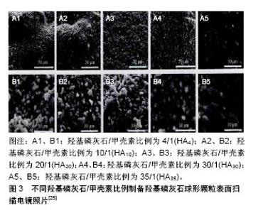
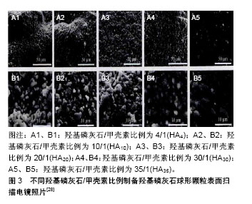
智伟等[28]通过调节溶胶-凝胶体系中羟基磷灰石粉末和甲壳素的质量比,制备出具有不同表面微形貌的羟基磷灰石球形颗粒。研究结果表明,羟基磷灰石所占比例越高,其孔隙率越高,在羟基磷灰石/甲壳素为4/1和20/1时(以HA4、HA20表示),BMSCs与其共培养后,球形颗粒表面均可见有大量均匀矿化层覆盖,且覆盖于HA4球形颗粒表面的晶片尺寸普遍大于HA20。而在羟基磷灰石/甲壳素为35/1(HA35)的致密光滑球形表面,只有局部区域观察到少量絮状矿化物覆盖,说明更高微孔率和粗糙度的羟基磷灰石球形颗粒表面具有更好的形成类骨磷灰石的能力,见图3。"
| [1] 李强,王建武,党建军,等.三维骨组织工程复合体修复兔股骨头坏死的实验研[J].陕西医药杂志,2017,12(3):275-277.[2] Kalita SJ, Bhardwaj A, Bhatt HA. Nanocrystalline calcium phosphate ceramics in biomedical engineering. Materials Science & Engineering C. 2007;27(3):441-449. [3] Goldner G, Pötter R.Radiotherapy in lymph node-positive prostate cancer patients - a potential cure? Single institutional experience regarding outcome and side effects.Front Radiat Ther Oncol. 2008;41:68-76.[4] 刘芳.纳米材料的结构与性质[J].光谱实验室, 2011,28(2): 735-738.[5] 赵玉岭.纳米材料性质及应用[J].煤炭技术,2009,28(8):149-151.[6] 朱婧.纳米材料在医学影像上的应用[D].苏州:苏州大学:2016.[7] Yang YC, Chao KS, Lin CP, et al. Oxaliplatin regulates DNA repair responding to ionizing radiation and enhances radiosensitivity of human cervical cancer cells.Int J Gynecol Cancer. 2009;19(4):782-786. [8] Braux J, Velard F, Guillaume C, et al. A new insight into the dissociating effect of strontium on bone resorption and formation.Acta Biomater. 2011;7(6):2593-2603. [9] 鹿鸣.碳纤维增强纳米羟基磷灰石/聚酰胺66复合材料力学性能和组织相容性研究[D].北京:中国人民解放军总医院,2015.[10] 陈津.间充质(干)细胞的定义变迁[J/CD].中华细胞与干细胞杂志(电子版),2017,7(4):247-250.[11] Dominici M, Le Blanc K, Mueller I, et al. Minimal criteria for defining multipotent mesenchymal stromal cells. The International Society for Cellular Therapy position statement. Cytotherapy. 2006;8(4):315-317. [12] Dulchavsky D, Gao X, Liu YB, et al. Bone marrow-derived stromal cells (BMSCs) interact with fibroblasts in accelerating wound healing.J Invest Surg. 2008;21(5):270-279. [13] Kumar S, Chanda D, Ponnazhagan S.Therapeutic potential of genetically modified mesenchymal stem cells.Gene Ther. 2008;15(10):711-715. [14] Liu Y, Luo D, Wang T.Hierarchical Structures of Bone and Bioinspired Bone Tissue Engineering.Small. 2016;12(34): 4611-4632. [15] 刘飚,李仕群,张宁,等.微量元素对羟基磷灰石晶体结构的影响[J].济南大学学报,2006,20(3):193-194.[16] 陈德敏.生物陶瓷材料[J].口腔材料器械杂志, 2007,15(2): 94-100.[17] 庞晓峰,曾红娟.纳米羟基磷灰石粉体的生物活性的研究[J].材料工程,2009,4:14-17,22.[18] 尹美林,赵善科,郑岩,等.羟基磷灰石纳米晶体的制备?表征及补钙效能研究[J].化学试剂,2018,40(3),212-216.[19] 徐俊昌,吴涛,吴桂华,等.人骨髓基质干细胞体外培养和生物学特性鉴定[J].中国现代医学杂志,2009,19(7):999-1002.[20] Liu Y, Wang G, Cai Y, et al. In vitro effects of nanophase hydroxyapatite particles on proliferation and osteogenic differentiation of bone marrow-derived mesenchymal stem cells.J Biomed Mater Res A. 2009;90(4):1083-1091. [21] Tomoaia G, Pop LB, Petean I, et al. Significance of Surface Structure on Orthopedic Materials. Materiale Plastice. 2012; 49(1):48-54. [22] Remya NS, Syama S, Gayathri V, et al. An in vitro study on the interaction of hydroxyapatite nanoparticles and bone marrow mesenchymal stem cells for assessing the toxicological behaviour.Colloids Surf B Biointerfaces. 2014; 117:389-397. [23] Zhu W, Guo D, Chen Y, et al. Cytocompatibility of PLA/Nano-HA composites for interface fixation.Artif Cells Nanomed Biotechnol. 2016;44(4):1122-1126. [24] He S, Lin KF, Sun Z, et al. Effects of Nano-hydroxyapatite/ Poly(DL-lactic-co-glycolic acid) Microsphere-Based Composite Scaffolds on Repair of Bone Defects: Evaluating the Role of Nano-hydroxyapatite Content.Artif Organs. 2016; 40(7):E128-135. [25] 谭羽莹,张舵,李玉新,等.改性纳米羟基磷灰石/PLGA同骨髓基质干细胞复合后相关生物学评价[J].中国组织工程研究与临床康复,2011,15(25):4619-4622.[26] Abdel-Gawad EI, Awwad SA. Biocompatibility of intravenous nano hydroxyapatite in male rats. Gawad. 2010;8(9):60-68. [27] 周琰春,蔡玉荣,刘丽,等.球形羟基磷灰石纳米颗粒的可控合成及其对间充质干细胞生长分化的影响[J].无机化学学报, 2007, 23(8):1335-1340.[28] 智伟,匙峰,李金雨,等.羟基磷灰石球形颗粒表面微型貌构建及其对干细胞生物学行为的调控[J].无机材料学学报, 2017,32(3): 319-325.[29] 杨华伟,陈凯,尚光伟,等.粗糙钛表面纳米掺锶羟基磷灰石涂层对BMSCs成骨分化的影响[J].同济大学学报,2015,36(1):13-17.[30] 郭凌云,张劲娥,袁建兵,等.快速原型技术制备Nano-HA/PCL支架与犬骨髓基质干细胞体外复合研究[J].口腔颌面外杂志, 2015, 25(1):34-38.[31] Fu DL, Jiang QH, He FM, et al. Adhesion of bone marrow mesenchymal stem cells on porous titanium surfaces with strontium-doped hydroxyapatite coating. J Zhejiang Univ Sci B. 2017;18(9):778-788. [32] 乔瑞红,王东,孙海钰,等.骨髓基质干细胞与负载去细胞基质的壳聚糖/纳米羟基磷灰石复合微球联合培养的生物相容性研究[J].中华创伤骨科杂志,2014,16(8):710-714.[33] 史月华,骨志远,郑园娜,等.掺镁羟基磷灰石涂层对种植体骨结合的影响[J].口腔医学,2014,34(4):249-252.[34] 付昆,孟志斌,邵增务,等.人骨髓基质干细胞与表面置换珊瑚羟基磷灰石培养的定向诱导分化[J].颈腰痛杂志, 2008,29(6): 519-522.[35] 薛震,牛丽媛,安刚,等.纳米羟基磷灰石/壳聚糖/半水硫酸钙为可注射骨组织工程支架材料的可行性[J].中国组织工程研究, 2015, 19(8):1169-1164.[36] 李家峰,徐金霞,管海虹,等.纳米羟基磷灰石/聚已内酯复合大鼠骨髓间充质干细胞的生物相容性[J].中国组织工程研究, 2012, 16(38):7042-7046.[37] 宋华,任向前,未东兴.纳米羟基磷灰石对缺损骨再生的影响[J].中国组织工程研究,2015,19(8):1155-1159.[38] 李昂,王晓宇,李泽成,等.人骨髓基质干细胞与支架材料纳米羟基磷灰石/聚酰胺66的生物相容性研究[J].中华创伤骨科杂志, 2016,18(3):241-246.[39] 王立新,袁峰,万怡灶,等.纳米羟基磷灰石/细菌纤维素复合组织工程支架的细胞毒性和生物相容性[J].中国组织工程研究, 2014, 18(47):7615-7620.[40] 李善昌,孔祥盼,李德超.纳米羟基磷灰石修复种植体周围骨缺损的实验研究[J].口腔医学研究,2008,24(5):537-539.[41] 王晓敏,李海坤,李旭东,等.胶原-羟基磷灰石复合骨组织引导再生性膜的细胞相容性实验研究[J].世界科技研究与发展, 2011, 33(6):1060-1062.[42] 刘彪,李运峰,胡静.含锶羟基磷灰石对骨质疏松症大鼠的影响[J].中国骨质疏松杂志,2015,21(5):524-530.[43] 徐学武,郭铁芳,韩雪峰,等.预制作生物工程性肌骨瓣的实验研究[J].哈尔滨医科大学学报,2007,41(4):327-332.[44] 徐勇,安良,曾丹林,等.羟基磷灰石纳米带作为蛋白药物载体的研究[J].功能材料,2018,49(3):03124-03129,03135.[45] 韩丽丽,张慧慧,马惠芳,等.纳米羟基磷灰石对暖巢癌细胞株的抑制作用研究[J].新医学,2017,48(11):775-778.[46] Gupta SK, Kumar R, Mishra NC.Influence of quercetin and nanohydroxyapatite modifications of decellularized goat-lung scaffold for bone regeneration.Mater Sci Eng C Mater Biol Appl. 2017;71:919-928. [47] 唐月军.纳米增韧(HA-ZrO2系)生物复合多孔陶瓷的制备及其颌骨缺损修复实验研究[D].上海:第二军医大学,2006.[48] Lai GJ, Shalumon KT, Chen JP.Response of human mesenchymal stem cells to intrafibrillar nanohydroxyapatite content and extrafibrillar nanohydroxyapatite in biomimetic chitosan/silk fibroin/nanohydroxyapatite nanofibrous membrane scaffolds.Int J Nanomedicine. 2015;10:567-584. [49] Luo Y, Lode A, Wu C, et al. Alginate/nanohydroxyapatite scaffolds with designed core/shell structures fabricated by 3D plotting and in situ mineralization for bone tissue engineering.ACS Appl Mater Interfaces. 2015;7(12): 6541-6549. [50] Zhu W, Guo D, Chen Y, et al. Cytocompatibility of PLA/Nano-HA composites for interface fixation.Artif Cells Nanomed Biotechnol. 2016;44(4):1122-1126. [51] Yang W, Both SK, Zuo Y, et al. Biological evaluation of porous aliphatic polyurethane/hydroxyapatite composite scaffolds for bone tissue engineering.J Biomed Mater Res A. 2015;103(7): 2251-2259. [52] Reddy S, Wasnik S, Guha A, et al. Evaluation of nano-biphasic calcium phosphate ceramics for bone tissue engineering applications: in vitro and preliminary in vivo studies.J Biomater Appl. 2013;27(5):565-575. [53] Yang HW, Lin MH, Xu YZ, et al. Osteogenesis of bone marrow mesenchymal stem cells on strontium-substituted nano-hydroxyapatite coated roughened titanium surfaces.Int J Clin Exp Med. 2015;8(1):257-264. [54] Johnson I, Wang SM, Silken C, et al. A systemic study on key parameters affecting nanocomposite coatings on magnesium substrates.Acta Biomater. 2016;36:332-349. [55] Minardi S, Corradetti B, Taraballi F, et al. Evaluation of the osteoinductive potential of a bio-inspired scaffold mimicking the osteogenic niche for bone augmentation.Biomaterials. 2015;62:128-137. [56] Morelli S, Salerno S, Holopainen J, et al. Osteogenic and osteoclastogenic differentiation of co-cultured cells in polylactic acid-nanohydroxyapatite fiber scaffolds.J Biotechnol. 2015;204:53-62. [57] Garai S, Sinha A.Biomimetic nanocomposites of carboxymethyl cellulose-hydroxyapatite: novel three dimensional load bearing bone grafts.Colloids Surf B Biointerfaces. 2014;115:182-190. [58] Shalumon KT, Lai GJ, Chen CH, et al. Modulation of Bone-Specific Tissue Regeneration by Incorporating Bone Morphogenetic Protein and Controlling the Shell Thickness of Silk Fibroin/Chitosan/Nanohydroxyapatite Core-Shell Nanofibrous Membranes.ACS Appl Mater Interfaces. 2015; 7(38):21170-21181. [59] Zhou C, Deng C, Chen X, et al. Mechanical and biological properties of the micro-/nano-grain functionally graded hydroxyapatite bioceramics for bone tissue engineering.J Mech Behav Biomed Mater. 2015;48:1-11. [60] Qian X, Yuan F, Zhimin Z, et al. Dynamic perfusion bioreactor system for 3D culture of rat bone marrow mesenchymal stem cells on nanohydroxyapatite/polyamide 66 scaffold in vitro.J Biomed Mater Res B Appl Biomater. 2013;101(6):893-901. [61] Qin J, Zhong Z, Ma J.Biomimetic synthesis of hybrid hydroxyapatite nanoparticles using nanogel template for controlled release of bovine serum albumin.Mater Sci Eng C Mater Biol Appl. 2016;62:377-383. [62] Zhang M, Wang D, Yin R. Histocompatibility of nano-hydroxyapatite/poly-co-glycolic acid tissue engineering bone modified by mesenchymal stem cells with vascular endothelial frowth factor. Zhonghua Yi Xue Za Zhi. 2015; 95(37):3061-3065. [63] Bodakhe S, Verma S, Garkhal K, et al. Injectable photocrosslinkable nanocomposite based on poly(glycerol sebacate) fumarate and hydroxyapatite: development, biocompatibility and bone regeneration in a rat calvarial bone defect model. Nanomedicine (Lond). 2013;8(11):1777-1795. [64] 李毅,陈槐卿,成敏,等.不同基因转染对骨髓基质干细胞成骨活性的影响[J].生物医学工程杂志,2016,23(1):153-158.[65] 周航宇,夏德林,甘生远,等.骨形态蛋白2和血管内皮生长因子165双基因转染骨髓基质干细胞的异位诱导成骨能力[J].中国组织工程研究,2017,21(9):1334-1339.[66] 史素琴,潘研,岳新,等.Egr-1通过上调NDRG1诱导骨髓间充质干细胞成骨分化[J].重庆医学,2017,46(4):442-445. |
| [1] | Pu Rui, Chen Ziyang, Yuan Lingyan. Characteristics and effects of exosomes from different cell sources in cardioprotection [J]. Chinese Journal of Tissue Engineering Research, 2021, 25(在线): 1-. |
| [2] | Lin Qingfan, Xie Yixin, Chen Wanqing, Ye Zhenzhong, Chen Youfang. Human placenta-derived mesenchymal stem cell conditioned medium can upregulate BeWo cell viability and zonula occludens expression under hypoxia [J]. Chinese Journal of Tissue Engineering Research, 2021, 25(在线): 4970-4975. |
| [3] | Zhang Tongtong, Wang Zhonghua, Wen Jie, Song Yuxin, Liu Lin. Application of three-dimensional printing model in surgical resection and reconstruction of cervical tumor [J]. Chinese Journal of Tissue Engineering Research, 2021, 25(9): 1335-1339. |
| [4] | Hou Jingying, Yu Menglei, Guo Tianzhu, Long Huibao, Wu Hao. Hypoxia preconditioning promotes bone marrow mesenchymal stem cells survival and vascularization through the activation of HIF-1α/MALAT1/VEGFA pathway [J]. Chinese Journal of Tissue Engineering Research, 2021, 25(7): 985-990. |
| [5] | Shi Yangyang, Qin Yingfei, Wu Fuling, He Xiao, Zhang Xuejing. Pretreatment of placental mesenchymal stem cells to prevent bronchiolitis in mice [J]. Chinese Journal of Tissue Engineering Research, 2021, 25(7): 991-995. |
| [6] | Liang Xueqi, Guo Lijiao, Chen Hejie, Wu Jie, Sun Yaqi, Xing Zhikun, Zou Hailiang, Chen Xueling, Wu Xiangwei. Alveolar echinococcosis protoscolices inhibits the differentiation of bone marrow mesenchymal stem cells into fibroblasts [J]. Chinese Journal of Tissue Engineering Research, 2021, 25(7): 996-1001. |
| [7] | Fan Quanbao, Luo Huina, Wang Bingyun, Chen Shengfeng, Cui Lianxu, Jiang Wenkang, Zhao Mingming, Wang Jingjing, Luo Dongzhang, Chen Zhisheng, Bai Yinshan, Liu Canying, Zhang Hui. Biological characteristics of canine adipose-derived mesenchymal stem cells cultured in hypoxia [J]. Chinese Journal of Tissue Engineering Research, 2021, 25(7): 1002-1007. |
| [8] | Geng Yao, Yin Zhiliang, Li Xingping, Xiao Dongqin, Hou Weiguang. Role of hsa-miRNA-223-3p in regulating osteogenic differentiation of human bone marrow mesenchymal stem cells [J]. Chinese Journal of Tissue Engineering Research, 2021, 25(7): 1008-1013. |
| [9] | Lun Zhigang, Jin Jing, Wang Tianyan, Li Aimin. Effect of peroxiredoxin 6 on proliferation and differentiation of bone marrow mesenchymal stem cells into neural lineage in vitro [J]. Chinese Journal of Tissue Engineering Research, 2021, 25(7): 1014-1018. |
| [10] | Zhu Xuefen, Huang Cheng, Ding Jian, Dai Yongping, Liu Yuanbing, Le Lixiang, Wang Liangliang, Yang Jiandong. Mechanism of bone marrow mesenchymal stem cells differentiation into functional neurons induced by glial cell line derived neurotrophic factor [J]. Chinese Journal of Tissue Engineering Research, 2021, 25(7): 1019-1025. |
| [11] | Duan Liyun, Cao Xiaocang. Human placenta mesenchymal stem cells-derived extracellular vesicles regulate collagen deposition in intestinal mucosa of mice with colitis [J]. Chinese Journal of Tissue Engineering Research, 2021, 25(7): 1026-1031. |
| [12] | Pei Lili, Sun Guicai, Wang Di. Salvianolic acid B inhibits oxidative damage of bone marrow mesenchymal stem cells and promotes differentiation into cardiomyocytes [J]. Chinese Journal of Tissue Engineering Research, 2021, 25(7): 1032-1036. |
| [13] | Wang Xianyao, Guan Yalin, Liu Zhongshan. Strategies for improving the therapeutic efficacy of mesenchymal stem cells in the treatment of nonhealing wounds [J]. Chinese Journal of Tissue Engineering Research, 2021, 25(7): 1081-1087. |
| [14] | Wang Shiqi, Zhang Jinsheng. Effects of Chinese medicine on proliferation, differentiation and aging of bone marrow mesenchymal stem cells regulating ischemia-hypoxia microenvironment [J]. Chinese Journal of Tissue Engineering Research, 2021, 25(7): 1129-1134. |
| [15] | Zeng Yanhua, Hao Yanlei. In vitro culture and purification of Schwann cells: a systematic review [J]. Chinese Journal of Tissue Engineering Research, 2021, 25(7): 1135-1141. |
| Viewed | ||||||
|
Full text |
|
|||||
|
Abstract |
|
|||||