Chinese Journal of Tissue Engineering Research ›› 2017, Vol. 21 ›› Issue (30): 4781-4786.doi: 10.3969/j.issn.2095-4344.2017.30.005
Previous Articles Next Articles
Bone marrow mesenchymal stem cells/biphasic calcium phosphate ceramics for cartilage repair in juvenile rats
- Department of Spinal Surgery, Fifth Affiliated Hospital of Sun Yat-sen University, Zhuhai 519000, Guangdong Province, China
-
Received:2017-08-07Online:2017-10-28Published:2017-11-07 -
Contact:Zhang Rong-kai, M.D., Associate chief physician, Department of Spinal Surgery, Fifth Affiliated Hospital of Sun Yat-sen University, Zhuhai 519000, Guangdong Province, China -
About author:Li Guo-wei, Master, Attending physician, Department of Spinal Surgery, Fifth Affiliated Hospital of Sun Yat-sen University, Zhuhai 519000, Guangdong Province, China
CLC Number:
Cite this article
Li Guo-wei, Guo Yuan-qing, Chen Tao, Zhang Kui-bo, Yu Bing, Zhang Da-wei, Zhang Rong-kai .
share this article
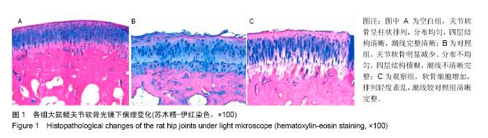
2.1 软骨组织苏木精-伊红染色倒置显微镜观察结果 空白组:大体观察关节软骨表面平滑、完整;显微镜下观察关节软骨呈柱状排列,分布均匀,四层结构清晰,潮线完整清晰。 对照组:大体观察关节软骨表面可见明显的缺损、凹凸不平;显微镜下观察关节软骨明显减少、分布不均匀,四层结构模糊,潮线不清晰完整。 观察组:大体观察关节软骨表面缺损有一定程度的修复;显微镜下观察软骨细胞增加,排列轻度紊乱,潮线的清晰完整程度较对照组改善。 对照组的Mankin’s评分明显高于空白组(P < 0.05);而观察组有所下降,明显低于模型组(P < 0.05),见图1,表1。"
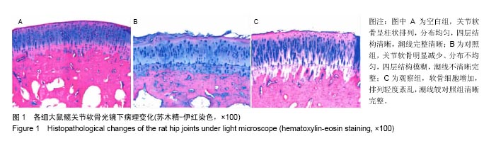

"

2.2 MTT检测结果 与空白组比较,对照组的吸光度值明显降低(P < 0.05),治疗后观察组吸光度值明显升高,与对照组比较,差异有显著性意义(P < 0.05),见图2。"

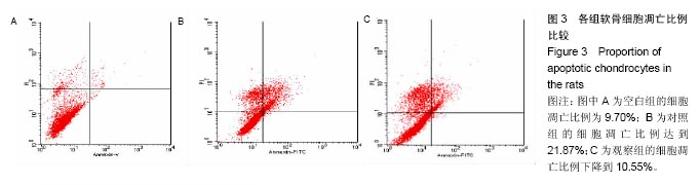
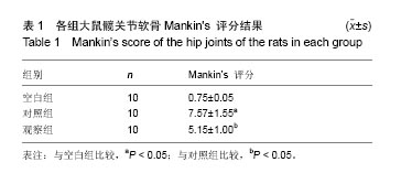
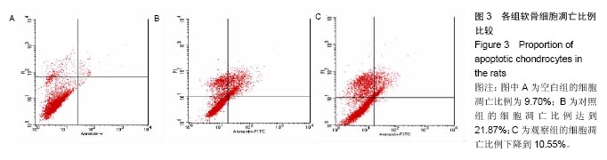
2.3 流式细胞仪检测结果 空白组的软骨细胞凋亡比例为9.70%(图3A),对照组的软骨细胞凋亡比例达到21.87%(图3B),与空白组相比较,对照组软骨细胞凋亡的比例明显升高(P < 0.05)。观察组的软骨细胞凋亡比例下降到10.55%(图3C)。"
| [1]Sen ES, Clarke SL, Ramanan AV. The child with joint pain in primary care. Best Pract Res Clin Rheumatol. 2014;28(6): 888-906.[2]Chambers HG. Update on neuromuscular disorders in pediatric orthopaedics: Duchenne muscular dystrophy, myelomeningocele, and cerebral palsy. J Pediatr Orthop. 2014;34 Suppl 1:S44-48.[3]Mazloumi SM, Ebrahimzadeh MH, Kachooei AR. Evolution in diagnosis and treatment of Legg-Calve-Perthes disease. Arch Bone Jt Surg. 2014;2(2):86-92. [4]Chaudhry S, Phillips D, Feldman D. Legg-Calvé-Perthes disease: an overview with recent literature. Bull Hosp Jt Dis (2013). 2014;72(1):18-27.[5]Johnson ZI, Shapiro IM, Risbud MV. Extracellular osmolarity regulates matrix homeostasis in the intervertebral disc and articular cartilage: evolving role of TonEBP. Matrix Biol. 2014; 40:10-16. [6]Demoor M, Ollitrault D, Gomez-Leduc T, et al. Cartilage tissue engineering: molecular control of chondrocyte differentiation for proper cartilage matrix reconstruction. Biochim Biophys Acta. 2014;1840(8):2414-2440.[7]O'Conor CJ, Case N, Guilak F. Mechanical regulation of chondrogenesis. Stem Cell Res Ther. 2013;4(4):61. [8]王永明,王峰,邵婷,等. MSCs复合双相磷酸钙陶瓷促进腰椎骨折患者脊柱融合[J]. 现代生物医学进展, 2015,15(33): 6484-6486.[9]傅淑平,张荣华.不同浓度梓醇对SD大鼠骨髓间充质干细胞增殖及骨向分化的影响[J]. 时珍国医国药, 2012,23(10): 2398-2400.[10]Liu XX, Li XH, Zhou JT. Experimental study on replicating knee osteoarthritis by modified Hulth's modeling method. Zhongguo Zhong Xi Yi Jie He Za Zhi. 2005;25(12): 1104-1108.[11]Gerlier D, Thomasset N. Use of MTT colorimetric assay to measure cell activation. J Immunol Methods. 1986;94(1-2): 57-63.[12]McCloskey TW, Oyaizu N, Coronesi M, et al. Use of a flow cytometric assay to quantitate apoptosis in human lymphocytes. Clin Immunol Immunopathol. 1994;71(1): 14-18.[13]van der Sluijs JA, Geesink RG, van der Linden AJ, et al. The reliability of the Mankin score for osteoarthritis. J Orthop Res. 1992;10(1):58-61.[14]Bahmanyar S, Montgomery SM, Weiss RJ, et al. Maternal smoking during pregnancy, other prenatal and perinatal factors, and the risk of Legg-Calvé-Perthes disease. Pediatrics. 2008;122(2):e459-464.[15]Mohammadi H, Mequanint K, Herzog W. Computational aspects in mechanical modeling of the articular cartilage tissue. Proc Inst Mech Eng H. 2013;227(4):402-420.[16]Mabvuure N, Hindocha S, Khan WS. The role of bioreactors in cartilage tissue engineering. Curr Stem Cell Res Ther. 2012;7(4):287-292.[17]Galle J, Bader A, Hepp P, et al. Mesenchymal stem cells in cartilage repair: state of the art and methods to monitor cell growth, differentiation and cartilage regeneration. Curr Med Chem. 2010;17(21):2274-2291.[18]Bao JP, Chen WP, Wu LD. Lubricin: a novel potential biotherapeutic approaches for the treatment of osteoarthritis. Mol Biol Rep. 2011;38(5):2879-2885.[19]Lee HS, Huang GT, Chiang H, et al. Multipotential mesenchymal stem cells from femoral bone marrow near the site of osteonecrosis. Stem Cells. 2003;21(2):190-199.[20]Wieczorek G, Steinhoff C, Schulz R, et al. Gene expression profile of mouse bone marrow stromal cells determined by cDNA microarray analysis. Cell Tissue Res. 2003;311(2): 227-237.[21]Abukawa H, Terai H, Hannouche D, et al. Formation of a mandibular condyle in vitro by tissue engineering. J Bone Joint Surg Am. 2005;87(5):936-944.[22]Jaiswal N, Haynesworth SE, Caplan AI, et al. Osteogenic differentiation of purified, culture-expanded human mesenchymal stem cells in vitro. J Cell Biochem. 1997; 64(2):295-312.[23]刘玉艳. N,O-羧甲基壳聚糖/纳米β-磷酸三钙复合材料的制备及其生物学性能的基础研究[D]. 长春:吉林大学, 2011.[24]Webster TJ, Ergun C, Doremus RH, et al. Specific proteins mediate enhanced osteoblast adhesion on nanophase ceramics. J Biomed Mater Res. 2000;51(3):475-483.[25]Schumacher M, Uhl F, Detsch R, et al. Static and dynamic cultivation of bone marrow stromal cells on biphasic calcium phosphate scaffolds derived from an indirect rapid prototyping technique.J Mater Sci Mater Med. 2010;21(11):3039-3048.[26]Park JC, Oh SY, Lee JS, et al. In vivo bone formation by human alveolar-bone-derived mesenchymal stem cells obtained during implant osteotomy using biphasic calcium phosphate ceramics or Bio-Oss as carriers. J Biomed Mater Res B Appl Biomater. 2016;104(3):515-524.[27]Lobo SE, Glickman R, da Silva WN, et al. Response of stem cells from different origins to biphasic calcium phosphate bioceramics. Cell Tissue Res. 2015;361(2):477-495.[28]Li B, Liao X, Zheng L, et al. Effect of nanostructure on osteoinduction of porous biphasic calcium phosphate ceramics. Acta Biomater. 2012;8(10):3794-3804.[29]Mitri F, Alves G, Fernandes G, et al. Cytocompatibility of porous biphasic calcium phosphate granules with human mesenchymal cells by a multiparametric assay. Artif Organs. 2012;36(6):535-542.[30]王涛,田卫东,刘磊,等.新型多孔纳米双相磷酸钙陶瓷支架体外细胞相容性的实验研究[J].华西口腔医学杂志,2005,23(2): 106-109.[31]Hu J, Yang Z, Zhou Y, et al. Porous biphasic calcium phosphate ceramics coated with nano-hydroxyapatite and seeded with mesenchymal stem cells for reconstruction of radius segmental defects in rabbits. J Mater Sci Mater Med. 2015;26(11):257.[32]Kim DH, Shin KK, Jung JS, et al. The Role of Magnesium Ion Substituted Biphasic Calcium Phosphate Spherical Micro-Scaffolds in Osteogenic Differentiation of Human Adipose Tissue-Derived Mesenchymal Stem Cells. J Nanosci Nanotechnol. 2015;15(8):5520-5523.[33]张晓强.骨髓间充质干细胞复合β-TCP生物陶瓷修复山羊骨软骨缺损的实验研究[D].广州:南方医科大学, 2009.[34]Talakoob S, Joghataei MT, Parivar K, et al. Capability of Cartilage Extract to In Vitro Differentiation of Rat Mesenchymal Stem Cells (MSCs) to Chondrocyte Lineage. Int J Mol Cell Med. 2015;4(1):9-21.[35]陆勇,陈克敏,丁晓毅. 关节软骨磁共振生理性成像临床应用[J]. 国际骨科学杂志,2006,27(4):200-202. [36]郝鹏,裴福兴. 关节软骨损伤后体外培养软骨细胞的功能变化[J]. 中国组织工程研究, 2011, 15(28):5131-5135. |
| [1] | Yao Xiaoling, Peng Jiancheng, Xu Yuerong, Yang Zhidong, Zhang Shuncong. Variable-angle zero-notch anterior interbody fusion system in the treatment of cervical spondylotic myelopathy: 30-month follow-up [J]. Chinese Journal of Tissue Engineering Research, 2022, 26(9): 1377-1382. |
| [2] | Wang Jing, Xiong Shan, Cao Jin, Feng Linwei, Wang Xin. Role and mechanism of interleukin-3 in bone metabolism [J]. Chinese Journal of Tissue Engineering Research, 2022, 26(8): 1260-1265. |
| [3] | Xiao Hao, Liu Jing, Zhou Jun. Research progress of pulsed electromagnetic field in the treatment of postmenopausal osteoporosis [J]. Chinese Journal of Tissue Engineering Research, 2022, 26(8): 1266-1271. |
| [4] | An Weizheng, He Xiao, Ren Shuai, Liu Jianyu. Potential of muscle-derived stem cells in peripheral nerve regeneration [J]. Chinese Journal of Tissue Engineering Research, 2022, 26(7): 1130-1136. |
| [5] | Tian Chuan, Zhu Xiangqing, Yang Zailing, Yan Donghai, Li Ye, Wang Yanying, Yang Yukun, He Jie, Lü Guanke, Cai Xuemin, Shu Liping, He Zhixu, Pan Xinghua. Bone marrow mesenchymal stem cells regulate ovarian aging in macaques [J]. Chinese Journal of Tissue Engineering Research, 2022, 26(7): 985-991. |
| [6] | Hu Wei, Xie Xingqi, Tu Guanjun. Exosomes derived from bone marrow mesenchymal stem cells improve the integrity of the blood-spinal cord barrier after spinal cord injury [J]. Chinese Journal of Tissue Engineering Research, 2022, 26(7): 992-998. |
| [7] | Wen Dandan, Li Qiang, Shen Caiqi, Ji Zhe, Jin Peisheng. Nocardia rubra cell wall skeleton for extemal use improves the viability of adipogenic mesenchymal stem cells and promotes diabetes wound repair [J]. Chinese Journal of Tissue Engineering Research, 2022, 26(7): 1038-1044. |
| [8] | Zhu Bingbing, Deng Jianghua, Chen Jingjing, Mu Xiaoling. Interleukin-8 receptor enhances the migration and adhesion of umbilical cord mesenchymal stem cells to injured endothelium [J]. Chinese Journal of Tissue Engineering Research, 2022, 26(7): 1045-1050. |
| [9] | Cui Xing, Sun Xiaoqi, Zheng Wei, Ma Dexin. Huangqin Decoction regulates autophagy to intervene with intestinal acute graft-versus-host disease in mice [J]. Chinese Journal of Tissue Engineering Research, 2022, 26(7): 1057-1062. |
| [10] | Fang Xiaolei, Leng Jun, Zhang Chen, Liu Huimin, Guo Wen. Systematic evaluation of different therapeutic effects of mesenchymal stem cell transplantation in the treatment of ischemic stroke [J]. Chinese Journal of Tissue Engineering Research, 2022, 26(7): 1085-1092. |
| [11] | Guo Jia, Ding Qionghua, Liu Ze, Lü Siyi, Zhou Quancheng, Gao Yuhua, Bai Chunyu. Biological characteristics and immunoregulation of exosomes derived from mesenchymal stem cells [J]. Chinese Journal of Tissue Engineering Research, 2022, 26(7): 1093-1101. |
| [12] | Zhang Jinglin, Leng Min, Zhu Boheng, Wang Hong. Mechanism and application of stem cell-derived exosomes in promoting diabetic wound healing [J]. Chinese Journal of Tissue Engineering Research, 2022, 26(7): 1113-1118. |
| [13] | Hou Jingying, Guo Tianzhu, Yu Menglei, Long Huibao, Wu Hao. Hypoxia preconditioning targets and downregulates miR-195 and promotes bone marrow mesenchymal stem cell survival and pro-angiogenic potential by activating MALAT1 [J]. Chinese Journal of Tissue Engineering Research, 2022, 26(7): 1005-1011. |
| [14] | Liang Xuezhen, Yang Xi, Li Jiacheng, Luo Di, Xu Bo, Li Gang. Bushen Huoxue capsule regulates osteogenic and adipogenic differentiation of rat bone marrow mesenchymal stem cells via Hedgehog signaling pathway [J]. Chinese Journal of Tissue Engineering Research, 2022, 26(7): 1020-1026. |
| [15] | Chen Xiaoxu, Luo Yaxin, Bi Haoran, Yang Kun. Preparation and application of acellular scaffold in tissue engineering and regenerative medicine [J]. Chinese Journal of Tissue Engineering Research, 2022, 26(4): 591-596. |
| Viewed | ||||||
|
Full text |
|
|||||
|
Abstract |
|
|||||