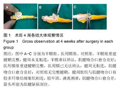
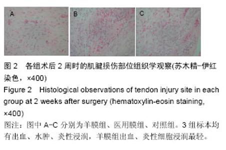
| [1] Khanna A,Friel M,Gougoulias N,et al.Prevention of adhesions insurgery of the fl exor tendons of the hand: what is the evidence? Br Med Bull.2009;90:85-109.[2] 顾玉东.我国手外科进展[J].中华手外科杂志, 1994,10(3): 29-132.[3] 石继祥.促进肌腱愈合及预防肌腱粘连的研究进展[J].中国修复重建外科杂志,2005,19(5): 400-403.[4] 徐梁,曹德君,刘伟,等.体内构建组织工程腱鞘的初步研究[J].组织工程与重建外科杂志,2008,4(6):308-311.[5] 罗静聪,李秀群,杨志明,等.脱细胞羊膜的制备及其生物相容性研究[J].中国修复重建外科杂志, 2004,18(2):108-111.[6] Lundburg G.Experimental flexor tendon healing without adhesion formation-A newconcept of tendon nutrition and intrinsic healing mechanisms.A preliminary report.The Hand.1976;8(3):235.[7] 郭效朋,沈尊理.屏障材料预防肌腱粘连的研究进展[J].现代生物医学进展杂志,2011,11(16): 3179-3182.[8] 高鸣,赵红芳,田德虎,等.人羊膜修复腱鞘缺损的实验研究[J].中国修复重建外科杂志,2013,27(3):335-339.[9] 郝鹏,项舟,罗静聪,等.脱细胞羊膜预防生物衍生肌腱修复鸡屈趾肌腱Ⅱ区缺损修复后的粘连[J].中国组织工程研究与临床康复,2011,15(34):6355-6359.[10] Atik B,Tan O,Dogan A,et al.A new method in tendon repair: angulartechnique ofi nterlocking (ATIK).Ann Hast Surg.2008;60(3):251-253.[11] 林立功,张澜,丰波,等.生物膜预防肌腱粘连的研究进展[J].医学综述,2014,20(14):2531-2534.[12] Hunritz PJ,Weizblit J, HeUer D.Experimental coating of flexortendons with Polyvinyl pyrrelidone to reduce postoperative adhesions. Plast Reconstr Surg. 1985; 76(5) :798-799.[13] Kessler FB.Fascia Patch graft for a digital flexr sheath defect over Primary tendon repair in the chicken.J Hand Surg Am.1986;11:241.[14] 鲍卫汉,朱洪荫.自体筋膜移植物理性状的实验研究[J].中华外科杂志,1987,25(3):601.[15] 汤锦波,侍德.手指屈肌腱鞘的基础和临床研究[J].手外科杂志,1990,6(21):39.[16] Strauch B.The fate of tendon healing after re-storation of theintegrity of the tendon sheath with autogenous vein grafts.J Hand Surg Am.1985;10:790.[17] 潘进社,凌 彤,王学札,等.生物膜修补腱鞘缺损的实验研究[J].中华手外科杂志,1994,10(2):111-113.[18] 邵新中,凌彤,张煜.生物膜防止屈肌腱粘连的实验研究[J].中华外科杂志,1993,31(1):56-59.[19] 范伟杰,杨志明,邓力,等.羊膜的基础和临床应用研究进展[J].中国修复重建外科杂志,2006, 20(1):65-68.[20] Niknejad H,Peirovi H,Jorjani M,et al.Properties of the amnioticmembrane for potential use in tissue engineering.Eur Cell Mater.2008;15:88-99.[21] Gris O,Lopez-Navidad A,Caballero F,et al.Amnio tic mem2brane transp lantation fo r ocular surface patho logy: long2term results.Transplant Proc. 2003;35(5): 203122035.[22] 董丽霞.两种阴道成形术式的临床应用比较[J].中国修复重建外科杂志,2006,20(8):829-831.[23] 潘险峰,张正治.无滑膜肌腱在体滑膜化的实验研究[J].第三军医大学学报,2004,26(3):234-237.[24] Manske PR,Gelbeman RH,Vande Berg JS,et al.Intrinsic flexor tendon repair:a morphological study in vitro.J Bone Joint Surger.1984;66A:385-396.[25] Abrahamsson SO,Lundborg G,Lohmander LS.Tendon healing invivo.An expenrimental model.Scand J Plasteconstr Surg Hand Surg.1989;23(3):199-205. |


