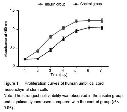
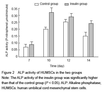
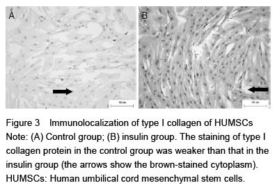
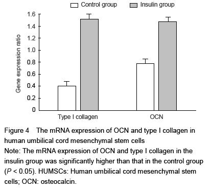
| [1] Nyenwe EA, Jerkins TW, Umpierrez GE, et al. Management of type 2 diabetes: evolving strategies for the treatment of patients with type 2 diabetes. Metabolism. 2011;60(1):1-23.[2] de Waard EA, van Geel TA, Savelberg HH, et al. Increased fracture risk in patients with type 2 diabetes mellitus: an overview of the underlying mechanisms and the usefulness of imaging modalities and fracture risk assessment tools. Maturitas. 2014;79(3):265-274.[3] Pietschmann P, Patsch JM, Schernthaner G. Diabetes and bone. Horm Metab Res. 2010;42(11):763-768.[4] Coe LM, Irwin R, Lippner D, et al. The bone marrow microenvironment contributes to type I diabetes induced osteoblast death. J Cell Physiol. 2011;226(2) 477-483.[5] Motyl KJ, Botolin S, Irwin R, et al. Bone inflammation and altered gene expression with type I diabetes early onset. J Cell Physiol. 2009;218(3):575-583.[6] Harris DT. Umbilical cord tissue mesenchymal stem cells: characterization and clinical applications. Curr Stem Cell Res Ther. 2013;8(5):394-399.[7] Shoji T, Li M, Mifune Y, et al. Local transplantation of human multipotent adipose-derived stem cells accelerates fracture healing via enhanced osteogenesis and angiogenesis. Lab Invest. 2010;90(4):637-649.[8] Mendes SC, Tibbe JM, Veenhof M, et al. Bone tissue-engineered implants using human bone marrow stromal cells: effect of culture conditions and donor age. Tissue Eng. 2002;8(6):911-920.[9] Majore I, Moretti P, Stahl F, et al. Growth and differentiation properties of mesenchymal stromal cell populations derived from whole human umbilical cord. Stem Cell Rev. 2011;7(1):17-31.[10] Park HJ, Bang G, Lee BR, et al. Neuroprotective effect of human mesenchymal stem cells in an animal model of double toxin-induced multiple system atrophy parkinsonism. Cell Transplant. 2011;20(6): 827-835.[11] Wang G, Li Y, Wang Y, et al. Roles of the co-culture of human umbilical cord Wharton’s jelly-derived mesenchymal stem cells with rat pancreatic cells in the treatment of rats with diabetes mellitus. Exp Ther Med. 2014;8(5):1389-1396.[12] Yang CC, Shih YH, Ko MH, et al. Transplantation of human umbilical mesenchymal stem cells from Wharton’s jelly after complete transection of the rat spinal cord. PLoS One. 2008;3(10):e3336.[13] Miranda HC, Herai RH, Thome CH, et al. A quantitative proteomic and transcriptomic comparison of human mesenchymal stem cells from bone marrow and umbilical cord vein. Proteomics. 2012;12(17): 2607-2617.[14] Creecy CM, O’Neill CF, Arulanandam BP, et al. Mesenchymal stem cell osteodifferentiation in response to alternating electric current. Tissue Eng Part A. 2013; 19(3-4):467-474.[15] Kang H, Sung J, Jung HM, et al. Insulin-like growth factor 2 promotes osteogenic cell differentiation in the parthenogenetic murine embryonic stem cells. Tissue Eng Part A. 2012;18(3-4):331-341.[16] Davey GC, Patil SB, O’Loughlin A, et al. Mesenchymal stem cell-based treatment for microvascular and secondary complications of diabetes mellitus. Front Endocrinol (Lausanne). 2014;5:86.[17] Ha C, Tian S, Sun K, et al. Hydrogen sulfide attenuates IL-1beta-induced inflammatory signaling and dysfunction of osteoarthritic chondrocytes. Int J Mol Medint. 2015;35(6):1657-1666.[18] Duya P, Bian Y, Chu X, et al. Stem cells for reprogramming: could hUMSCs be a better choice? Cytotechnology. 2013;65(3):335-345.[19] Marcus AJ, Woodbury D. Fetal stem cells from extra-embryonic tissues: do not discard. J Cell Mol Med. 2008;12(3):730-742.[20] Baksh D, Yao R, Tuan RS. Comparison of proliferative and multilineage differentiation potential of human mesenchymal stem cells derived from umbilical cord and bone marrow. Stem cells. 2007;25(6):1384-1392.[21] Hickman J, McElduff A. Insulin promotes growth of the cultured rat osteosarcoma cell line UMR-106-01: an osteoblast-like cell. Endocrinology. 1989;124(2): 701-706.[22] Kayal RA, Alblowi J, McKenzie E, et al. Diabetes causes the accelerated loss of cartilage during fracture repair which is reversed by insulin treatment. Bone. 2009; 44(2):357-363.[23] Qu Z, Mi S, Fang G. Clinical study on treatment of bone nonunion with MSCs derived from human umbilical cord. Zhongguo Xiu Fu Chong Jian Wai Ke Za Zhi. 2009; 23(3): 345-347.[24] Huang Z, Ren PG, Ma T, et al. Modulating osteogenesis of mesenchymal stem cells by modifying growth factor availability. Cytokine. 2010;51(3):305-310.[25] Li Y, Yu X, Lin S, et al. Insulin-like growth factor 1 enhances the migratory capacity of mesenchymal stem cells. Biochem Biophys Res Commun. 2007;356(3): 780-784.[26] Dillin A, Crawford DK, Kenyon C. Timing requirements for insulin/IGF-1 signaling in C. elegans. Science. 2002; 298(5594):830-834.[27] Wlodarski KH. Properties and origin of osteoblasts. Clin Orthop Relat Res 1990;(252):276-293.[28] Yin H, Cui L, Liu G, et al. Vitreous cryopreservation of tissue engineered bone composed of bone marrow mesenchymal stem cells and partially demineralized bone matrix. Cryobiology. 2009;59(2):180-187.[29] Booth SL, Centi A, Smith SR, et al. The role of osteocalcin in human glucose metabolism: marker or mediator? Nat Rev Endocrinol. 2013;9(1):43-55.[30] Ma Y, Fen LW, Chen H. Effect of insulin and IGF-1on expression of typeⅠcollagen of human osteosarcoma cell line MG63. Zhong Guo Gu Zhi Shu Song Za Zhi. 2006;12(5):466-469 [31] Ma Y, Fen LW, Chen H. The effects of insulin and IGF-1 on expression of bone gla protein of human osteoblast like cell line MG63. Zhong Guo Gu Zhi Shu Song Za Zhi. 2007;13(6):398-401.[32] Gopalakrishnan V, Vignesh RC, Arunakaran J, et al. Effects of glucose and its modulation by insulin and estradiol on BMSC differentiation into osteoblastic lineages. Biochem Cell Biol. 2006;84(1):93-101. |