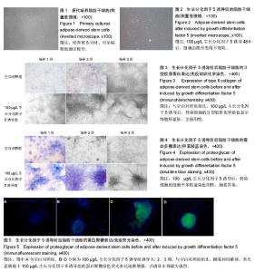
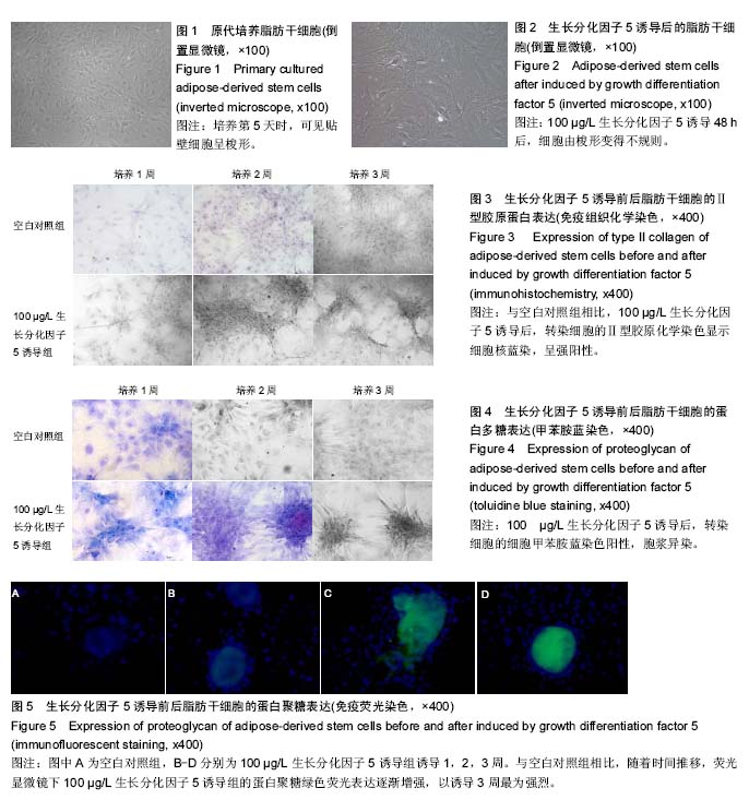
| [1] Murphy MK,Huey DJ,Hu JC,et al.TGF-β1, GDF-5, and BMP-2 stimulation induces chondrogenesis in expanded human articular chondrocytes and marrow-derived stromal cells.Stem Cells.2015;33(3): 762-773. [2] Bigot N,Mouche A,Preti M,et al. Hypoxia Differentially Modulates the Genomic Stability of Clinical-Grade ADSCs and BM-MSCs in Long-Term Culture.Stem Cells.2015;33(12):3608-3620.[3] Suzuki E,Fujita D,Takahashi M,et al.Adipose tissue-derived stem cells as a therapeutic tool for cardiovascular disease.World J Cardiol.2015; 7(8): 454-465.[4] Daniunaite K,Serenaite I,Misgirdaite R,et al.Epigenetic regulation of human adipose-derived stem cells differentiation.Mol Cell Biochem.2015;410(1-2):111-120.[5] Tsai SY,Huang YC,Chueh LL,et al.Intra-articular transplantation of porcine adipose-derived stem cells for the treatment of canine osteoarthritis: A pilot study.World J Transplant.2014;4(3):196-205.[6] Kleinschmidt K,Ploeger F, Nickel J, et al. Enhanced reconstruction of long bone architecture by a growth factor mutant combining positive features of GDF-5 and BMP-2. Biomaterials.2013;34(24):5926-5936.[7] Enochson L,Stenberg J,Brittberg M,et al.GDF5 reduces MMP13 expression in human chondrocytes via DKK1 mediated canonical Wnt signaling inhibition.Osteoarthritis Cartilage.2014;22(4):566-577.[8] Feng C,Liu H,Yang Y,et al. Growth and differentiation factor-5 contributes to the structural and functional maintenance of the intervertebral disc.Cell Physiol Biochem.2015;35(1):1-16. [9] Coleman CM,Vaughan EE,Browe DC,et al.Growth differentiation factor-5 enhances in vitro mesenchymal stromal cell chondrogenesis and Hypertrophy.Stem Cells Dev.2013;22(13):1968-1976. [10] 席彦东,李双,徐钧,等.GDF-5对大鼠肌腱细胞及腱鞘滑膜细胞增殖及凋亡的影响[J].华中科技大学学报(医学版), 2009,38(6):800-811.[11] Mayer JE,Iatridis JC,Chan D,et al.Genetic polymorphisms associated with intervertebral disc degeneration.Spine J.2013;13(3):299-317.[12] Jin L,Li X.Growth differentiation factor 5 regulation in bone regeneration.Curr Pharm Des.2013;19(19): 3364-3373.[13] Karimi H,Soudmand A,Orouji Z,et al.Burn wound healing with injection of adipose-derived stem cells: a mouse model study.Ann Burns Fire Disasters. 2014; 27(1):44-49.[14] Yang X,Shang H,Katz A,et al.A modified aggregate culture for chondrogenesis of human adipose-derived stem cells genetically modified with growth and differentiation factor 5.Biores Open Access.2013;2(4): 258-265. [15] Yang X, Li XD.Nucleus pulposus tissue engineering: a brief review.Eur Spine J.2009;18(11):1564-1572.[16] Osório C,Chacón PJ,Kisiswa L,et al.Growth differentiation factor 5 is a key physiological regulator of dendrite growth during development.Development. 2013;140(23):4751-4762.[17] 张长春,陆泉秀,周新社,等.生长分化因子-5及碱性成纤维细胞生长因子诱导家兔椎间盘髓核细胞成骨作用的研究[J].中国组织化学与细胞化学杂志,2014,23(3):227-232. [18] 林智军,刘航涛,王万明.椎间盘内注射生长分化因子5及成骨蛋白1对椎间盘退变影响的研究进展[J].中国修复重建外科杂志,2008,22(4):435-438.[19] Myers TJ,Granero-Molto F,Longobardi L,et al. Mesenchymal Stem Cells at the Intersection of Cell and Gene Therapy.Expert Opin Biol Ther.2010;10(12): 1663-1679.[20] Chen YL,Sun CK,Tsai TH,et al.Adipose-derived mesenchymal stem cells embedded in platelet-rich fibrin scaffolds promote angiogenesis, preserve heart function, and reduce left ventricular remodeling in rat acute myocardial infarction.Am J Transl Res.2015; 7(5): 781-803. [21] Kim JY,Kim MR,Kim SJ.Modulation of osteoblastic/odontoblastic differentiation of adult mesenchymal stem cells through gene introduction: a brief review.J Korean Assoc Oral Maxillofac Surg.2013; 39(2):55-62.[22] Zuk PA,Zhu M,Ashjian P,et al.Human adipose tissue is a source of multipotent stem cells.Mol Biol Cell.2002; 13(12):4279-4295.[23] Abdel Aziz Aly L,El-Menoufy H,Ragae A,et al.Adipose Stem Cells as Alternatives for Bone Marrow Mesenchymal Stem Cells in Oral Ulcer Healing.Int J Stem Cells.2012;5(2):104-114.[24] Gao S,Zheng Y,Cai Q,et al.Different methods for inducing adipose-derived stem cells to differentiate into Schwann-like cells.Arch Med Sci.2015;11(4):886-892.[25] Zhu P,Liu J,Shi J,et al.Melatonin protects ADSCs from ROS and enhances their therapeutic potency in a rat model of myocardial infarction.J Cell Mol Med.2015; 19(9):2232-2243.[26] Zhang Y,Xu L,Wang S,et al.Concise Review: Differentiation of Human Adult Stem Cells Into Hepatocyte-like Cells In vitro.Int J Stem Cells.2014; 7(2):49-54.[27] Yan Y,Ma T,Gong K,et al.Adipose-derived mesenchymal stem cell transplantation promotes adult neurogenesis in the brains of Alzheimer's disease Mice. Neural Regen Res.2014;9(8):798-805.[28] Prins HJ,Braat AK,Gawlitt AD,et al.In vitro induction of alkaline phosphatase levels predicts in vivo bone forming capacity of human bone marrow stromal cells.Stem Cells Res.2013;12(12):428-440 |