[1] KOVAL K J, SANDERS R. The radiologic evaluation of calcaneal fractures. Clin Orthop Relat Res. 1993;(290):41-46.
[2] RODRIGUEZ SR, GARDUNO RB, RAYGOZA CO. Surgical treatment of calcaneal fractures with a special titanium AO plate. Acta Ortopedica Mexicana. 2004; 18(s1):34-38.
[3] TOSOUNIDIS TH, POUNTOS I, GIANNOUDIS PV. Calcaneal fractures//Fracture Reduction and Fixation Techniques.Springer, Cham, 2020:417-426.
[4] JANDOVA S, PAZOUR J. Limited versus Extended Lateral Approach for Osteosynthesis of Calcaneal Fractures - Comparison of Temporal and Dynamic Parameters of the Gait Cycle. Acta Chir Orthop Traumatol Cech. 2018;85(1):57-61.
[5] XIA S, LU Y, WANG H, et al. Open reduction and internal fixation with conventional plate via L-shaped lateral approach versus internal fixation with percutaneous plate via a sinus tarsi approach for calcaneal fractures-a randomized controlled trial. Int J Surg. 2014;12(5):475-480.
[6] MEHTA CR, AN VVG, PHAN K, et al. Extensile lateral versus sinus tarsi approach for displaced, intra-articular calcaneal fractures:a meta-analysis. J Orthop Surg Res. 2018;13(1):243.
[7] ZHOU H, YU T, REN H, et al. Clinical Comparison of Extensile Lateral Approach and Sinus Tarsi Approach Combined with Medial Distraction Technique for Intra‐Articular Calcaneal Fractures. Orthop Surg. 2017;9(1):77-85.
[8] SU J, CAO X. Risk factors of wound infection after open reduction and internal fixation of calcaneal fractures. Medicine. 2017;96(44):e8411.
[9] 伍凯,林健,黄建华,等.扩大L型切口治疗闭合性跟骨骨折伤口并发症的相关因素分析[J].上海交通大学学报(医学版),2014,34(7):1043-1048.
[10] YEO JH, CHO HJ, LEE KB. Comparison of two surgical approaches for displaced intra-articular calcaneal fractures: sinus tarsi versus extensile lateral approach. BMC Musculoskelet Disord. 2015;16(1):1-7.
[11] LI S. Wound and sural nerve complications of the sinus tarsi approach for calcaneus fractures. Foot Ankle Int. 2018;39(9):1106-1112.
[12] MENG Q, WANG Q, WU X, et al. Clinical application of the sinus tarsi approach in the treatment of intra-articular calcaneal fracture. Medicine. 2018;97(13):e0175.
[13] WEBER M, LEHMANN O, SAGESSER D, et al. Limited open reduction and internal fixation of displaced intra-articular fractures of the calcaneum. Bone Joint Surg Br. 2008;90(12):1608-1616.
[14] COSKUN S,BUYUKCERAN I,PISKIN A. Comparison of Conservative and Surgical Treatment of Displaced Intra-Articular Calcaneal Fractures. 2018.
[15] ASLAN A, SARGIN S, GULCU A, et al.Clinical, radiological and patient-reported outcomes in intra-articular calcaneal fractures:Comparison of conservative and surgical treatment. Joint Dis Relat Surg. 2019;30(2):143-148.
[16] RAMMELT S, AMLANG M, BARTHEL S, et al. Percutaneous treatment of less severe intraarticular calcaneal frctures. Clin Orthop Relat Res. 2010;(468):983-990.
[17] BARRICK B, JOYCE DA, WERNER FW, et al. Effect of Calcaneus Fracture Gap Without Step-Off on Stress Distribution Across the Subtalar Joint. Foot Ankle Int. 2017;38(3):298-303.
[18] 董福,陆春.距下关节后侧入路关节镜辅助下微创治疗跟骨骨折[J].中国重建修复外科,2017,31(1):36-37.
[19] YAO H, LIANG T, XU Y, et al. Sinus tarsi approach versus extensile lateral approach for displaced intra-articular calcaneal fracture:a meta-analysis of current evidence base. J Orthop Surg Res. 2017;12(1):1-9.
[20] FOLK JW, STARR AJ, EARLY JS. Early wound complications of operative treatment of calcaneus fractures: analysis of 190frctures. J Orthop Trauma. 1999;13(5):369-372.
[21] KEENER BJ, SIZENSKY JA. The anatomy of the calcaneus and surrounding structures. Foot Ankle Clin. 2005;10(3):413-424.
[22] GAVLIK JM, RAMMELT S, ZWIPP H. The use of subtalar arthroscopy in open reduction and internal fixation of intra-articular calcaneal fractures. Injury. 2002;33(1):63-71.
[23] DEWALL M, HENDERSON CE, MCKINLEY TO, et al. Percutaneous reduction and fixation of displaced intra-articular calcaneus fractures. J Orthop Trauma. 2010;24:466-472.
[24] 俞光荣,周家钤,燕晓宇,等.距下关节镜辅助下闭合复位经皮螺钉内固定治疗跟骨关节内骨折[J].中华骨科杂志,2006,26(2):73-75.
[25] 张雁儒.关节镜辅助下有限切口内固定治疗Sanders Ⅱ-Ⅲ型跟骨关节内骨折[J].中国矫形外科杂志,2019,27(12):1141-1143.
[26] 郝巍琳,周家悦.关节镜辅助下复位空心螺钉内固定治疗跟骨骨折的临床效果[J].临床医学研究与实践,2019,4(13):96-97.
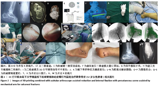
|